Loading subject page…


37,301 views

Video Summary: What are Structural Joints Cartilaginous Joints
Did you know that your ribcage can expand during breathing thanks to specialized cartilaginous joints anatomy types that connect your ribs to your sternum? Cartilaginous joints in anatomy represent a unique class of connections where bones unite through cartilage rather than bone tissue, creating structures that balance stability with controlled movement. A prime example is the pubic symphysis in the human pelvis, which slightly separates during childbirth to accommodate delivery. Watch the full video on JoVE Coach to master this concept with expert-led visuals and step-by-step explanations.
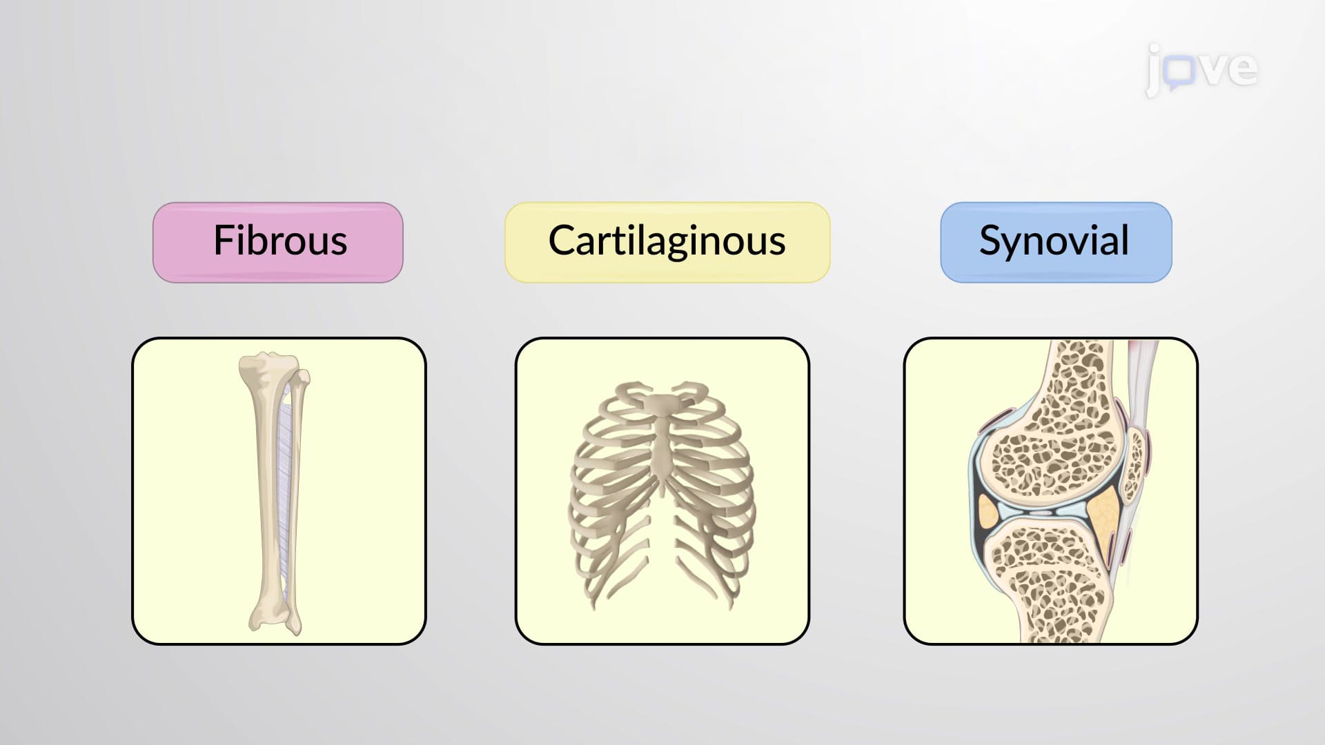
Cartilaginous joints represent a fascinating compromise between stability and mobility in human anatomy. Unlike synovial joints with their fluid-filled cavities or fibrous joints with direct bone-to-bone connections, cartilaginous joints use cartilage as an intermediary tissue. This design creates joints that resist dislocation while permitting limited, controlled movement essential for functions like breathing and childbirth.
The absence of a synovial cavity distinguishes these joints from more mobile articulations. Instead, the cartilaginous material acts as both connector and shock absorber, distributing mechanical forces across the joint surface. This arrangement proves particularly valuable in weight-bearing areas and regions requiring structural integrity with slight flexibility.
Synchondroses utilize hyaline cartilage, the body's most common cartilage type, to unite bones. These joints typically exhibit minimal to no movement, earning classification as synarthrotic joints. The smooth, glassy appearance of hyaline cartilage provides excellent compression resistance while maintaining structural integrity.
Temporary synchondroses play crucial roles in bone development. The epiphyseal plates in growing children represent classic examples, where hyaline cartilage separates the bone shaft (diaphysis) from the bone ends (epiphyses). As adolescents complete puberty, these growth plates ossify, permanently fusing the bone segments. This process, monitored through X-rays in pediatric medicine, indicates skeletal maturity.
Permanent synchondroses persist throughout life, such as the connection between the first rib and manubrium (upper sternum). This joint allows slight movement during deep inspiration while maintaining chest wall stability-essential for respiratory mechanics taught in AP Biology and college anatomy courses.
Symphyses employ fibrocartilage, a tougher, more flexible cartilage type containing dense collagen fibers. This composition creates amphiarthrotic joints capable of limited movement while absorbing significant mechanical stress. The fibrocartilaginous disc acts like a biological shock absorber, compressing under load and returning to original shape when pressure releases.
The pubic symphysis exemplifies symphysis function perfectly. During pregnancy, hormones like relaxin soften this joint, allowing pelvic expansion during delivery. This adaptation, studied extensively in obstetrics and tested on MCAT examinations, demonstrates how cartilaginous joints respond to physiological demands.
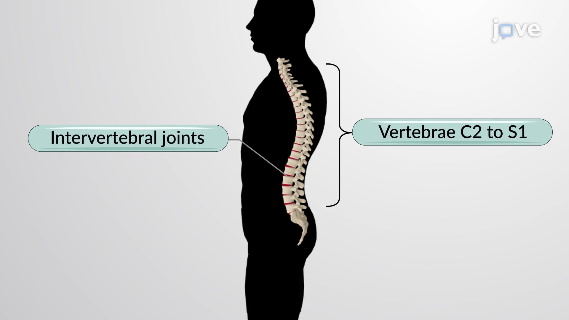
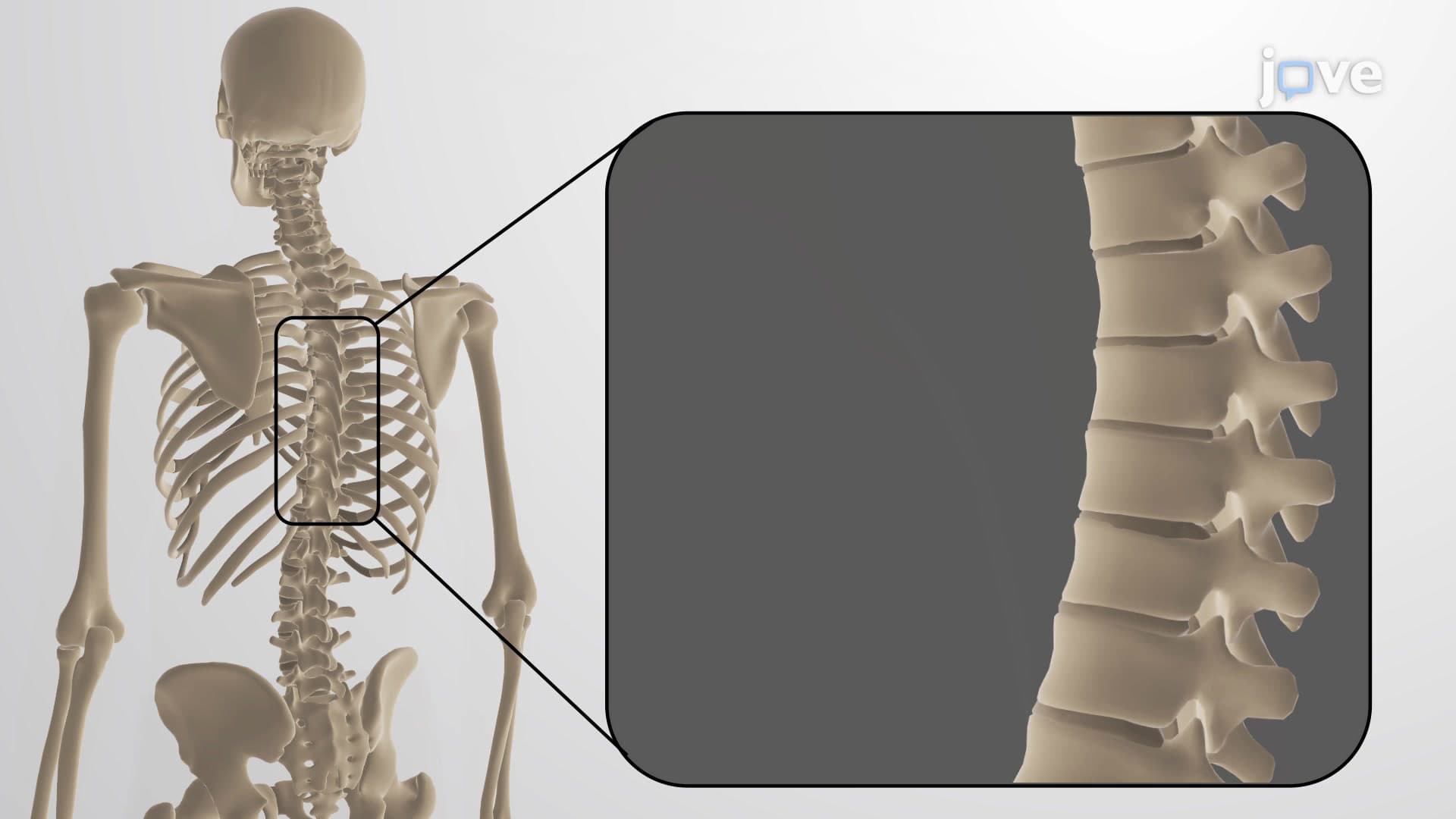
Intervertebral discs represent another symphysis type, though more complex. These structures enable spinal flexibility while supporting body weight-concepts frequently examined in USMLE Step 1 and college biomechanics courses.
Related Micro-courses