Loading subject page…

- Anatomy and Physiology
- The Joints
- Structural Joints: Fibrous Joints
Micro-course: The Joints
37,300 views
Learn key concepts and earn your certificate.




Structural Joints: Fibrous Joints

00:00
01:03
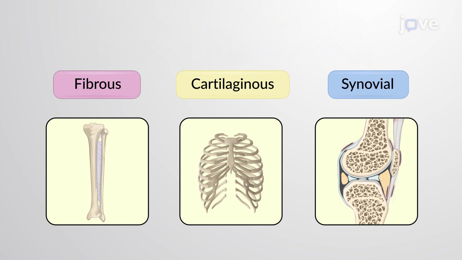
Video Summary: What are Structural Joints Fibrous Joints
Did you know that your skull contains over 20 immovable joints that protect your brain like interlocking puzzle pieces? Fibrous joints anatomy types represent one of the body's most fascinating structural adaptations, connecting bones through dense connective tissue without any joint cavity. From the sutures in a newborn's skull that allow for brain growth to the periodontal ligaments anchoring your teeth during orthodontic treatment, What are Structural Joints Fibrous Joints demonstrates how form follows function in human anatomy. Watch the full video on JoVE Coach to master this concept with expert-led visuals and step-by-step explanations.
- Understand What are Structural Joints Fibrous Joints and their role in connecting bones through dense fibrous connective tissue without synovial cavities
- Identify the three distinct types of fibrous joints: syndesmoses, sutures, and gomphoses, and their specific anatomical locations
- Analyze how ligament length determines mobility in syndesmoses, from slightly movable tibiofibular joints to flexible interosseous membranes
- Compare the structural differences between immovable sutures in the skull versus movable syndesmoses in the forearm
- Evaluate the functional significance of gomphoses in dental health and how periodontal ligaments maintain tooth stability
- Apply knowledge of fibrous joint classification to identify joint types in radiographic images and anatomical specimens
- Recognize clinical implications of fibrous joint dysfunction, including craniosynostosis and periodontal disease
- Explore the relationship between collagen fiber arrangement and joint mobility across different fibrous joint types
What are Structural Joints Fibrous Joints: Foundation of Skeletal Stability
Fibrous joints anatomy types represent a crucial category of articulations where bones unite through dense fibrous connective tissue, creating connections without synovial cavities or joint spaces. Unlike synovial joints that prioritize movement, fibrous joints emphasize structural integrity and protection. These joints contain predominantly Type I collagen fibers arranged in specific patterns that determine their mechanical properties and functional capabilities.
The absence of a synovial cavity means these joints lack the lubricating synovial fluid found in movable joints. Instead, the fibrous connective tissue itself serves as both the connecting medium and the limiting factor for any potential movement. This design principle appears throughout the human body wherever stability takes precedence over mobility.
Syndesmoses: The Flexible Stabilizers
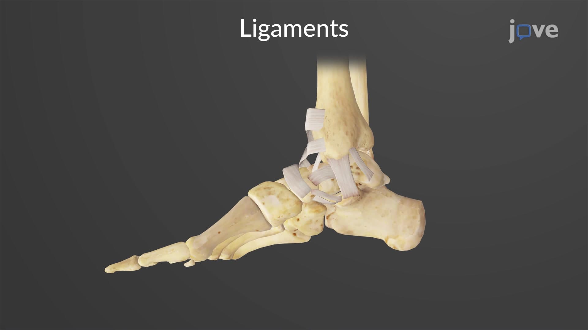
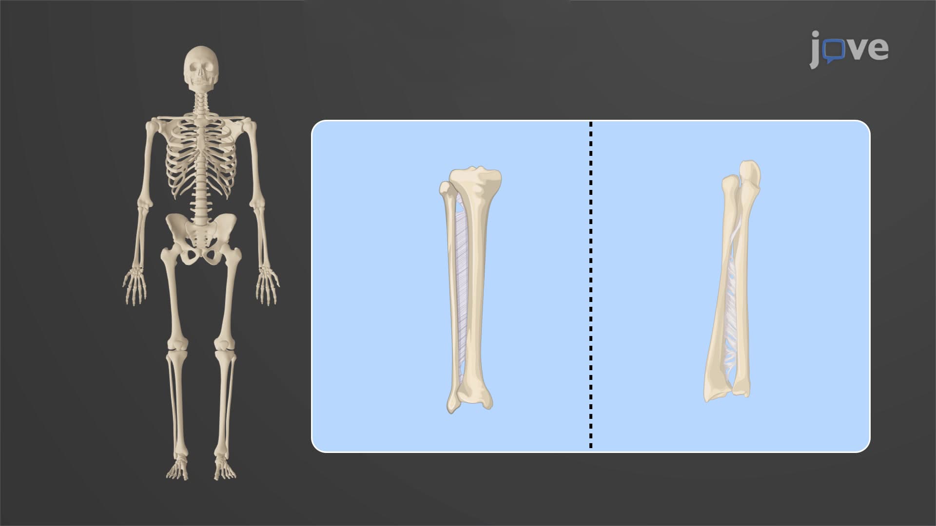
Syndesmoses represent the most mobile category within fibrous joint classification explained. These joints connect bones through ligaments or interosseous membranes, with movement potential directly proportional to ligament length. The classic example involves the distal tibiofibular joint, where ligaments allow slight separation during ankle dorsiflexion-a movement essential for normal walking mechanics.
The radioulnar interosseous membrane provides another excellent illustration. This fibrous sheet connects the radius and ulna throughout their length, permitting pronation and supination while maintaining structural integrity. Medical students studying for the USMLE Step 1 frequently encounter questions about how injuries to this membrane can compromise forearm rotation, particularly in cases of Monteggia or Galeazzi fractures.
Sutures: Cranial Protection Through Interlocking Design
Sutures exemplify suture skull fibrous joint architecture, featuring short collagen fibers that create essentially immovable connections between cranial bones. The serrated edges of sutural bones interlock like jigsaw puzzle pieces, distributing mechanical stress across wide surface areas. This design prevents localized pressure points that could compromise brain protection during impact.
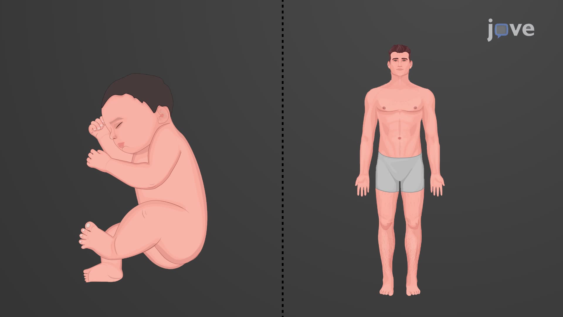
During infancy, sutures remain flexible to accommodate rapid brain growth. The anterior fontanelle doesn't close until 18-24 months, allowing for this critical expansion. Premature suture fusion (craniosynostosis) represents a serious condition requiring surgical intervention, commonly tested on the MCAT and in pediatric medicine curricula.
Gomphoses: Specialized Dental Anchoring Systems
Gomphosis tooth fibrous joints create unique peg-and-socket connections between teeth and their alveolar bone sockets. The periodontal ligament, composed of organized collagen fiber bundles, suspends each tooth root within its bony socket while allowing microscopic movements that facilitate orthodontic treatment and normal mastication forces.
These joints demonstrate remarkable adaptation to functional demands. Heavy chewing forces stimulate periodontal ligament remodeling, while orthodontic forces can gradually move teeth through controlled tissue reorganization. Dental hygiene programs and pre-dental students studying for the DAT examination must understand how bacterial inflammation disrupts this delicate fibrous joint mobility system, leading to periodontitis and tooth loss.
Frequently Asked Questions
Fibrous joints are immovable or slightly movable connections where bones unite through dense fibrous connective tissue without any synovial cavity. They include sutures in the skull, syndesmoses between long bones, and gomphoses anchoring teeth. These joints prioritize stability and protection over movement, using collagen fibers to create strong mechanical connections.
MCAT questions often focus on structure-function relationships, asking students to predict joint mobility based on collagen fiber length or identify joint types from anatomical descriptions. AP Biology emphasizes classification systems and evolutionary adaptations. Both exams may include clinical scenarios involving craniosynostosis, dental health, or fracture patterns affecting interosseous membranes.
Syndesmoses allow slight movement through long ligaments (tibiofibular joint), sutures create immovable skull connections through short fibers, and gomphoses form specialized tooth-socket joints. Remember: longer fibers = more movement. Focus on anatomical locations and functional significance rather than just memorizing definitions for better exam performance.
Emergency room physicians assess skull fractures by understanding sutural anatomy, orthodontists rely on gomphosis flexibility for teeth alignment, and orthopedic surgeons consider syndesmosis integrity in ankle injuries. Physical therapists working with forearm fractures must understand how interosseous membrane damage affects rotation. These concepts directly impact patient care and treatment decisions.
Not at all-basic biology and anatomy knowledge suffices for most educational levels. Understanding collagen as a structural protein and recognizing that "fibrous" means "made of fibers" provides the foundation. High school students can master this topic by focusing on the three types, their locations, and movement capabilities rather than complex biochemical details.
Use location-based mnemonics: "Skull Sutures Stay Still" for immovable sutures, "Syndesmosis Slightly Sways" for moveable syndesmoses, and "Gomphosis Grips teeth" for dental joints. Create visual associations with real body parts you can touch, and practice identifying joint types on anatomical models or diagrams before exams.
Explore synovial joint classification to understand the mobility spectrum, investigate bone remodeling processes affected by mechanical stress, and examine pathological conditions like arthritis affecting different joint types. Understanding embryological joint development also provides valuable context for developmental biology and medical school preparation.
Fractures often occur at or near fibrous joints due to stress concentration patterns. Le Fort facial fractures follow sutural lines, ankle fractures frequently involve the syndesmotic ligaments, and dental trauma affects gomphoses. Understanding normal fibrous joint anatomy helps predict injury mechanisms and guides treatment approaches in orthopedics and oral surgery.
Related Micro-courses

22 Concepts

20 Concepts

26 Concepts