Loading subject page…

- Anatomy and Physiology
- The Joints
- Structural Joints: Synovial Joints
Micro-course: The Joints
37,300 views
Learn key concepts and earn your certificate.



Structural Joints: Synovial Joints


00:00
01:16
Video Summary: What are Structural Joints Synovial Joints
Did you know that every time you swing a baseball bat or bend your knee to climb stairs, you're relying on synovial joints anatomy types working in perfect harmony? These remarkable structures allow bones to move freely against each other, like the ball-and-socket joint in your shoulder that enables a pitcher at Yankee Stadium to throw a 95-mph fastball. Understanding what are structural joints synovial joints reveals the intricate engineering behind human movement, from the protective cartilage to the lubricating fluid that keeps everything running smoothly. Watch the full video on JoVE Coach to master this concept with expert-led visuals and step-by-step explanations.
- Understand what are structural joints synovial joints and identify their role as freely movable articulations in the human skeletal system
- Identify the key anatomical components including articular cartilage, joint capsule, synovial membrane, and synovial fluid
- Analyze how synovial fluid functions as both a lubricant and shock absorber while distributing nutrients to surrounding tissues
- Compare the structural differences between the fibrous outer capsule and the loose inner synovial membrane
- Recognize how capsular ligaments strengthen joints and contain sensory nerves for movement monitoring and pain detection
- Apply knowledge of synovial joint structure to explain common joint injuries and their underlying anatomical causes
- Evaluate the relationship between blood vessel networks and synovial fluid production through plasma filtration
- Explore how different types of synovial joints enable specific movement patterns in various body regions
Understanding What are Structural Joints Synovial Joints
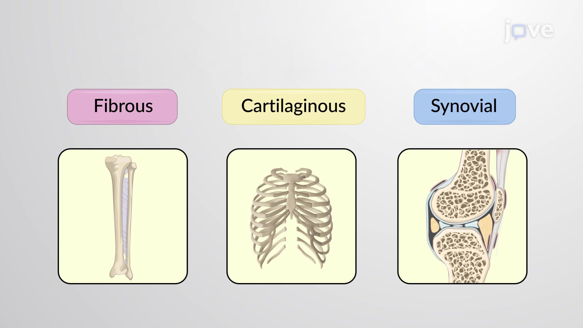
Synovial joints represent the most complex and functionally diverse category of joints in the human body. Unlike fibrous or cartilaginous joints that provide stability with limited movement, synovial joint structure explained reveals a sophisticated system designed for maximum mobility. These joints, classified as diarthroses, enable the fluid movements essential for everything from a basketball player's jump shot at Duke University to a surgeon's precise hand movements during operations at Johns Hopkins Hospital.
Anatomical Components and Synovial Joint Structure Explained
The foundation of every synovial joint begins with articular cartilage, a specialized type of hyaline cartilage that caps the ends of articulating bones. This smooth, glassy surface reduces friction to nearly zero-more efficient than ice on ice. The joint capsule forms a fibrous sleeve around the joint, with its dense outer layer anchoring directly to the periosteum of each bone, creating a sealed environment crucial for joint function.
Within this capsule lies the synovial membrane, a thin tissue layer that serves as the joint's metabolic powerhouse. This membrane contains an extensive network of blood vessels that filter blood plasma to produce synovial fluid-a viscous, egg-white-like substance that fills the joint cavity. Students preparing for the MCAT will recognize this as an example of selective filtration, similar to glomerular filtration in the kidneys but specialized for joint lubrication.
Functional Significance of Synovial Fluid and Joint Mechanics
What are synovial joints and how are they structured becomes clearer when examining their functional design. The synovial fluid serves triple duty: lubricating moving surfaces, absorbing shock during impact (think of a football player's knee hitting the ground), and transporting nutrients to the avascular articular cartilage. This nutrient transport occurs through diffusion, making joint movement essential for cartilage health-a concept frequently tested on AP Biology exams.
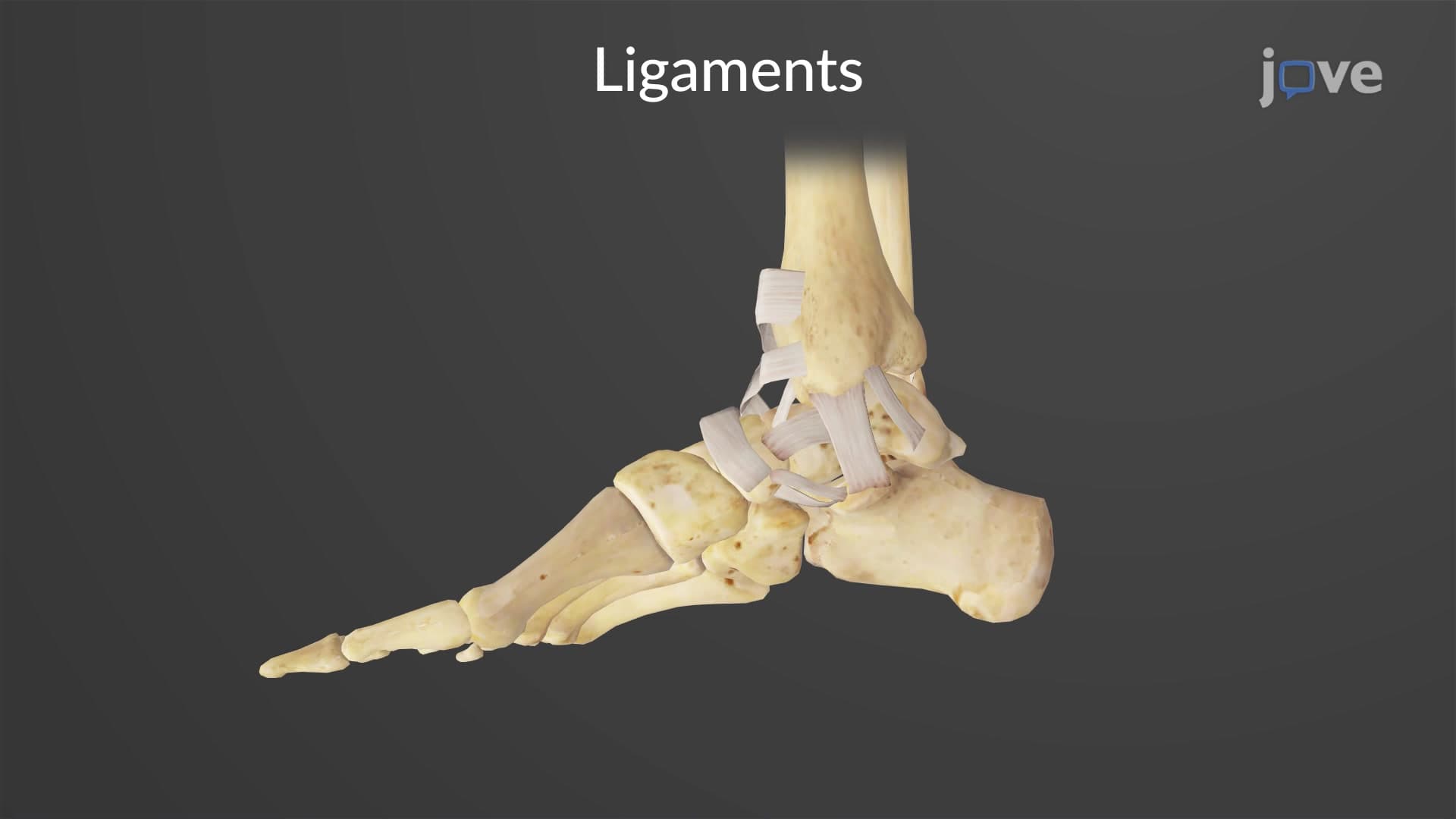
The capsular ligaments provide structural reinforcement while housing sensory nerve endings that create proprioception-your body's awareness of joint position. This sensory feedback prevents injury and enables complex coordinated movements. Physical therapy students at universities like USC and NYU study how damage to these proprioceptive systems affects rehabilitation outcomes.
Clinical Applications and Exam Relevance
Understanding synovial joint anatomy proves essential for students pursuing healthcare careers. NCLEX and HESI A2 exams frequently test knowledge of joint structure when addressing conditions like rheumatoid arthritis, where synovial membrane inflammation leads to joint destruction. Similarly, USMLE Step 1 examines how synovial fluid analysis helps diagnose joint infections or crystal arthropathies like gout.
For students taking college anatomy courses, recognizing the relationship between joint structure and function helps explain why certain movements cause specific injury patterns-knowledge that's invaluable whether you're headed to medical school or pursuing careers in athletic training, physical therapy, or biomedical engineering.
Frequently Asked Questions
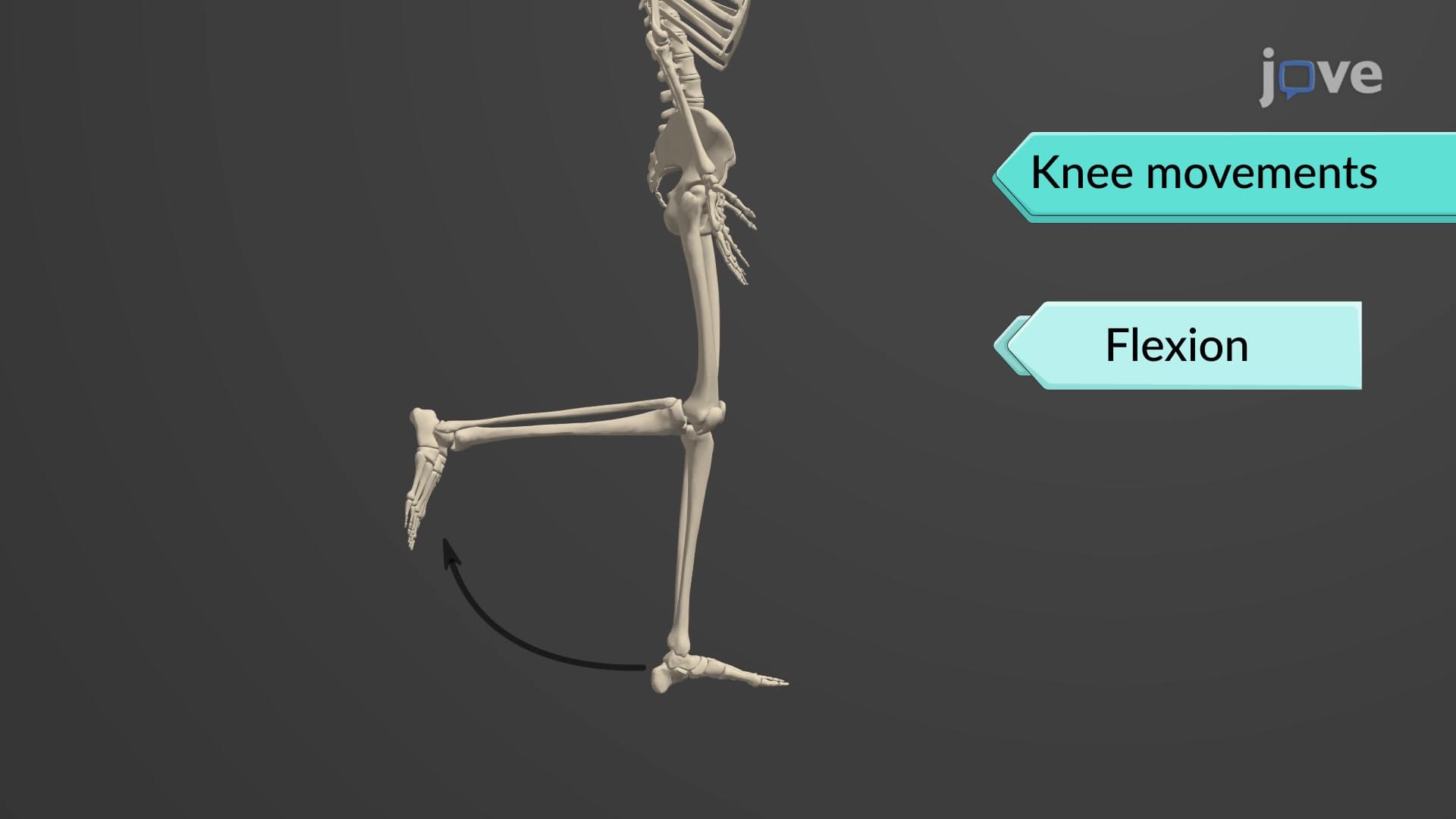
Synovial joints are freely movable articulations characterized by a fluid-filled cavity between bones, unlike immovable fibrous joints or slightly movable cartilaginous joints. They feature articular cartilage, joint capsules, and synovial fluid that enables smooth, friction-free movement. Examples include your knee, shoulder, and hip joints that allow complex movements like rotation, flexion, and extension.
These exams frequently test synovial fluid production through plasma filtration, cartilage nutrition via diffusion, and joint classification systems. You'll encounter questions about how synovial membrane blood vessels filter plasma to create lubricating fluid. Practice identifying joint components in diagrams and explaining how structure relates to function, especially regarding movement types and injury prevention.
NCLEX commonly tests synovial joint knowledge through arthritis care, joint replacement nursing, and mobility assessment scenarios. Questions may ask about synovial fluid analysis in joint infections or how inflammatory conditions affect joint capsules and cartilage. Understanding normal joint structure helps you recognize abnormal findings and appropriate nursing interventions for joint-related conditions.
Professional athletes like NBA players rely on optimal synovial joint function for explosive movements-the synovial fluid in Steph Curry's ankle joints lubricates every pivot and jump shot. Tennis players like Serena Williams depend on shoulder joint synovial fluid for powerful serves, while NFL quarterbacks need perfectly functioning elbow and wrist synovial joints for accurate throws.
Synovial joint concepts are highly accessible because they relate directly to your daily movement experiences. You can feel these joints working when you bend your fingers or rotate your shoulders. Starting with familiar joints like your knee or elbow makes the anatomy more relatable, and the logical structure-function relationships help concepts stick naturally.
Create layered diagrams starting with basic joint capsule structure, then add synovial membrane, cartilage, and ligaments progressively. Use movement-based mnemonics-remember that synovial fluid "flows freely for frictionless function." Practice with unlabeled diagrams and explain each component's role aloud, focusing on how blood vessels create synovial fluid through filtration processes.
Synovial joints integrate with the muscular system for movement generation, nervous system for proprioception and pain sensing, and circulatory system for synovial fluid production and cartilage nutrition. Advanced students explore endocrine effects on joint health, immune system involvement in arthritis, and biomechanical engineering principles that inform joint replacement design.
Lab practicals often require identifying synovial joint components on preserved specimens or models while explaining their functions. Focus on distinguishing articular cartilage's smooth appearance from surrounding bone, locating synovial membrane within joint capsules, and recognizing how capsular ligaments reinforce joint structure. Practice describing the pathway from blood vessel to synovial fluid production.
Related Micro-courses

22 Concepts

20 Concepts

26 Concepts