Loading subject page…

- Anatomy and Physiology
- The Joints
- Development of the Limb Synovial Joints
Micro-course: The Joints
37,300 views
Learn key concepts and earn your certificate.




Development of the Limb Synovial Joints

00:00
01:07
Video Summary: What Is Development Limb Synovial Joints
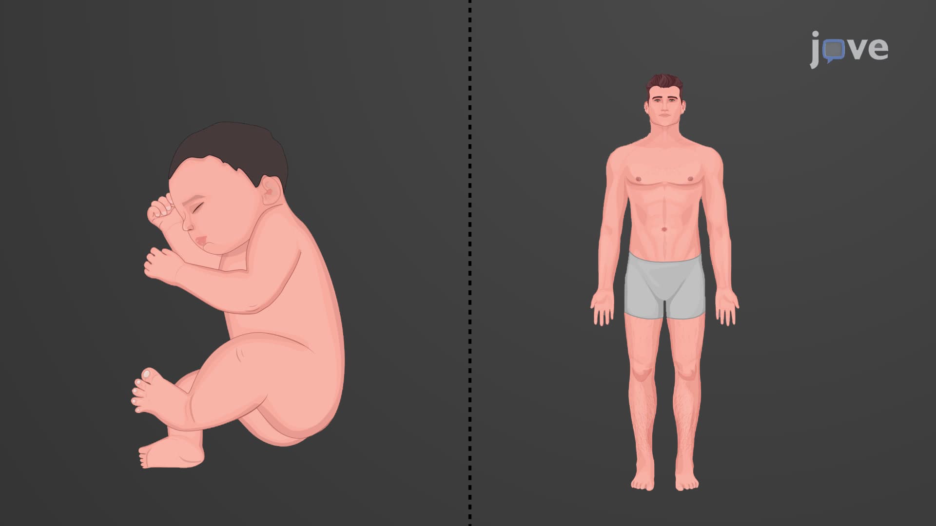
Ever wonder how your shoulder, knee, and elbow joints form before you're even born? Limb synovial joint development begins just four weeks after conception, transforming simple tissue clusters into the complex, movable joints that allow a newborn to grasp their first toy or kick their legs. This intricate process involves precise cellular death, cartilage formation, and bone development that creates the joints essential for human movement. Watch the full video on JoVE Coach to master this concept with expert-led visuals and step-by-step explanations.
- Understand what is development limb synovial joints and the timeline from embryonic limb buds to functional adult-like joints by birth
- Identify the key developmental stages: limb bud formation (4 weeks), cartilaginous modeling (6 weeks), joint cavitation (8-12 weeks), and ossification completion
- Analyze how mesenchymal cell death in the joint interzone creates synovial joint cavities through programmed cell death mechanisms
- Compare the transformation from hyaline cartilage models to mineralized bone through endochondral ossification processes
- Evaluate the role of joint interzone formation in separating adjacent developing bones and establishing future joint boundaries
- Apply knowledge of chondrogenesis and joint cavitation to explain common developmental joint disorders and malformations
- Recognize how supporting structures like articular capsules and ligaments develop from surrounding mesenchymal tissue
- Explore the connection between embryonic joint development and postnatal joint function in human movement
What is Development Limb Synovial Joints: The Foundation of Human Movement
Limb synovial joint development represents one of the most sophisticated processes in human embryology, creating the mobile connections between bones that enable everything from a pianist's finger movements to a basketball player's jump shot. This developmental process transforms simple clusters of embryonic cells into the complex, fluid-filled joints that define human mobility.
Early Limb Bud Formation and Mesenchymal Differentiation
The journey begins at just four weeks of embryonic development when limb buds emerge from mesenchymal tissue on either side of the developing embryo. These paddle-like structures contain multipotent cells that will eventually form bones, joints, muscles, and connective tissues. By the sixth week, these mesenchymal cells undergo chondrogenesis joint formation, differentiating into specialized cartilage-producing cells called chondrocytes.
This transformation creates hyaline cartilage models that serve as templates for future bones. Students preparing for the AP Biology exam should note that this process demonstrates key principles of cellular differentiation and morphogenesis that appear frequently in developmental biology questions.
Joint Interzone Formation and Cavitation Mechanisms
Perhaps the most fascinating aspect of joint development embryology occurs in the spaces between developing cartilaginous bone models. The joint interzone-a specialized region of densely packed mesenchymal cells-forms the boundary between future articulating bones. Within this interzone, a remarkable process called joint interzone cavitation begins around the eighth week.
During cavitation, mesenchymal cells in the center of the interzone undergo programmed cell death (apoptosis), creating the synovial cavity that will eventually fill with lubricating synovial fluid. This process exemplifies how controlled cell death contributes to normal development-a concept that frequently appears on MCAT biology sections and college anatomy exams.
Endochondral Ossification and Structural Maturation
The final phase involves endochondral ossification, beginning around the twelfth week of development. The cartilaginous bone models gradually transform into mineralized bone tissue while preserving the cartilaginous articular surfaces necessary for smooth joint movement. Simultaneously, mesenchymal cells surrounding the joint interzone differentiate into the articular capsule, synovial membrane, and supporting ligaments.
By birth, this process produces synovial joints that closely resemble adult structures in both form and function. This remarkable transformation from simple cell clusters to sophisticated mechanical systems illustrates principles of biomechanics and developmental biology that students encounter in advanced placement courses and undergraduate anatomy programs.
Frequently Asked Questions
Development limb synovial joints refers to the embryonic process that creates movable joints like your knees, elbows, and shoulders. It begins with limb buds at 4 weeks and involves cartilage formation, controlled cell death to create joint spaces, and bone development. By birth, these joints function similarly to adult joints, enabling full range of motion for activities like grasping and kicking.
MCAT biology sections frequently test joint development through questions about cell differentiation, apoptosis in cavitation, and endochondral ossification. Students should understand the timeline from limb buds (4 weeks) to functional joints (birth) and recognize how developmental biology principles apply to joint formation. Practice questions often focus on the role of programmed cell death in creating synovial cavities.
AP Biology emphasizes cellular differentiation during chondrogenesis, the role of apoptosis in joint cavitation, and morphogenesis principles. Students should understand how mesenchymal cells differentiate into specialized tissue types and how cell death contributes to normal development. Free response questions may ask students to explain developmental timelines or compare embryonic and adult joint structures.
Most undergraduate anatomy and developmental biology courses include embryonic joint development as part of musculoskeletal system units. Pre-med students encounter this material in courses like Human Anatomy, Developmental Biology, and Embryology before seeing clinical applications in medical school. The topic often appears on midterm exams alongside bone development and muscle formation concepts.
Orthopedic surgeons and pediatricians use knowledge of normal joint development to diagnose developmental dysplasia, arthrogryposis, and other congenital joint disorders. Understanding normal embryonic development helps clinicians distinguish between developmental variations and pathological conditions. This knowledge guides treatment decisions for conditions like hip dysplasia in newborns at hospitals like Boston Children's Hospital.
Basic high school biology provides sufficient background for understanding joint development concepts. Students need familiarity with cell types, tissue differentiation, and basic embryology terminology. The timeline and key processes can be mastered through careful study of developmental stages and vocabulary. Most students find the visual aspects of development easier to understand than complex molecular mechanisms.
Create timeline diagrams showing development from 4 weeks through birth, including key milestones like limb bud formation, cartilage modeling, and ossification. Practice drawing cross-sections of developing joints and labeling structures like the joint interzone and articular capsule. Use active recall to test your understanding of each developmental stage and its cellular mechanisms.
Explore bone development through intramembranous and endochondral ossification, muscle development and innervation patterns, and postnatal joint growth and remodeling. Consider studying joint pathology, including developmental disorders and degenerative diseases. Understanding normal development provides foundation for appreciating how joints respond to injury, disease, and aging throughout life.
Related Micro-courses

22 Concepts

20 Concepts

26 Concepts