Loading subject page…

- Anatomy and Physiology
- The Joints
- Knee Joint
Micro-course: The Joints
37,300 views
Learn key concepts and earn your certificate.




Knee Joint

00:00
01:23
Video Summary: What Is Knee Joint
Every time you walk, run, or squat, your knee joint anatomy structure performs one of biomechanics' most complex movements. This synovial hinge joint connects your thighbone to your shinbone while allowing flexion, extension, and slight rotation-movements essential for activities from climbing stairs to playing basketball at your local high school gym. Understanding what is knee joint mechanics reveals how three bones, multiple ligaments, and specialized cartilage work together to support your body weight while maintaining mobility. Watch the full video on JoVE Coach to master this concept with expert-led visuals and step-by-step explanations.
- Understand the knee joint as a complex synovial hinge joint connecting the femur, tibia, and patella through multiple articulation points
- Identify the two main joint types within the knee: tibiofemoral joints (hinge movement) and patellofemoral joint (gliding movement)
- Analyze how the cruciate and collateral ligaments work together to stabilize knee movement and prevent excessive motion in multiple planes
- Compare the roles of medial and lateral menisci in shock absorption, weight distribution, and joint protection during physical activity
- Recognize the anatomical features that enable the knee's unique combination of stability and mobility during daily movements
- Evaluate how ligament injuries (ACL, MCL tears) affect joint function and athletic performance
- Apply knowledge of knee anatomy to understand common sports injuries and rehabilitation principles
- Explore the relationship between knee joint structure and movement patterns in human locomotion
Understanding the Knee Joint: A Biomechanical Marvel
The knee joint anatomy structure represents one of the human body's most sophisticated mechanical systems. Unlike simple hinge joints, the knee combines multiple articulations within a single joint capsule, creating a compound joint that balances stability with remarkable mobility. This complexity becomes evident when studying for the MCAT or AP Biology exams, where understanding joint classification and function is essential.
Bone Components and Joint Classifications
The knee's structural foundation involves three primary bones forming distinct articulations. The tibiofemoral patellofemoral joint system includes two tibiofemoral joints where the femur's medial and lateral condyles interact with the tibial plateau, plus one patellofemoral joint where the patella glides along the femur's patellar groove. This arrangement explains why knee joint explained anatomy resources emphasize the joint's compound nature-it's technically three joints functioning as one integrated system.
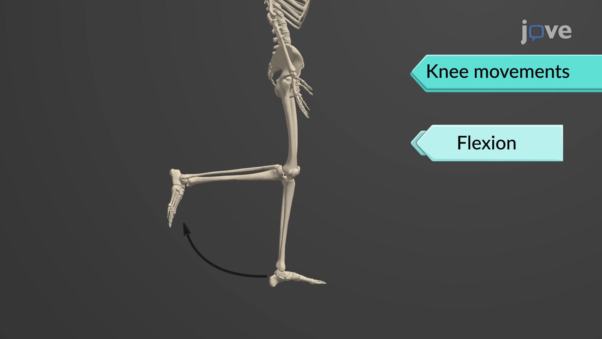
During normal movement, the femoral condyles perform a complex rolling and gliding motion across the tibial plateau. This mechanism allows the 140-degree range of motion necessary for activities like deep squats or climbing bleachers at a football stadium. The patellofemoral component functions as a plane joint, with the patella serving as a mobile fulcrum that increases the quadriceps muscle's mechanical advantage.
Ligamentous Support System
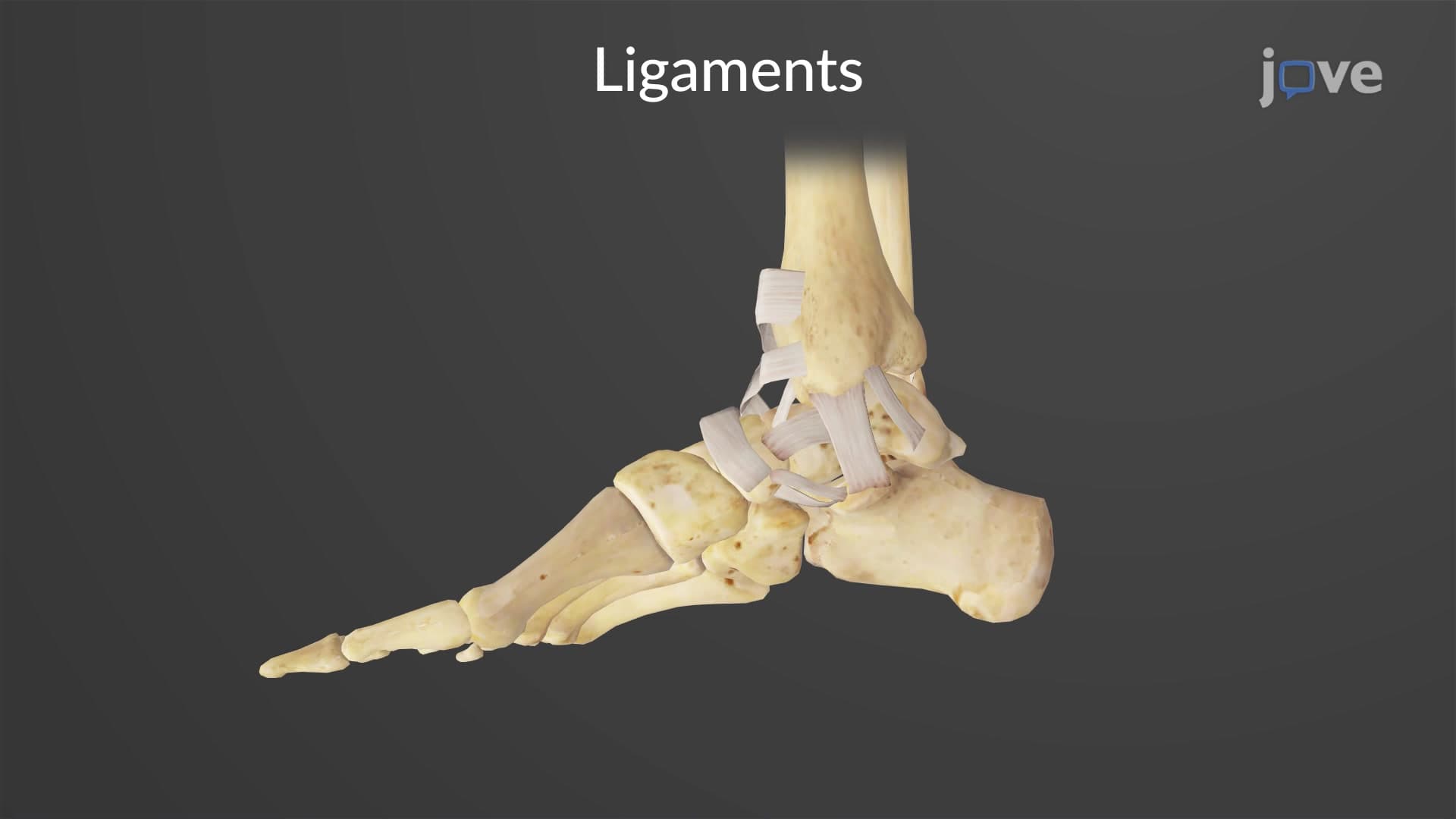
The knee's stability depends on a sophisticated cruciate collateral ligament network. The medial and lateral collateral ligaments provide valgus and varus stability, preventing excessive side-to-side movement that could damage the joint during sports like soccer or basketball. Within the joint capsule, the anterior cruciate ligament (ACL) and posterior cruciate ligament (PCL) cross each other, forming an X-pattern that controls anterior-posterior translation and rotational stability.
Understanding this ligament arrangement proves crucial for college anatomy courses and professional school preparation. The ACL prevents anterior tibial translation and is commonly torn during pivoting sports, while the PCL resists posterior displacement during activities like landing from jumps. These concepts frequently appear on USMLE Step 1 examinations and nursing school entrance exams like the HESI A2.
Meniscal Function and Synovial Knee Joint Cavity
The meniscus cartilage knee structures-medial and lateral menisci-function as biomechanical shock absorbers and load distributors. These crescent-shaped fibrocartilage structures increase the contact area between femur and tibia, reducing peak pressures that could damage articular cartilage. The synovial knee joint cavity produces synovial fluid that lubricates these surfaces and provides nutrients to avascular cartilage structures.
The menisci's unique C-shaped and O-shaped configurations (medial and lateral respectively) reflect their different attachment patterns and injury mechanisms. This anatomical detail becomes clinically relevant when studying orthopedic cases or preparing for clinical rotations in medical school.
Frequently Asked Questions
The knee joint is a compound synovial joint combining hinge and plane joint characteristics, making it unique among body joints. Unlike simple hinge joints such as the elbow, the knee allows flexion, extension, and slight rotation while supporting significant body weight. Its complex structure includes multiple bones (femur, tibia, patella), extensive ligament systems, and specialized cartilage structures that work together to provide both stability and mobility during daily activities.
MCAT questions often focus on joint classification, ligament function, and biomechanical principles of knee movement. AP Biology exams may test synovial joint structure, including the role of synovial fluid and cartilage in joint function. Students should understand the difference between tibiofemoral and patellofemoral articulations, know the four main knee ligaments (ACL, PCL, MCL, LCL), and be able to explain how menisci contribute to joint stability and shock absorption.
College courses typically cover detailed bone landmarks, ligament attachments, and movement analysis in multiple planes. Students learn to identify femoral condyles, tibial plateau features, and patellar groove anatomy on both models and imaging studies. Emphasis is placed on understanding injury mechanisms, particularly ACL tears in athletes, and how anatomical structure relates to clinical presentations commonly seen in orthopedic medicine.
Knee anatomy knowledge directly explains injury patterns seen in American sports like football, basketball, and soccer. ACL tears occur when the ligament cannot control excessive anterior tibial translation during pivoting movements, while meniscal tears result from rotational forces that exceed the cartilage's structural limits. Understanding these mechanisms helps explain why certain movements are high-risk and how rehabilitation protocols target specific anatomical structures.
Knee anatomy is approachable for motivated high school students, especially those taking AP Biology or planning health science careers. The key is connecting anatomical structures to familiar movements like walking, jumping, or cycling. Visual aids, anatomical models, and relating concepts to sports activities make the material more accessible and memorable for students at this level.
Create a systematic approach by first mastering bone identification, then adding ligaments, and finally understanding movement patterns. Use active recall techniques by drawing and labeling knee anatomy from memory, then checking accuracy against reliable sources. Practice identifying structures on both anterior and posterior views, and always connect anatomical features to their functional roles in movement and stability.
Progress to studying knee biomechanics and gait analysis, which builds on anatomical foundations to explain movement patterns. Explore comparative anatomy to understand how human knee structure differs from other mammals, or investigate clinical topics like osteoarthritis and joint replacement surgery. Students interested in sports medicine might examine advanced topics like proprioception and neuromuscular control of knee stability.
Related Micro-courses

22 Concepts

20 Concepts

26 Concepts