Loading subject page…

- Anatomy and Physiology
- The Lymphatic and Immune System
- Antigen Processing Pathways
Micro-course: The Lymphatic and Immune System
218,420 views
Learn key concepts and earn your certificate.




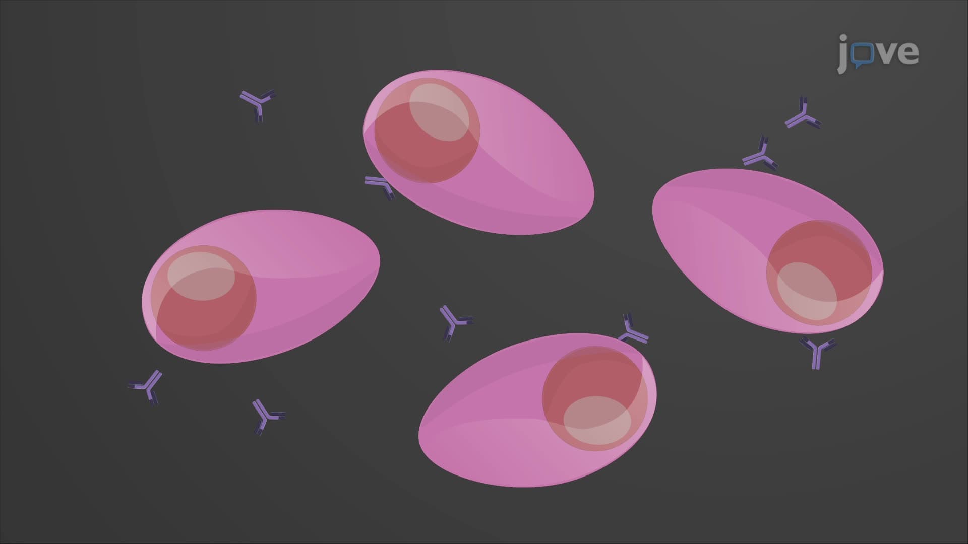

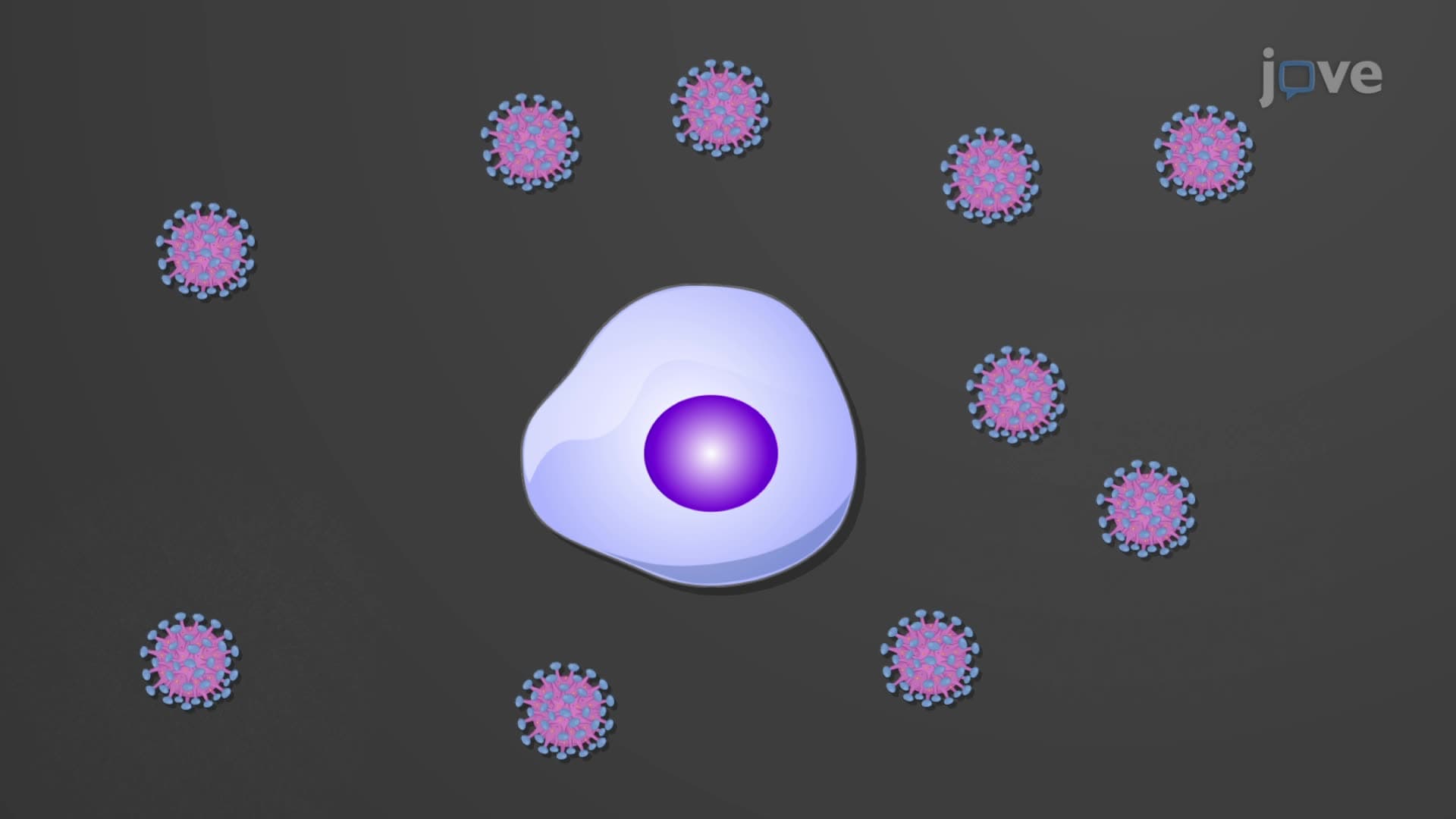
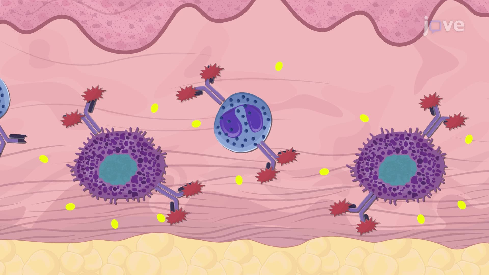
Antigen Processing Pathways


00:00
01:31
Video Summary: What are Antigen Processing Pathways
Ever wonder how your immune system recognizes the difference between a healthy cell and one infected with flu virus? Antigen processing pathways immunity mechanisms enable this crucial distinction by breaking down foreign proteins into recognizable fragments. For instance, when you receive the annual flu vaccine at your local CVS, these pathways help your dendritic cells present viral fragments to activate your immune response. What are antigen processing pathways exactly, and how do they protect you from disease? Watch the full video on JoVE Coach to master this concept with expert-led visuals and step-by-step explanations.
- Understand what are antigen processing pathways and their critical role in immune system function and pathogen recognition
- Identify the two main pathways: endogenous MHC class I pathway for intracellular threats and exogenous MHC class II pathway for extracellular pathogens
- Compare the cellular locations and molecular machinery involved in proteasome antigen processing versus lysosome antigen processing
- Analyze how different antigen-presenting cells utilize MHC antigen processing explained mechanisms to activate specific immune responses
- Recognize the importance of cross presentation antigen mechanisms in viral immunity and cancer surveillance
- Apply knowledge of pathway dysfunction to understand autoimmune diseases and immunodeficiency disorders
- Evaluate how vaccines exploit these natural processing pathways to generate protective immunity
- Explore the clinical significance of these pathways in organ transplant rejection and therapeutic interventions
Understanding What are Antigen Processing Pathways
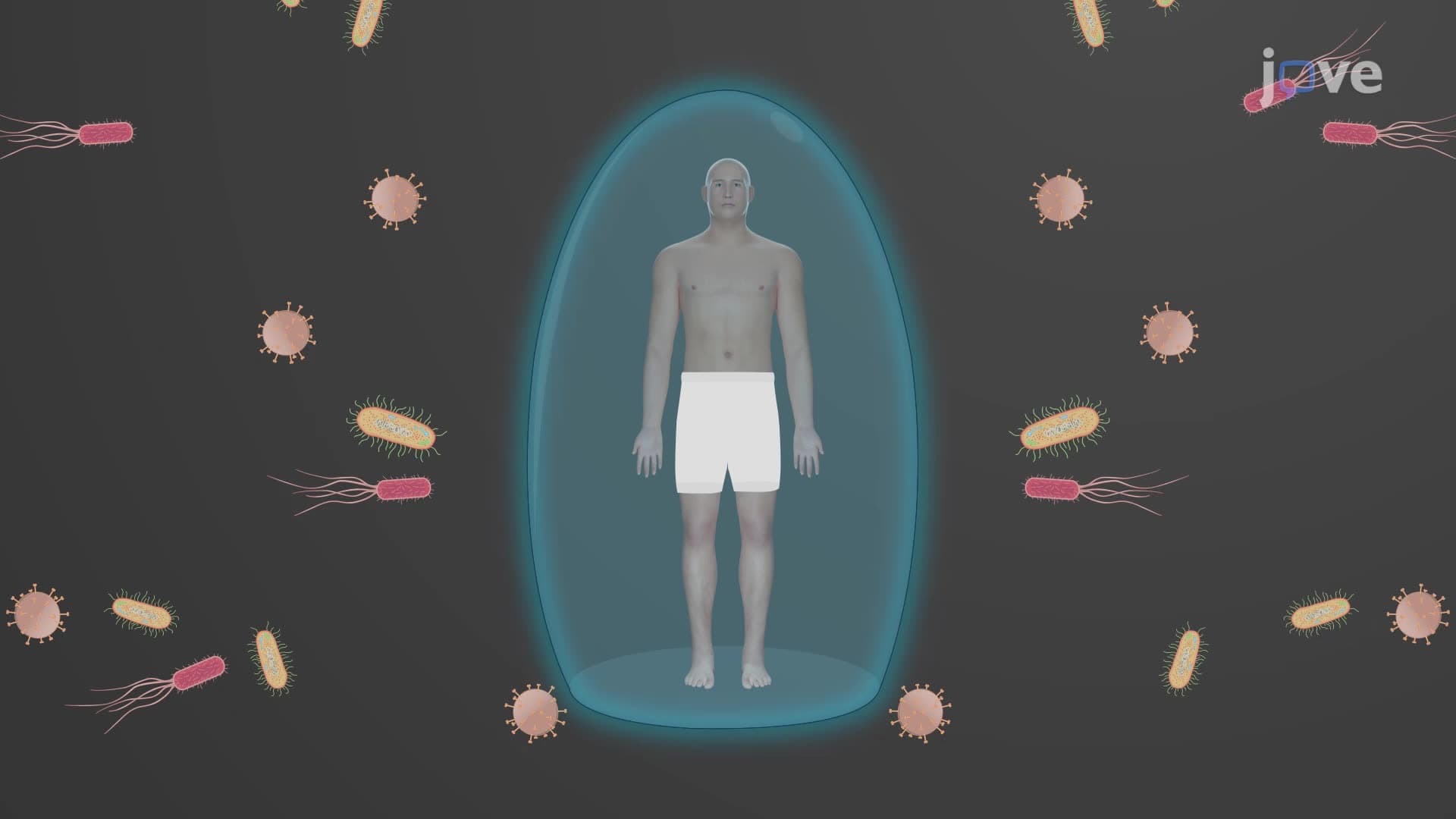
Antigen processing pathways immunity systems represent sophisticated cellular mechanisms that enable our immune system to survey both internal and external threats. These pathways function as molecular quality control systems, continuously sampling proteins from different cellular compartments and presenting suspicious fragments to immune cells for inspection. Think of them as security checkpoints that screen molecular "identification cards" to distinguish friend from foe.
The Endogenous MHC Class I Pathway: Internal Surveillance
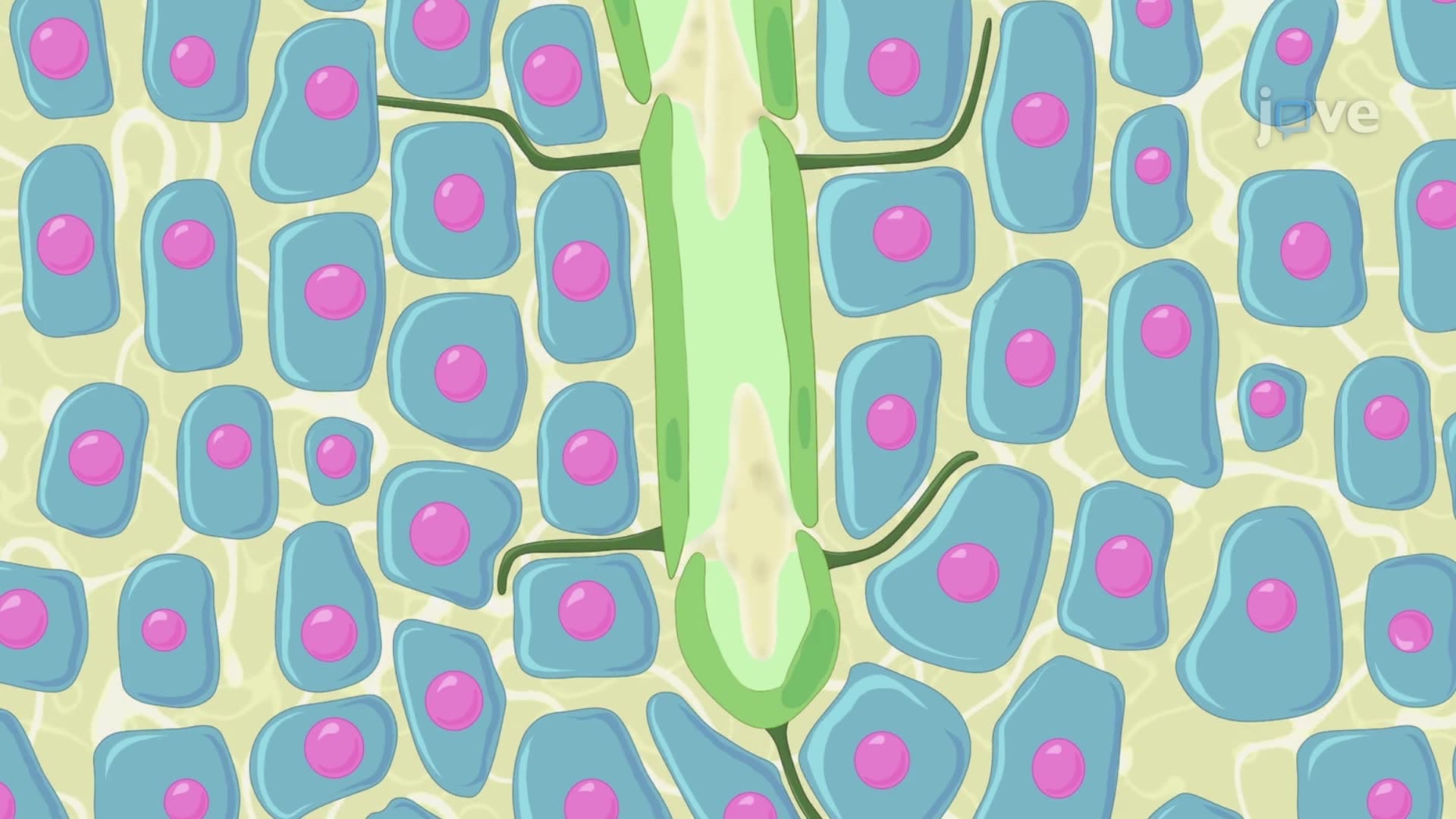
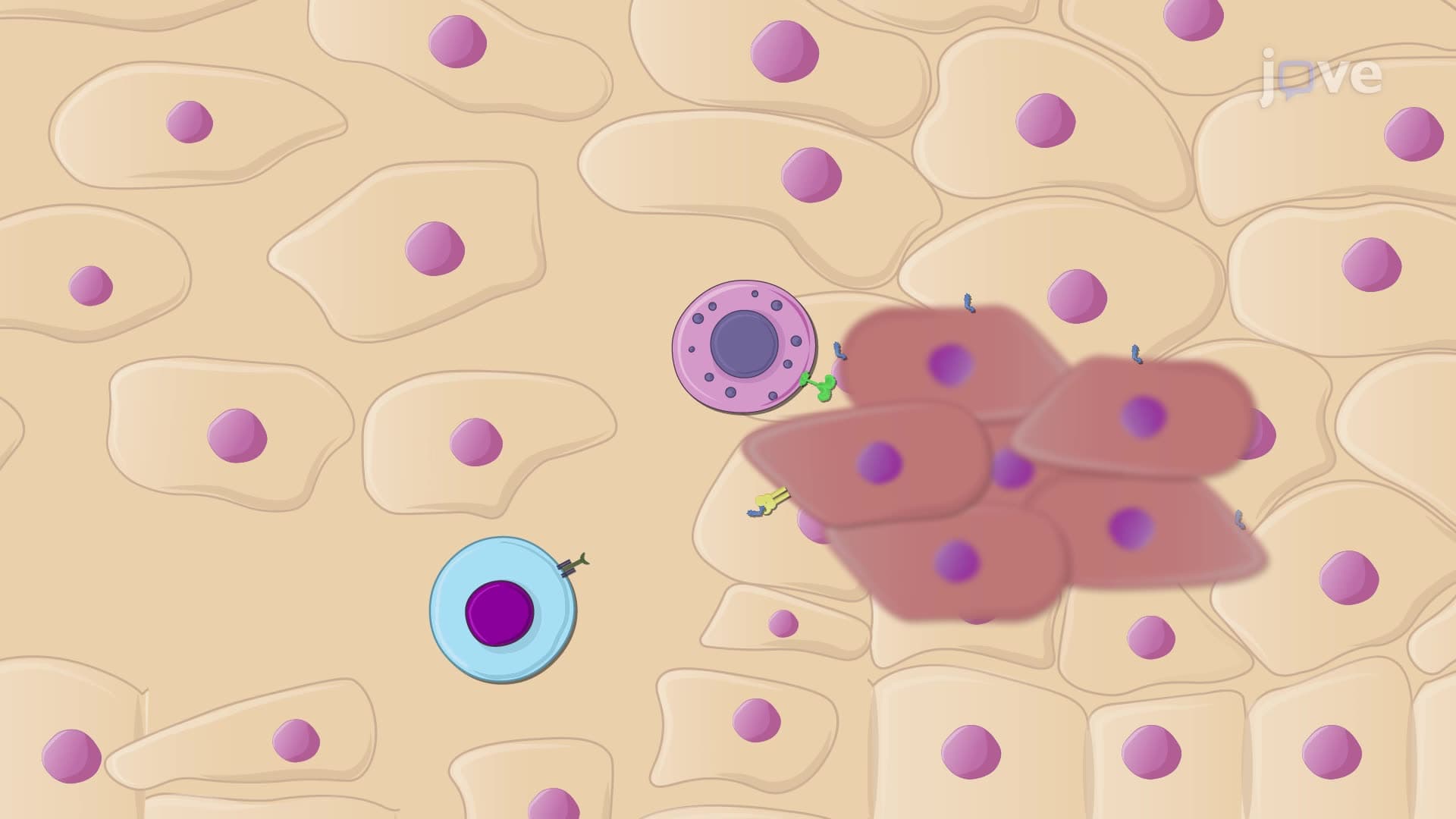
The endogenous MHC class I pathway operates as your body's internal security system, monitoring what's happening inside every nucleated cell. When cells become infected with viruses like influenza or develop into cancer cells, they produce abnormal proteins that trigger this pathway. The proteasome antigen processing machinery acts like a molecular shredder, breaking down these suspicious proteins into 8-11 amino acid fragments called peptides.
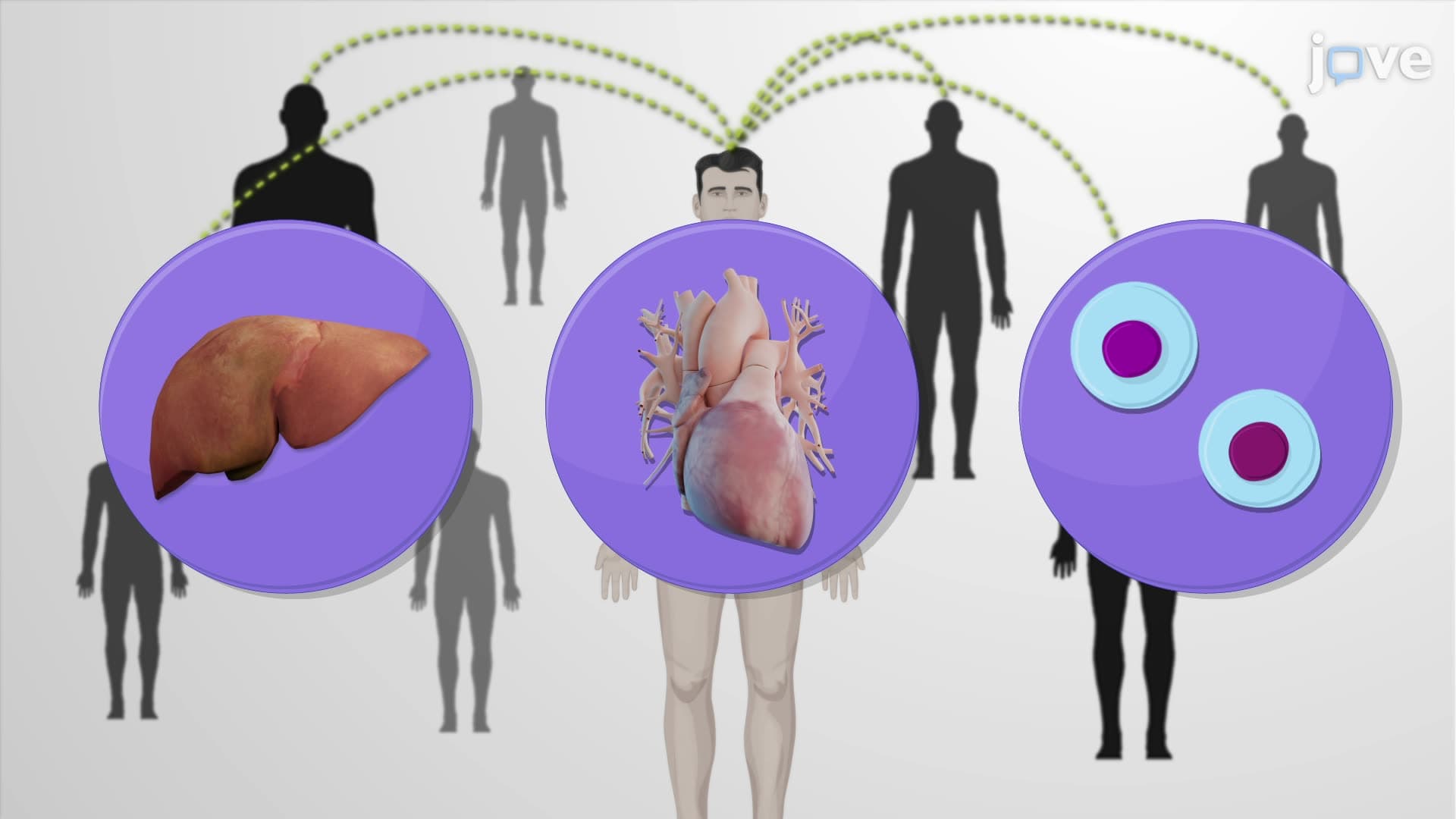
These peptide fragments then travel to the endoplasmic reticulum, where they encounter newly synthesized MHC class I molecules. The peptides bind to these MHC molecules, forming stable complexes that journey to the cell surface. Once displayed on the cell membrane, CD8+ T cells (cytotoxic T lymphocytes) can recognize these foreign peptides and eliminate the compromised cell. This pathway explains why organ transplant patients require immunosuppressive drugs-their immune system recognizes donor tissues as foreign through MHC class I presentation.
The Exogenous MHC Class II Pathway: External Threat Detection
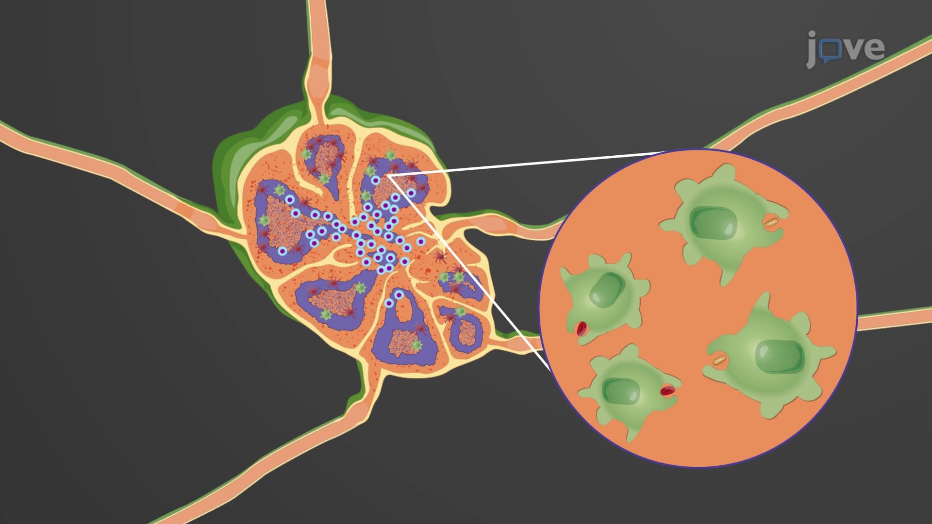
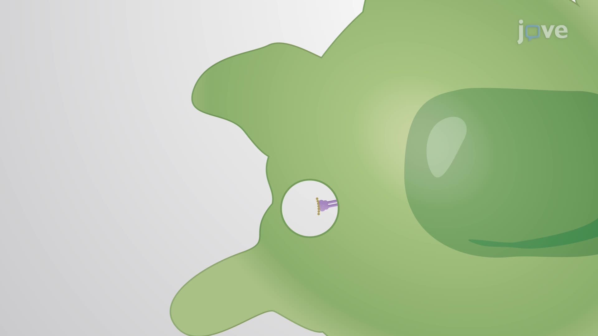
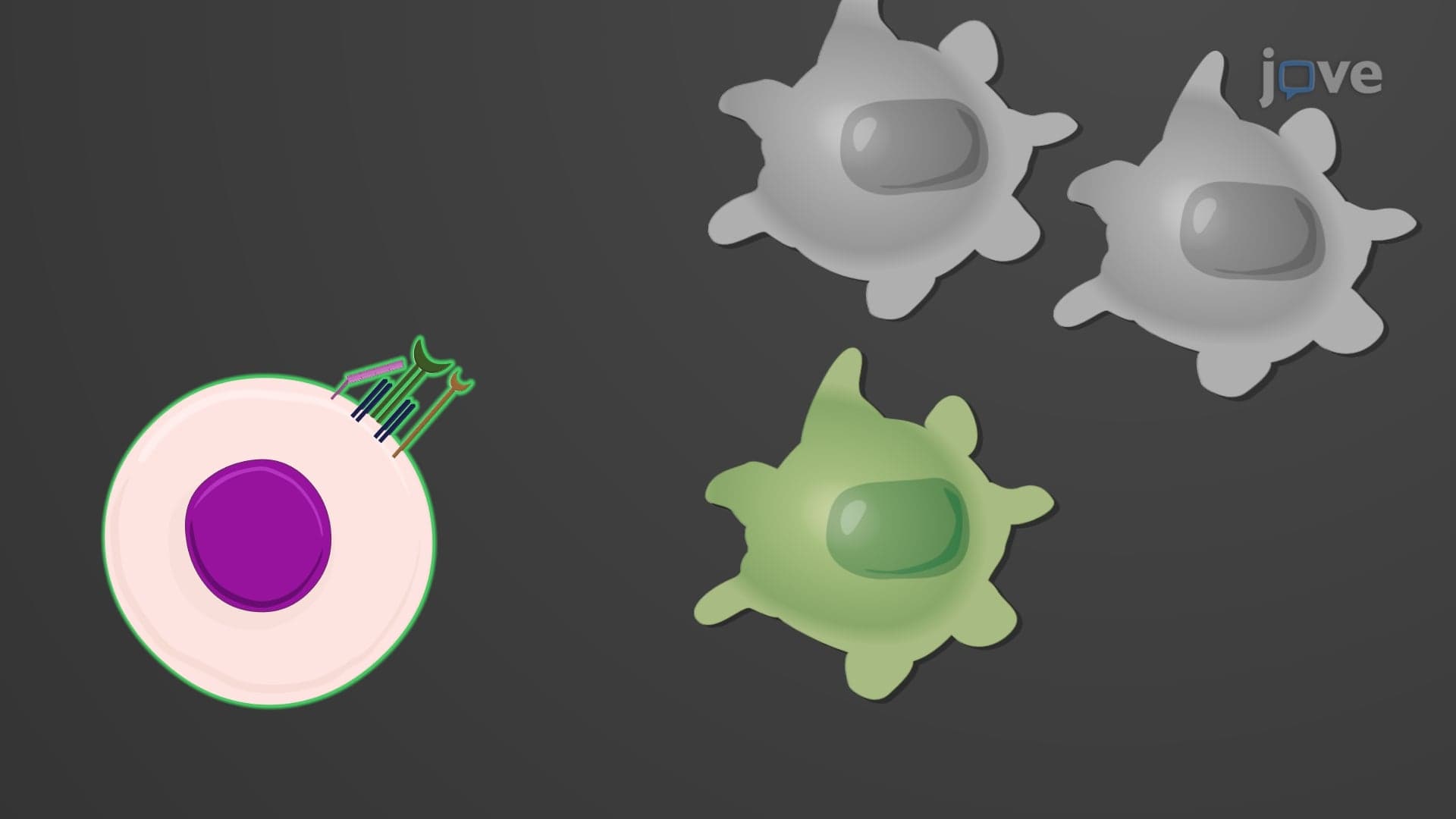
Professional antigen-presenting cells, including dendritic cells found in tissues throughout your body, specialize in the exogenous MHC class II pathway. These cellular sentries patrol tissues, capturing external threats like bacteria, fungi, and other pathogens through phagocytosis. Once internalized, pathogens become trapped in specialized vesicles called phagosomes.
The lysosome antigen processing phase begins when lysosomes fuse with these pathogen-containing vesicles, creating acidic environments rich in proteolytic enzymes. These enzymes dismantle pathogens into peptide fragments of 12-25 amino acids. Meanwhile, MHC class II molecules, synthesized in the endoplasmic reticulum, travel in separate vesicles that eventually fuse with the antigen-containing compartments. The resulting MHC II-peptide complexes migrate to the cell surface, where they present foreign antigens to CD4+ T helper cells, initiating adaptive immune responses.
Clinical Significance and Cross Presentation
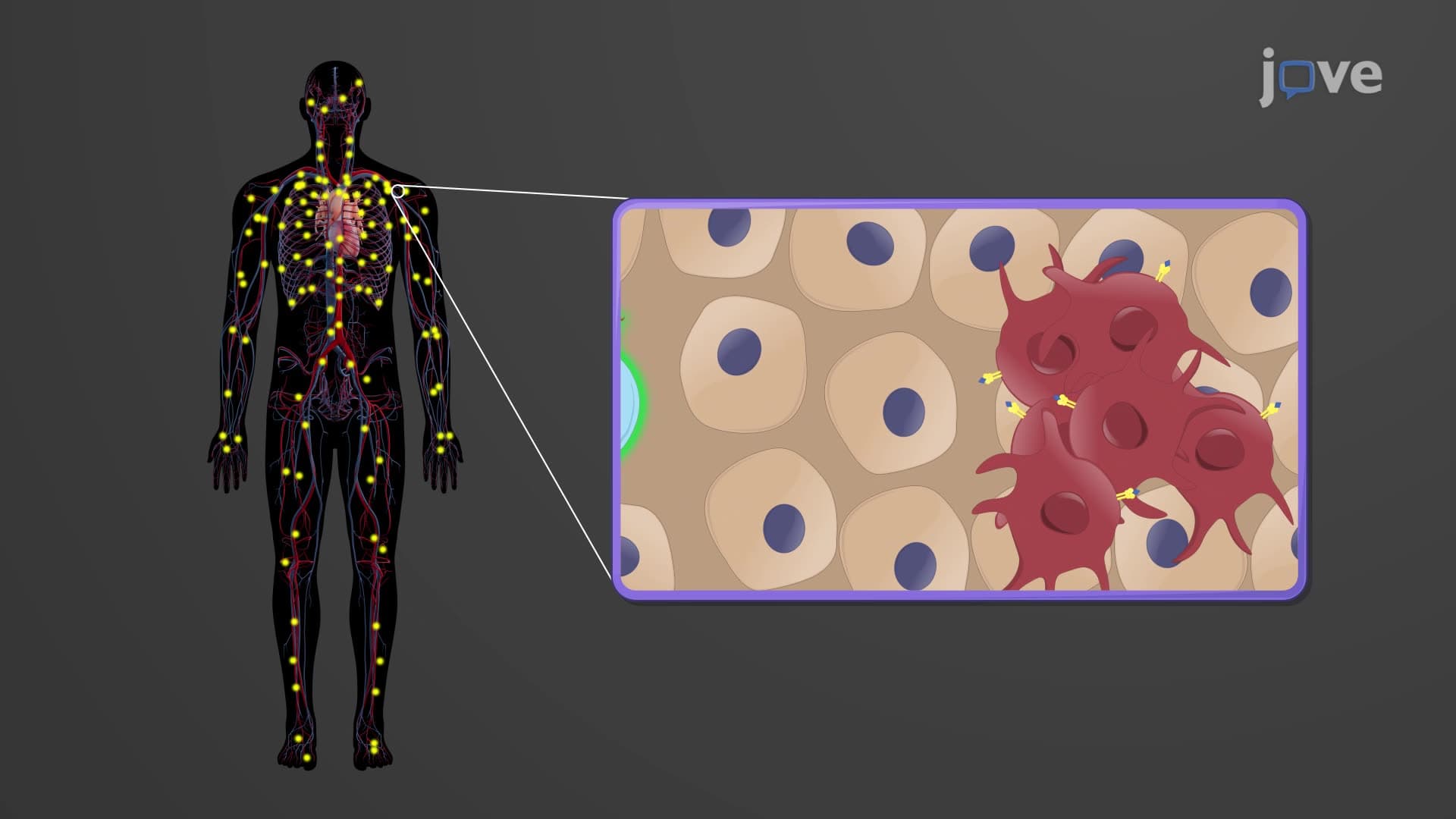
Cross presentation antigen mechanisms allow certain dendritic cells to present extracellular antigens on MHC class I molecules, bridging the gap between these pathways. This process proves crucial for generating immune responses against viruses that don't directly infect antigen-presenting cells and for cancer immunotherapy effectiveness.
Students preparing for the MCAT or AP Biology exams should understand how genetic defects in these pathways cause primary immunodeficiencies, while autoimmune diseases often result from inappropriate antigen presentation. These concepts frequently appear in college immunology courses and medical school curricula, forming the foundation for understanding vaccine mechanisms and therapeutic interventions.
Frequently Asked Questions
Antigen processing pathways are cellular mechanisms that break down proteins into small fragments and present them on cell surfaces for immune system recognition. They're essential because they allow your immune system to distinguish between normal cellular proteins and foreign or abnormal ones, enabling targeted responses against infections, cancer, and other threats while maintaining tolerance to healthy tissues.
The endogenous pathway uses proteasomes to process intracellular proteins and presents fragments on MHC class I molecules to CD8+ T cells, while the exogenous pathway uses lysosomes to process extracellular antigens and presents them on MHC class II molecules to CD4+ T cells. This division allows comprehensive immune surveillance of both internal cellular health and external environmental threats.
MCAT questions often focus on comparing pathway mechanisms, predicting outcomes when specific components are defective, and analyzing experimental scenarios involving antigen presentation. You might encounter passage-based questions about viral immune evasion strategies, autoimmune disease mechanisms, or vaccine development that require understanding both endogenous and exogenous processing pathways.
AP Biology exams frequently include free-response questions asking students to explain how antigen processing contributes to immune specificity and memory. Expect to diagram pathway steps, predict consequences of genetic mutations affecting processing components, or explain how vaccines utilize natural antigen processing mechanisms to generate protective immunity.
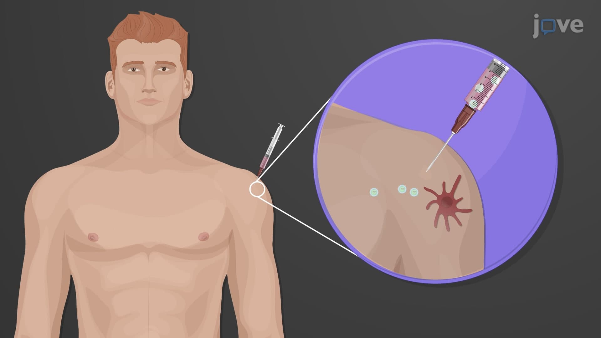
When you receive the annual flu vaccine at your local pharmacy, the vaccine components are processed through the exogenous MHC class II pathway by dendritic cells in your arm muscle. These cells present flu protein fragments to CD4+ T helper cells, which then activate B cells to produce antibodies and help generate memory cells that provide protection against future flu infections.
No advanced chemistry background is required to grasp these concepts. Basic understanding of protein structure, enzyme function, and cellular organelles from introductory biology courses provides sufficient foundation. The key is focusing on the biological logic of how cells sample, process, and present molecular information rather than memorizing complex biochemical details.
Create comparison charts highlighting pathway differences, practice drawing both pathways from memory, and work through clinical scenarios that illustrate pathway dysfunction. Focus on understanding the biological purpose of each step rather than just memorizing sequences, and use active recall techniques to test your ability to explain concepts without referring to notes.
Understanding these pathways prepares you for studying T cell development and selection, autoimmune disease mechanisms, cancer immunotherapy strategies, and transplant immunology. These foundational concepts also support learning about immune system evolution, comparative immunology, and emerging immunotherapeutic approaches in clinical medicine.
Related Micro-courses