Loading subject page…


986,640 views

Video Summary: What are G Protein Coupled Receptors
Did you know that caffeine's energizing effects happen because it blocks G protein coupled receptors in your brain? G protein coupled receptors (GPCRs) are specialized membrane proteins that detect external signals and convert them into cellular responses. From the adrenaline rush during a Starbucks run to how your eyes adjust to darkness, GPCRs control countless biological processes by activating complex signaling cascades inside cells. Watch the full video on JoVE Coach to master this concept with expert-led visuals and step-by-step explanations.
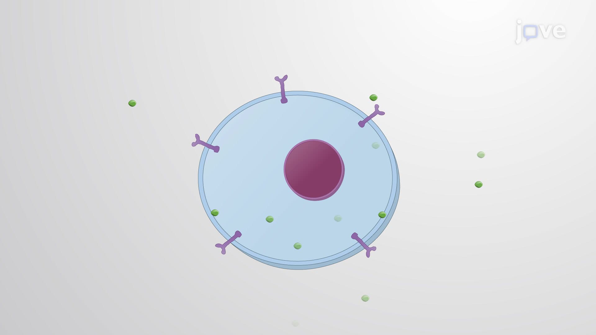
G protein coupled receptors represent one of biology's most elegant communication systems, allowing cells to detect and respond to their environment with remarkable precision. These sophisticated molecular machines span cell membranes seven times, creating a unique structure that bridges the gap between external signals and internal cellular machinery.
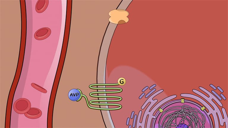
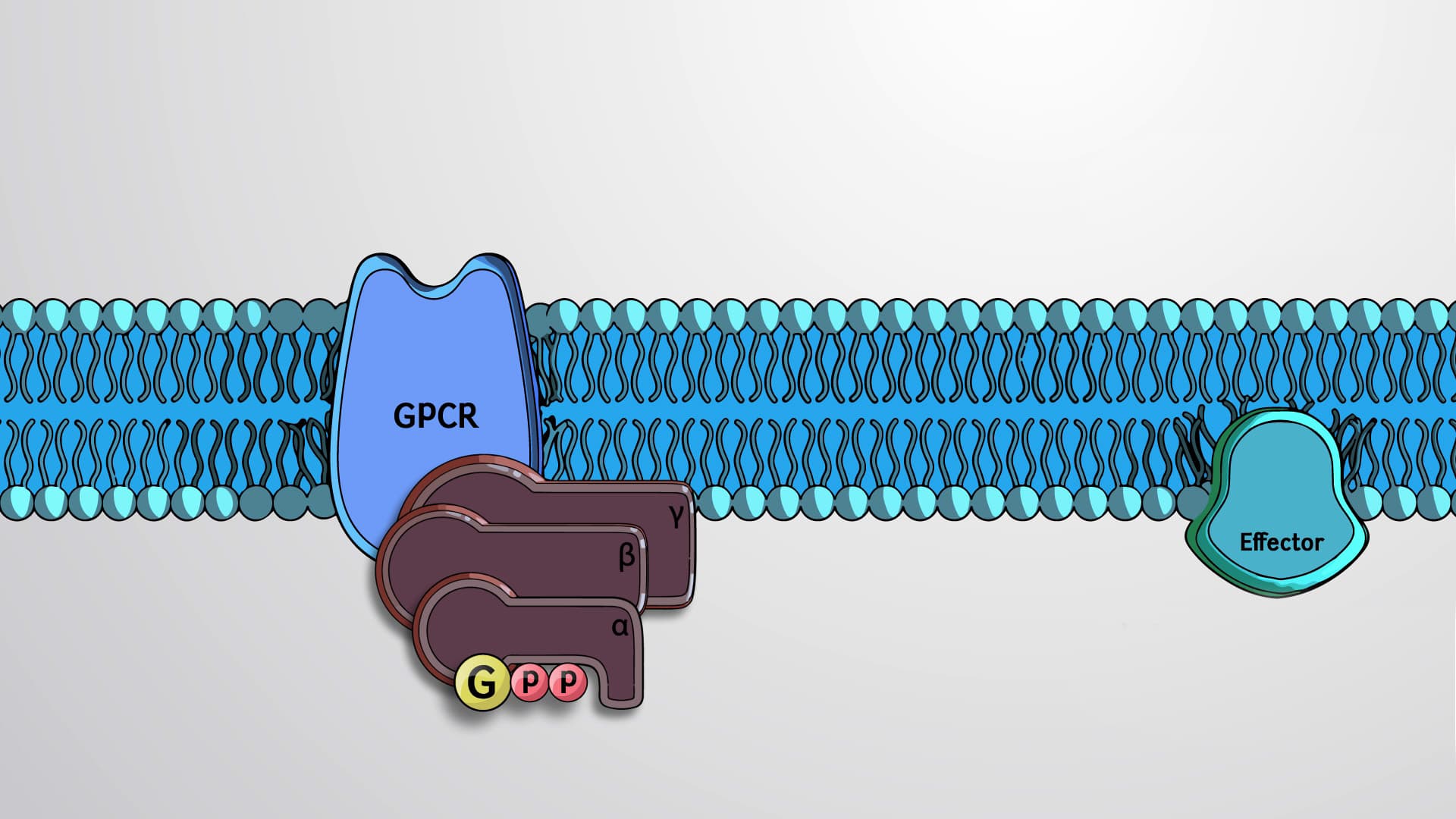
The GPCR structure and function relationship exemplifies biological efficiency. Each receptor contains three critical regions: an extracellular domain that recognizes specific signaling molecules (ligands), seven alpha-helical segments that thread through the membrane like a molecular accordion, and an intracellular domain that couples with G proteins. This seven transmembrane GPCR architecture creates a conformational switch-when ligands bind externally, the entire protein shifts shape, triggering internal changes.
Consider how your body responds to stress. When you're startled during a pop quiz, your adrenal glands release epinephrine (adrenaline). This hormone travels through your bloodstream until it encounters beta-adrenergic GPCRs on heart muscle cells. The moment epinephrine binds to these receptors, your heart rate increases within seconds-a perfect example of GPCR-mediated rapid response.
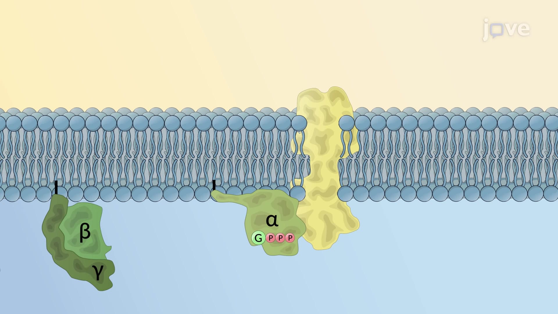
Understanding how G protein coupled receptors work in signaling requires following the molecular domino effect. In the resting state, the G protein alpha subunit holds GDP like a molecular key in the wrong position. When a ligand binds, the GPCR changes shape and acts as a guanine nucleotide exchange factor, prompting the alpha subunit to release GDP and grab GTP instead. This GTP binding causes the G protein alpha subunit to separate from its beta and gamma partners, creating two active signaling units.
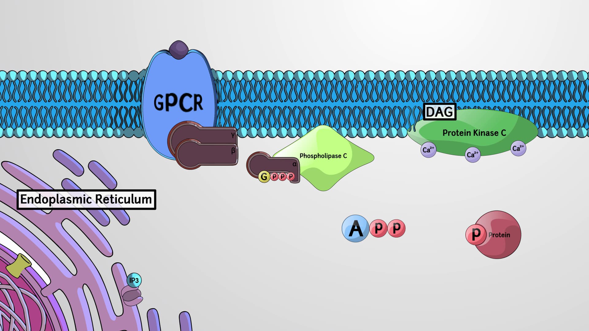
Both fragments-the GTP-bound alpha subunit and the beta-gamma dimer-then activate different effector proteins. For instance, in the GPCR second messenger pathway involving adenylyl cyclase GPCR systems, the alpha subunit activates adenylyl cyclase, which converts ATP into cyclic AMP (cAMP). This demonstrates GPCR signal amplification: one hormone molecule can generate thousands of cAMP molecules, amplifying the original signal exponentially.
GPCRs dominate pharmaceutical targets-over 40% of all prescription drugs target these receptors. Beta-blockers prescribed for high blood pressure work by blocking beta-adrenergic GPCRs, preventing excessive heart stimulation. This clinical relevance makes GPCRs essential topics for MCAT Cell Biology sections and AP Biology Unit 4 (Cell Communication and Cell Cycle).
For exam success, focus on the GTP/GDP cycle as the molecular timer that controls signal duration. Remember that GTP hydrolysis acts like a built-in off switch, ensuring cellular responses don't continue indefinitely. This regulatory mechanism prevents cellular chaos and maintains homeostasis-a concept frequently tested in college biochemistry courses.
Related Micro-courses