Loading subject page…


35,067 views


Video Summary: Somatosensory Motor and Association Cortex Explained
Ever wonder why stubbing your toe sends signals to a specific brain region while planning your next chess move activates completely different areas? The spinal cord serves as the critical highway connecting your brain's somatosensory motor and association cortex to the rest of your body. When University of California researchers studied paralyzed patients, they discovered how damage to spinal cord pathways disrupts the precise communication between these specialized brain regions and peripheral nerves. Watch the full video on JoVE Coach to master this concept with expert-led visuals and step-by-step explanations.
The human brain's ability to process sensations, control movement, and execute complex thoughts relies on three interconnected cortical systems that communicate through the spinal cord. This neurological network forms the foundation for everything from reflexively pulling your hand away from a hot stove to solving calculus problems on the AP exam.
Located in the parietal lobe's postcentral gyrus, the somatosensory cortex creates a detailed sensory map called the sensory homunculus. This organization follows a counterintuitive pattern: neurons processing foot sensations occupy the top regions, while facial sensations activate lower areas. The spinal cord carries these sensory signals through specific pathways - the dorsal column-medial lemniscal system handles fine touch and proprioception, while the spinothalamic tract processes pain and temperature.
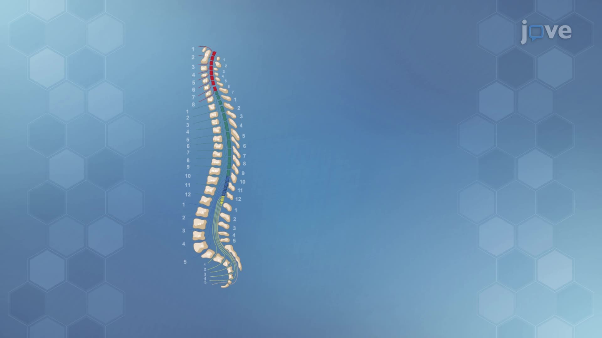
Students studying for the MCAT should understand that this topographic organization explains why certain spinal cord injuries affect specific body regions. For example, damage at the cervical level (neck region) can impair hand sensation while preserving leg function, as demonstrated in cases treated at Johns Hopkins Hospital's spinal cord injury center.
The primary motor cortex in the frontal lobe's precentral gyrus mirrors the somatosensory organization but controls voluntary movement instead of sensation. Motor neurons here send signals through the corticospinal tract, which travels down the spinal cord to activate specific muscle groups. This pathway explains why spinal cord injuries often cause paralysis below the injury site - the communication highway between brain commands and muscle targets gets severed.
College anatomy students frequently encounter this concept when studying conditions like amyotrophic lateral sclerosis (ALS), where motor cortex degeneration gradually eliminates voluntary muscle control while preserving cognitive function.
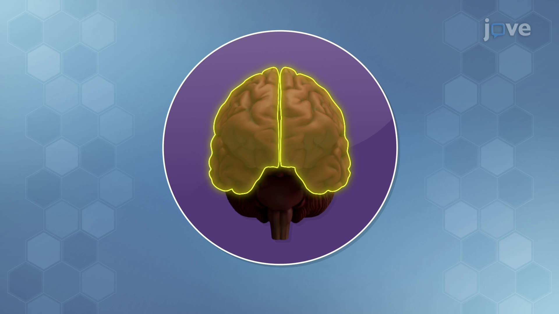
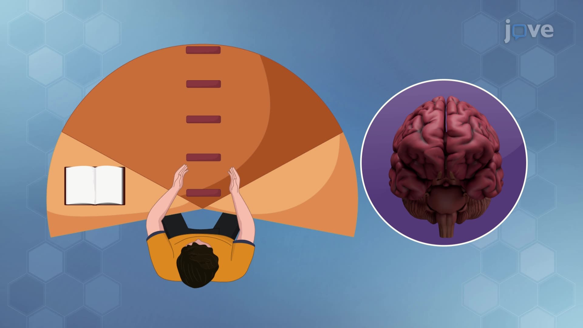
Unlike sensory and motor regions, association cortex areas don't directly process sensations or control movements. Instead, they integrate information from multiple sources to enable higher cognitive functions. Broca's area (typically in the left frontal lobe) specializes in speech production, while Wernicke's area (left temporal lobe) handles language comprehension. These regions communicate through association fibers and demonstrate how complex behaviors require coordination between specialized brain areas.
The association cortex becomes particularly relevant for students preparing for neuroscience sections of standardized tests, as questions often focus on how brain injuries affect specific cognitive abilities while leaving others intact.
Related Micro-courses