Loading subject page…


35,067 views


Video Summary: What Is Brain Imaging
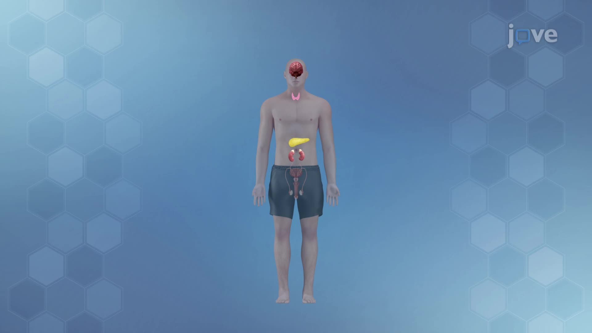
Did you know that doctors can watch your brain think in real-time? The endocrine system and brain work together through sophisticated imaging techniques that reveal both structure and function. What is Brain Imaging encompasses methods like MRI, fMRI, CT, and PET scans that help Stanford University researchers study everything from hormone regulation to cognitive processes. These powerful tools allow scientists to observe neural activity as it happens, revolutionizing our understanding of how the brain controls bodily functions. Watch the full video on JoVE Coach to master this concept with expert-led visuals and step-by-step explanations.
What is Brain Imaging represents one of modern medicine's greatest achievements, allowing us to peer inside the living brain without invasive procedures. These techniques have transformed neuroscience, enabling researchers to study the endocrine system definition through neural pathways and understand what is the endocrine system in detail by observing brain-hormone interactions. Brain imaging encompasses both structural and functional approaches, each serving distinct purposes in medical diagnosis and scientific research.
Computed Tomography (CT) scans create detailed three-dimensional images using X-rays from multiple angles. Widely used in emergency departments across the United States, CT scans excel at detecting brain hemorrhages, tumors, and structural abnormalities. The Cleveland Clinic uses CT scans as first-line imaging for stroke patients because of their speed and accuracy in identifying bleeding.
Magnetic Resonance Imaging (MRI) produces the highest resolution structural images of the brain using powerful magnetic fields and radio waves. Unlike CT scans, MRI provides excellent soft tissue contrast without ionizing radiation. Johns Hopkins University researchers frequently use MRI to study the endocrine system overview by examining hypothalamic and pituitary structures that control hormone production.
Functional MRI (fMRI) revolutionized neuroscience by measuring blood flow changes that indicate neural activity. Based on the principle that active brain regions require more oxygenated blood, fMRI enables real-time observation of brain function. Harvard Medical School researchers use fMRI to study how stress hormones affect brain activity, providing insights into the endocrine system basics.
Positron Emission Tomography (PET) scans use radioactive tracers to highlight metabolically active brain regions. The Mayo Clinic employs PET scans to diagnose Alzheimer's disease and study how neurotransmitters interact with endocrine function, advancing our understanding the endocrine system concept.
Brain imaging concepts frequently appear on the MCAT, particularly in psychology and biology sections. AP Psychology students encounter these techniques when studying biological bases of behavior. College neuroanatomy courses use brain imaging as practical examples of how the endocrine system study guide principles apply to real research. Understanding these imaging modalities prepares students for careers in medicine, neuroscience, and psychology while providing concrete examples of how scientific principles translate to clinical practice.
Related Micro-courses