Loading subject page…


35,067 views


Video Summary: What Is Neuroplasticity
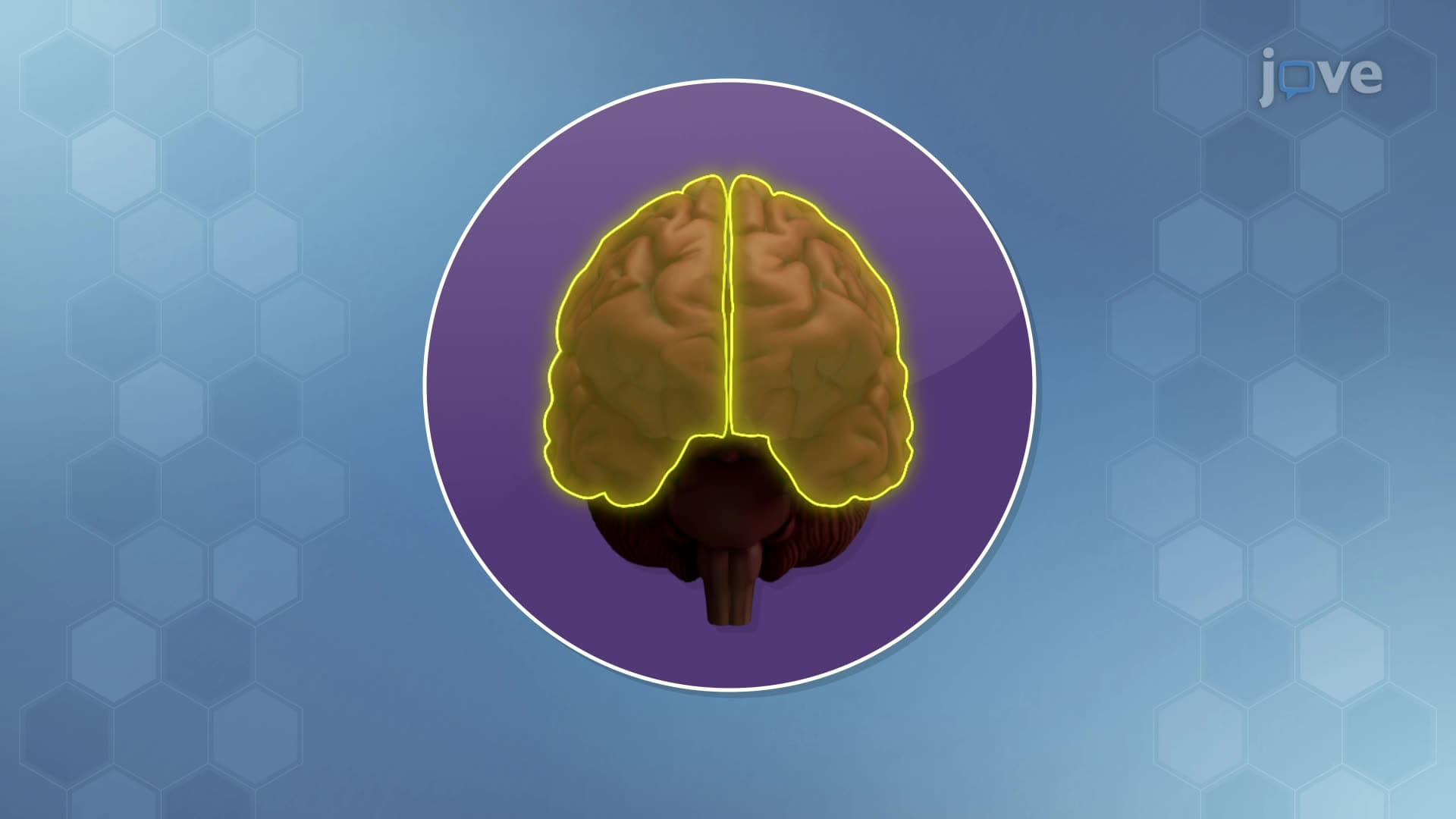
Brain imaging reveals that musicians' brains show dramatically different structures compared to non-musicians, with enlarged motor and auditory regions visible on MRI scans. This brain imaging definition demonstrates neuroplasticity-the brain's remarkable ability to reorganize and form new neural connections throughout life in response to learning, experience, or injury. Stanford researchers use brain imaging to track how London taxi drivers develop enlarged hippocampi from memorizing the city's complex street layout. What is neuroplasticity becomes clear when we see these structural changes happening in real-time through advanced brain imaging techniques. Watch the full video on JoVE Coach to master this concept with expert-led visuals and step-by-step explanations.
Neuroplasticity represents one of the most remarkable discoveries in modern neuroscience-the brain's lifelong capacity to rewire itself. Unlike earlier beliefs that adult brains were fixed structures, research from institutions like Harvard Medical School and the National Institutes of Health demonstrates that our neural networks remain dynamic throughout life. This brain imaging overview shows how experiences literally reshape brain structure and function.
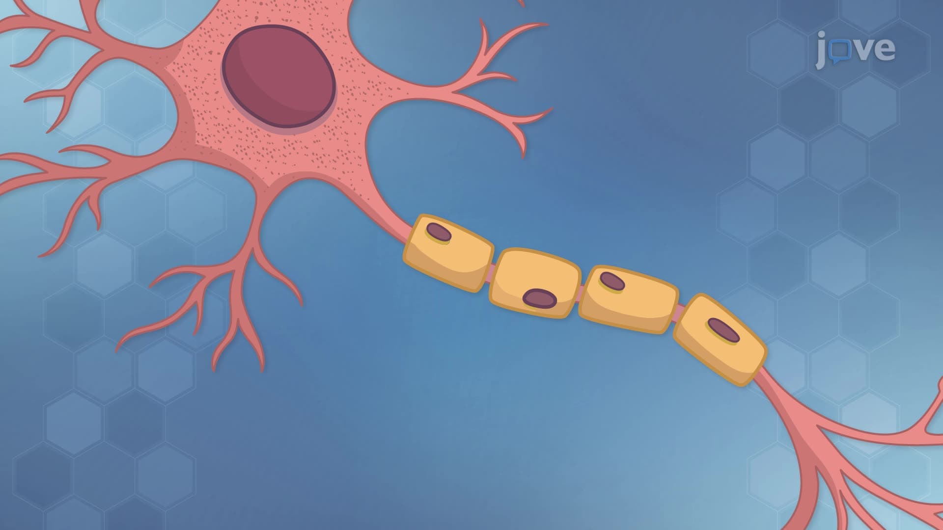
The brain accomplishes neuroplastic changes through four interconnected processes. Dendritic and axonal growth increases the physical complexity of neural networks, creating new branches that extend a neuron's reach. Brain imaging studies at UCLA show that college students learning new languages develop measurably thicker cortical regions within months.
Synaptogenesis forms entirely new synaptic connections, expanding communication pathways between neurons. This brain imaging concept becomes evident when researchers track violin players-their motor cortex regions controlling finger movements show dramatically increased synaptic density compared to control groups.
Synaptic pruning eliminates underused connections, optimizing neural efficiency. This process, extensively studied at Johns Hopkins, follows the "use it or lose it" principle. Brain imaging reveals that adolescent brains undergo massive pruning, eliminating up to 50% of synaptic connections while strengthening frequently used pathways.
Myelination wraps neural pathways in fatty sheaths, dramatically increasing signal transmission speed. Understanding brain imaging shows that practiced skills-from basketball free throws to mathematical calculations-develop thicker myelin, enabling faster, more automatic responses.
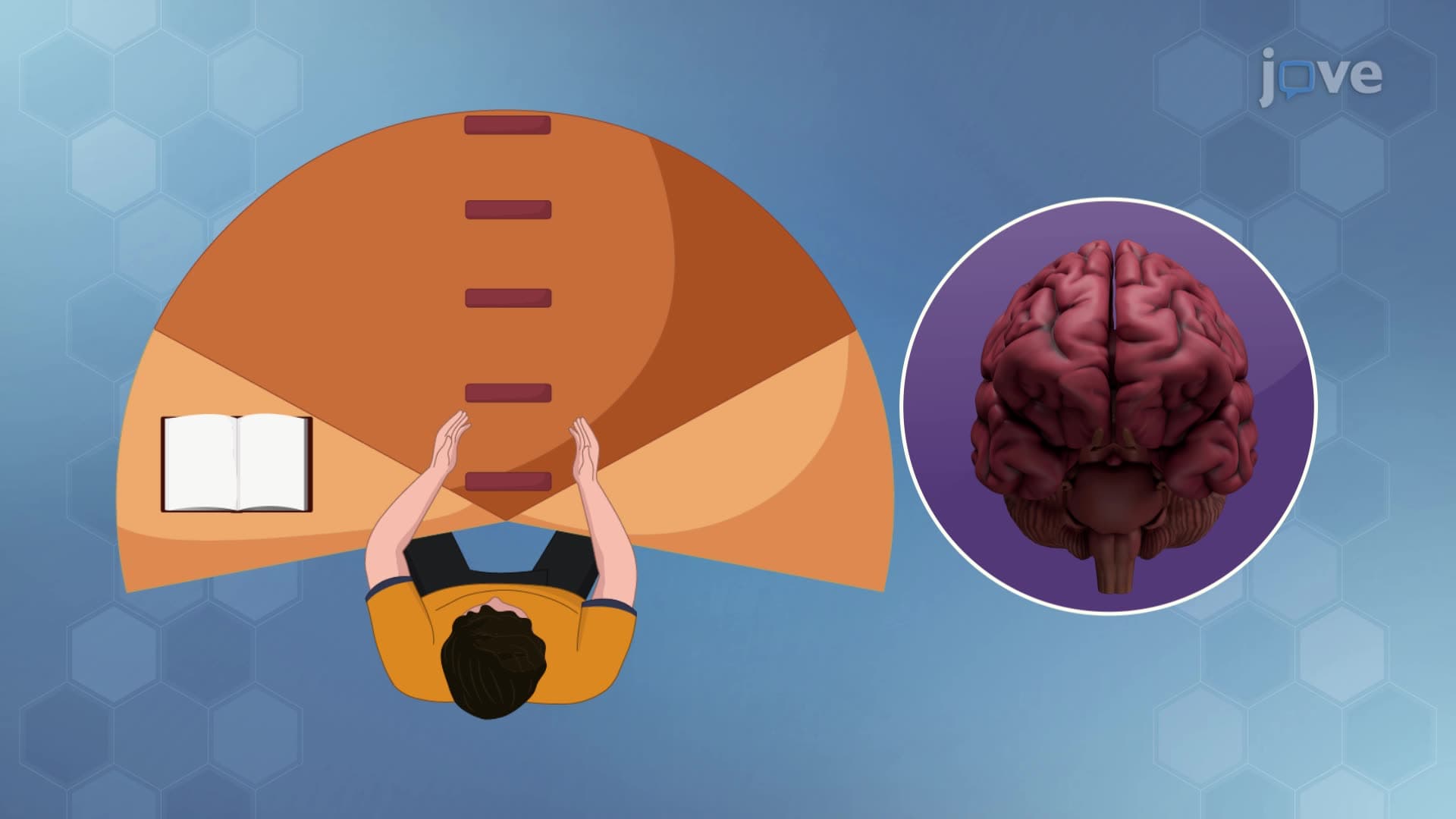
Neuroplasticity research revolutionizes rehabilitation medicine across US hospitals. Stroke patients at Mayo Clinic undergo constraint-induced movement therapy, forcing damaged brain regions to develop new neural pathways. Similarly, this brain imaging study guide principle explains why intensive practice sessions prove more effective than distributed learning for AP exam preparation.
For college undergraduates, neuroplasticity concepts frequently appear on MCAT questions, particularly in biological sciences sections. Students should understand how repetitive practice-whether learning organic chemistry reactions or perfecting laboratory techniques-creates measurable brain changes that enhance performance and retention.
Related Micro-courses