Loading subject page…


35,067 views

Video Summary: What Is Spinal Cord
Ever wonder how your hand jerks away from a hot stove before you even realize it's burning? This lightning-fast response demonstrates lateralization - the specialized organization of neural pathways that enables rapid, coordinated responses. The spinal cord, a critical highway of nerve tissue extending from your brainstem to your lower back, exemplifies how lateralization allows different sides and regions to process distinct types of information. In US emergency rooms, doctors routinely test spinal reflexes to assess neurological function and lateralization patterns. Watch the full video on JoVE Coach to master this concept with expert-led visuals and step-by-step explanations.
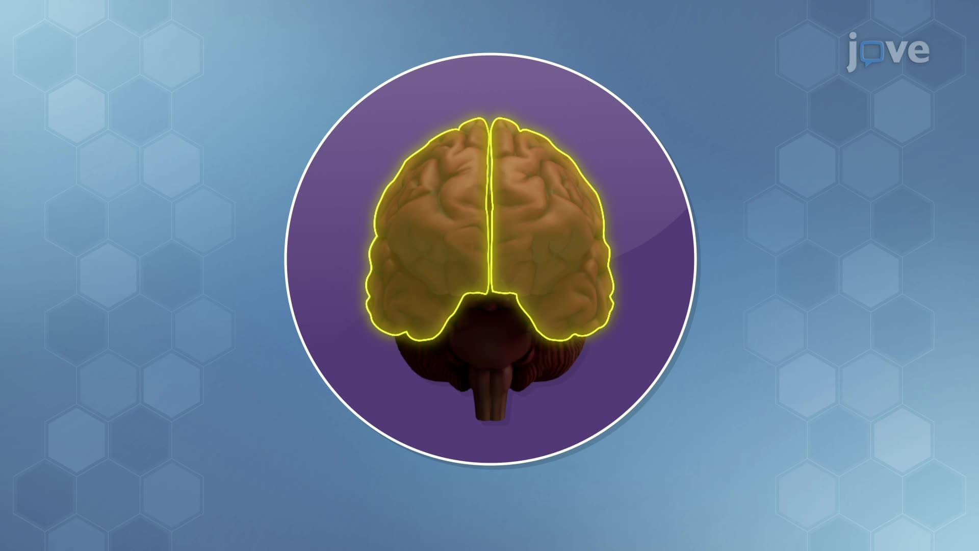
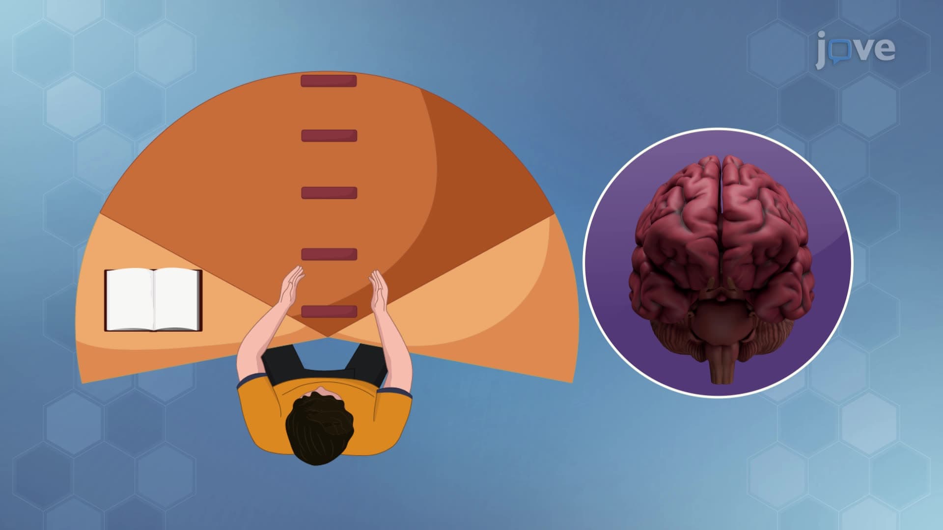
The spinal cord exemplifies one of neuroscience's most fundamental principles: lateralization. This 18-inch column of neural tissue demonstrates how biological systems organize functions across different sides and regions to maximize efficiency and control. Unlike the brain's dramatic left-right hemispheric specialization, spinal cord lateralization operates through segmented organization and bilateral coordination systems that ensure precise motor control and sensory processing.
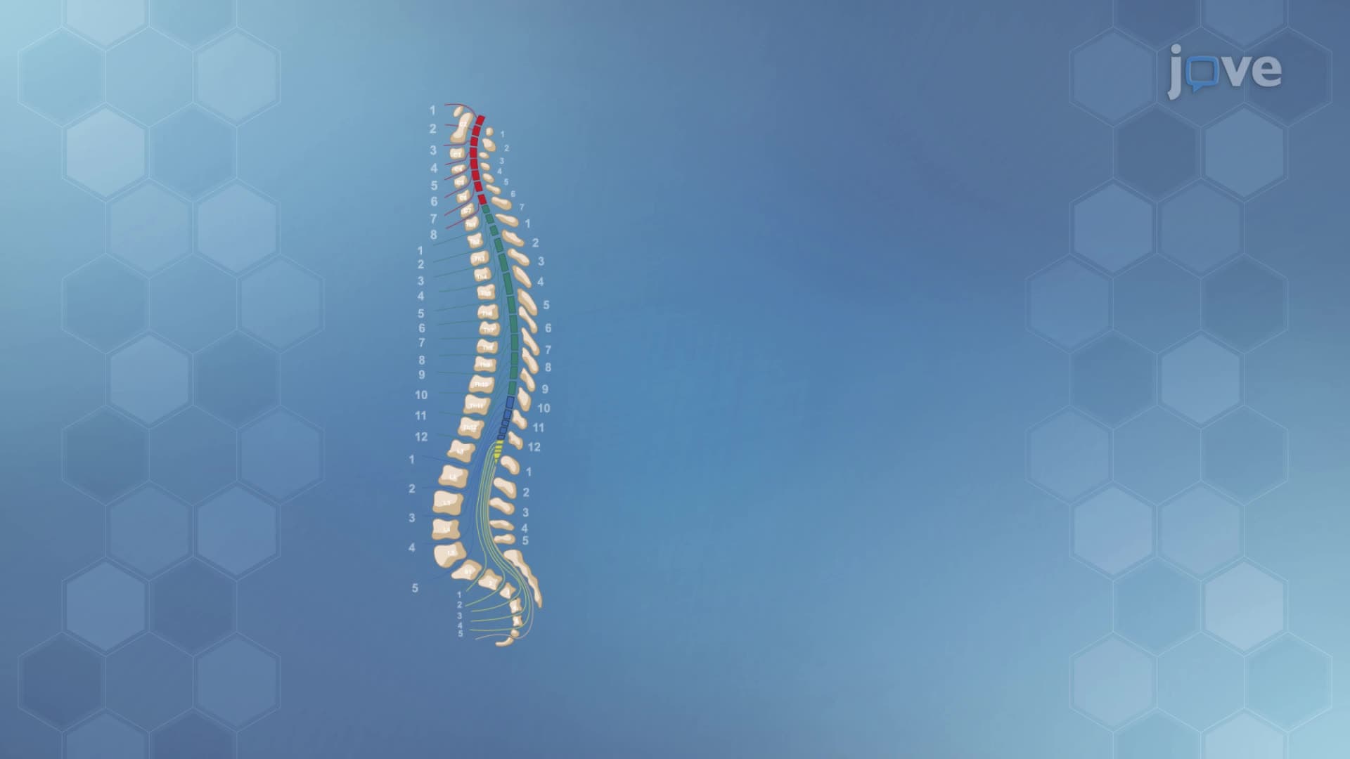
The spinal cord's 31 segments each correspond to specific body regions, creating a lateralized map of human anatomy. The eight cervical segments control neck and arm functions, with C5-C8 specifically managing hand and finger movements crucial for activities like texting or playing instruments. Thoracic segments T1-T12 coordinate trunk muscles and breathing, while lumbar segments L1-L5 control hip and leg movements essential for walking and running. This segmental lateralization allows targeted medical interventions - when UCLA Medical Center treats spinal injuries, doctors can predict functional impacts based on which segments are affected.
Spinal reflexes demonstrate lateralization at the cellular level through specialized neural circuits. When you step on a sharp object, sensory neurons on that foot's side rapidly communicate with interneurons, which then activate motor neurons controlling withdrawal muscles while simultaneously inhibiting opposing muscle groups. This process, called reciprocal inhibition, occurs within milliseconds and represents perfect lateralized coordination. Advanced Placement Biology students frequently encounter these concepts when studying homeostasis and neural regulation.
Healthcare professionals routinely assess spinal cord lateralization through standardized reflex testing. The patellar reflex (knee-jerk) tests L3-L4 segments, while Achilles reflex evaluation examines S1-S2 function. Abnormal lateralization patterns often indicate specific pathologies - Multiple Sclerosis patients may show asymmetric reflex responses, while stroke survivors might exhibit altered spinal processing affecting one body side more than the other. Medical students preparing for USMLE Step 1 must master these lateralization concepts to interpret neurological examinations accurately.
Physical therapy programs at institutions like Johns Hopkins utilize spinal cord lateralization principles when designing rehabilitation protocols. Therapists target specific segments and sides to restore balanced function, demonstrating how understanding lateralization translates into practical healthcare applications that improve patient outcomes.
Related Micro-courses