Loading subject page…

- Organic Chemistry
- Radical Chemistry
- Electron Paramagnetic Resonance (EPR) Spectroscopy: Organic Radicals
Micro-course: Radical Chemistry
85,200 views
Learn key concepts and earn your certificate.
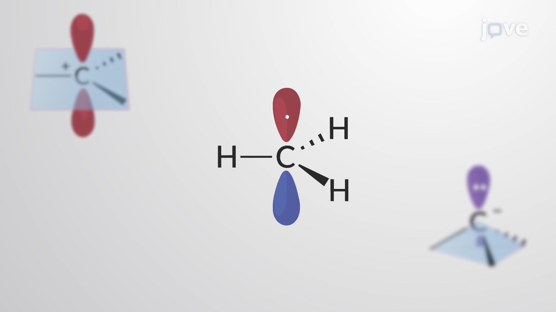


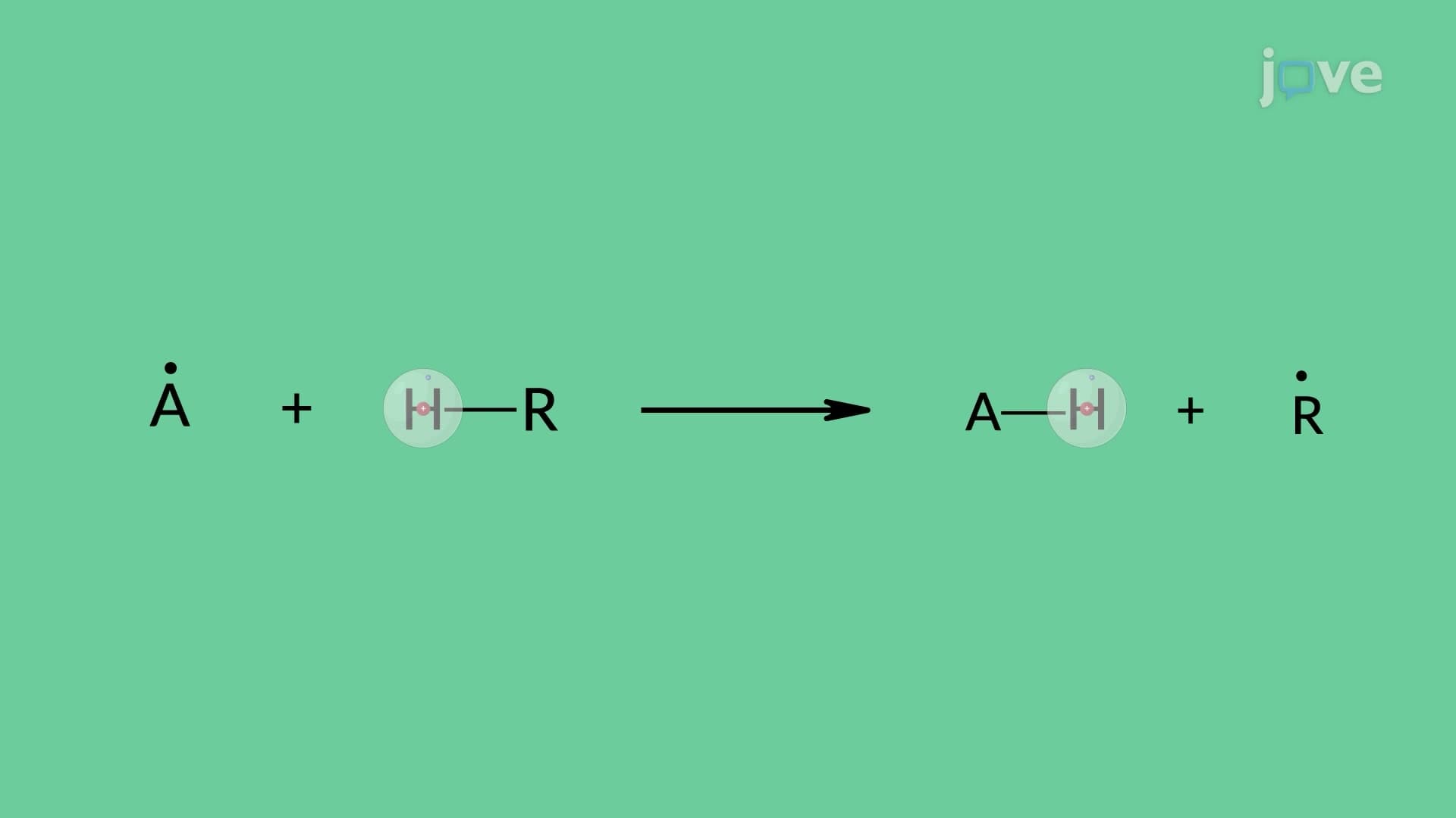
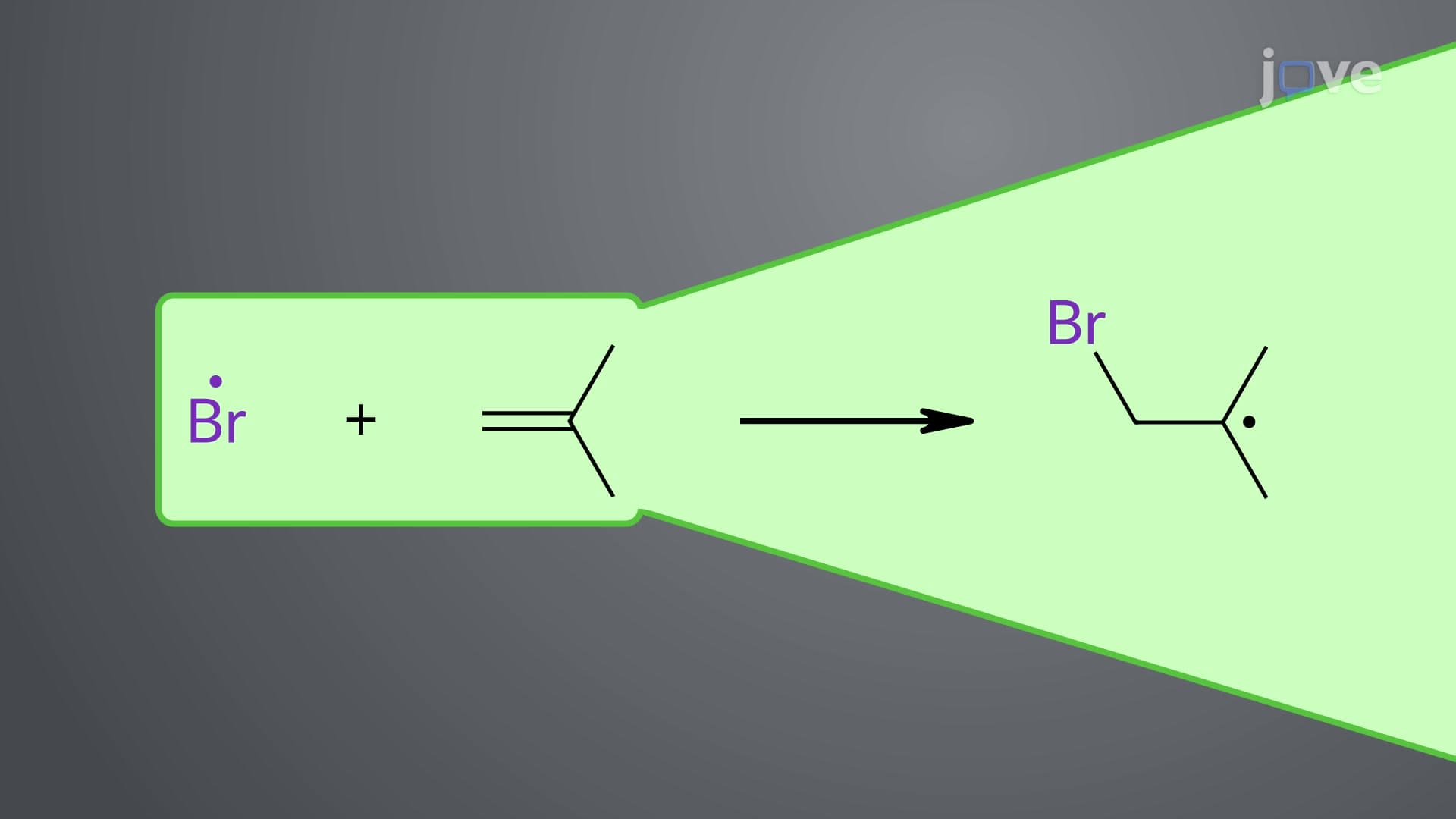
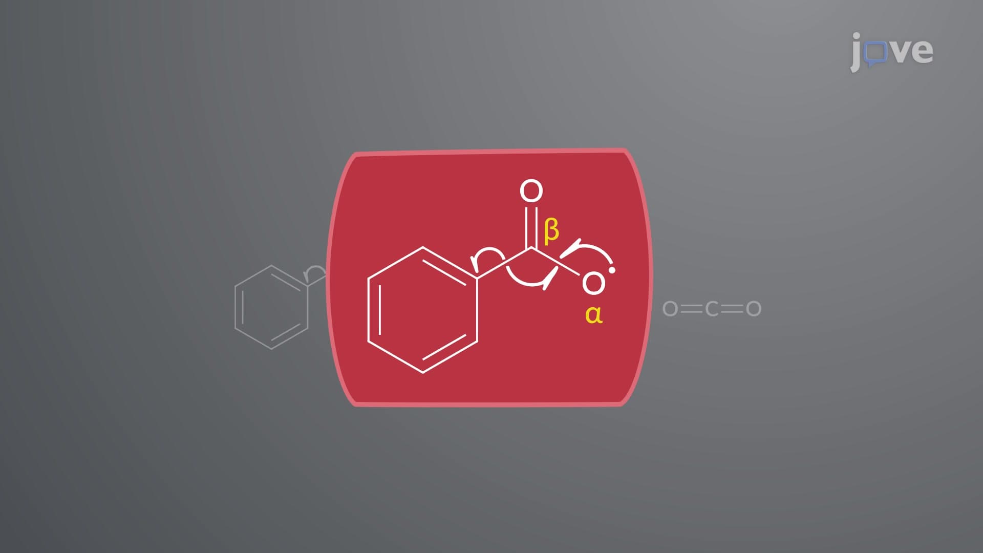








Electron Paramagnetic Resonance (EPR) Spectroscopy: Organic Radicals

00:00
01:17
Video Summary: Electron Paramagnetic Resonance Epr Spectroscopy Explained
Did you know that electron paramagnetic resonance EPR can detect single unpaired electrons in materials, making it invaluable for studying everything from vitamin deficiencies to semiconductor defects? This powerful analytical technique reveals how unpaired electrons interact with nearby atomic nuclei, creating distinctive spectral patterns that scientists at institutions like MIT and Stanford use to investigate radical chemistry and materials science. Understanding Electron Paramagnetic Resonance Epr Spectroscopy Explained opens doors to advanced research in biochemistry, materials engineering, and pharmaceutical development. Watch the full video on JoVE Coach to master this concept with expert-led visuals and step-by-step explanations.
- Understand the fundamental principles of Electron Paramagnetic Resonance Epr Spectroscopy Explained and how unpaired electrons create detectable signals in magnetic fields
- Identify hyperfine splitting patterns using the formula 2nI + 1 to predict the number of peaks in EPR spectra based on neighboring spin-active nuclei
- Analyze how nuclear-electronic spin coupling creates characteristic spectral fingerprints for different radical structures and molecular geometries
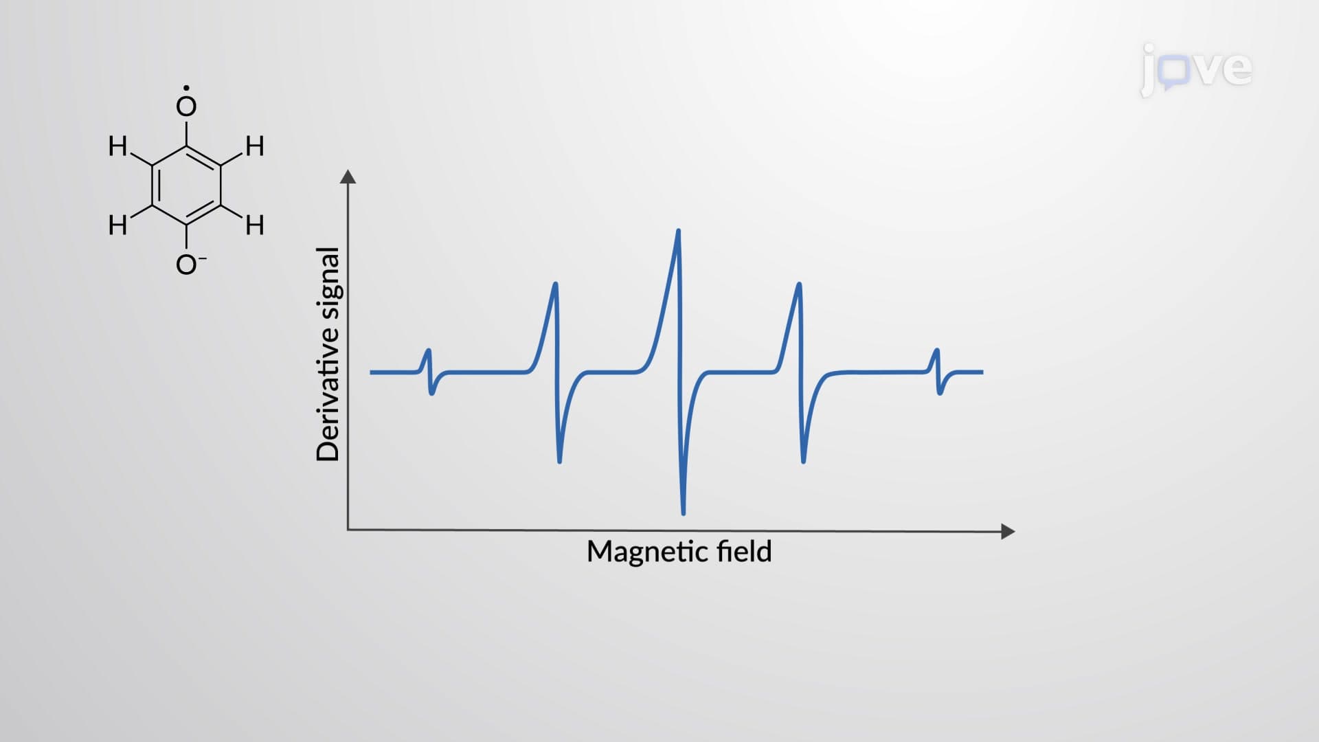
- Compare peak intensities and splitting patterns between simple radicals like methyl (1:3:3:1 quartet) and more complex systems like benzosemiquinone radicals
- Evaluate hyperfine splitting constants measured in millitesla or gauss units to determine molecular geometry and electron delocalization patterns
- Apply EPR spectroscopy principles to interpret real spectra and identify unknown radical species in laboratory and research settings
- Recognize the relationship between electron delocalization and hyperfine coupling strength in aromatic radical systems
- Explore how EPR spectroscopy complements other analytical techniques like NMR and UV-Vis spectroscopy in structural determination
Understanding Electron Paramagnetic Resonance EPR Spectroscopy
Electron Paramagnetic Resonance Epr Spectroscopy Explained begins with the fundamental concept that unpaired electrons possess magnetic moments due to their intrinsic spin. When placed in a strong magnetic field, these electrons can occupy two distinct energy states, creating the foundation for EPR detection. Unlike paired electrons that cancel each other's magnetic effects, unpaired electrons in free radicals, transition metals, and defect centers produce measurable signals that reveal crucial structural information.
Hyperfine Splitting: The Key to Molecular Structure
The most powerful aspect of EPR spectroscopy lies in hyperfine splitting-the interaction between unpaired electrons and nearby atomic nuclei with non-zero nuclear spin. This phenomenon transforms a simple two-level system into complex, information-rich spectra. The mathematical relationship 2nI + 1 predicts spectral complexity, where n represents equivalent nuclei and I denotes nuclear spin quantum number. For example, a methyl radical (CH3•) interacting with three equivalent protons (I = 1/2) produces 2(3)(1/2) + 1 = 4 peaks with characteristic 1:3:3:1 intensity ratios, reflecting statistical distributions of nuclear spin orientations.
Real-World Applications in US Research and Industry
EPR spectroscopy plays crucial roles across American universities and industries. At Harvard Medical School, researchers use EPR to study oxidative stress in biological systems, while semiconductor companies in Silicon Valley employ it to detect defects in computer chips. The technique proves invaluable for pharmaceutical companies developing antioxidant drugs, as it directly measures free radical scavenging activity. Students preparing for advanced placement chemistry exams or MCAT sections on analytical techniques should recognize EPR as complementary to NMR spectroscopy, providing unique insights into paramagnetic species that NMR cannot detect.
Interpreting EPR Spectra and Splitting Constants
Hyperfine splitting constants, measured in millitesla (mT) or gauss (G), quantify the strength of electron-nucleus interactions. Larger coupling constants indicate closer proximity or greater orbital overlap between unpaired electrons and magnetic nuclei. In benzosemiquinone radicals, electron delocalization across the aromatic ring creates coupling with multiple hydrogen atoms, producing five-line spectra that reveal electron distribution patterns. Understanding these relationships enables students to predict molecular geometries and electron localization, skills essential for success in advanced organic chemistry courses and graduate school entrance exams like the GRE Chemistry subject test.
Frequently Asked Questions
Electron paramagnetic resonance EPR spectroscopy is an analytical technique that detects and analyzes materials containing unpaired electrons. It works by placing samples in strong magnetic fields and measuring microwave absorption as electrons transition between spin energy states. This method provides detailed information about molecular structure, electron distribution, and chemical environments that other techniques cannot reveal.
EPR spectroscopy builds naturally on concepts you already know from general chemistry, particularly electron configuration and magnetic properties. While the mathematical aspects may seem complex, the core principles follow logical patterns similar to NMR spectroscopy. Most students find EPR accessible after mastering basic quantum mechanics and molecular orbital theory, typically covered in second-semester general chemistry.
MCAT questions typically focus on identifying paramagnetic species, predicting spectral patterns from molecular structures, and comparing EPR with other analytical techniques. Graduate exams like the GRE Chemistry test may include calculations using the 2nI + 1 formula and interpretation of hyperfine splitting patterns. Practice problems often involve determining the number of equivalent nuclei from spectral data or predicting relative peak intensities.
EPR spectroscopy is extensively used across US pharmaceutical companies for drug development, particularly studying antioxidant mechanisms and radical-mediated reactions. Semiconductor manufacturers like Intel and AMD employ EPR to detect defects in silicon wafers, while petroleum companies use it to analyze catalysts and study reaction mechanisms. Medical research institutions utilize EPR for investigating oxidative stress, cancer metabolism, and aging processes.
Focus on understanding the relationship between molecular structure and spectral patterns rather than memorizing individual spectra. Practice applying the 2nI + 1 formula with different molecular examples, and work through problems that connect hyperfine coupling constants to molecular geometry. Create comparison charts showing how EPR complements NMR and other analytical techniques, as this integrated understanding frequently appears on comprehensive exams.
EPR spectroscopy specifically detects only paramagnetic species-molecules or materials with unpaired electrons. This includes free radicals, transition metal complexes, and defect centers in crystals, but excludes diamagnetic molecules with all paired electrons. This selectivity actually represents a strength, allowing EPR to focus exclusively on reactive species that often play crucial roles in chemical and biological processes.
EPR knowledge provides essential foundation for advanced topics including radical chemistry, organometallic chemistry, and biochemical reaction mechanisms. It directly connects to physical chemistry concepts like molecular orbital theory and quantum mechanics, while supporting organic chemistry understanding of reaction intermediates. Many graduate programs expect students to interpret EPR data alongside NMR, IR, and mass spectrometry results for complete structural determination.
Build on EPR fundamentals by exploring advanced topics like electron-nuclear double resonance (ENDOR) spectroscopy and pulse EPR techniques. Study how EPR integrates with other analytical methods in structure determination, and investigate specific applications in your field of interest, whether materials science, biochemistry, or pharmaceutical chemistry. Consider practical experience with EPR instrumentation through undergraduate research opportunities at universities with strong chemistry programs.
Related Micro-courses
20 Concepts
15 Concepts

12 Concepts