Loading subject page…


96,702 views

Video Summary: Transducer Mechanism G Protein Coupled Receptors Explained
Did you know that over one-third of FDA-approved medications target the same cellular pathway? The transducer mechanism g protein system represents one of biology's most crucial communication networks, with G protein-coupled receptors (GPCRs) serving as molecular switches that control everything from your heart rate to pain perception. Consider how morphine relieves pain by activating opioid GPCRs in your nervous system. Transducer Mechanism G Protein Coupled Receptors Explained reveals how these seven-pass membrane proteins convert external signals into cellular responses through sophisticated molecular machinery. Watch the full video on JoVE Coach to master this concept with expert-led visuals and step-by-step explanations.
The transducer mechanism g protein system represents cellular biology's most versatile communication network. GPCRs function as sophisticated molecular switches, converting external chemical signals into intracellular responses that govern virtually every physiological process. From the adrenaline rush during a medical emergency to the calming effects of anesthesia during surgery, these receptors orchestrate our body's responses to countless stimuli.
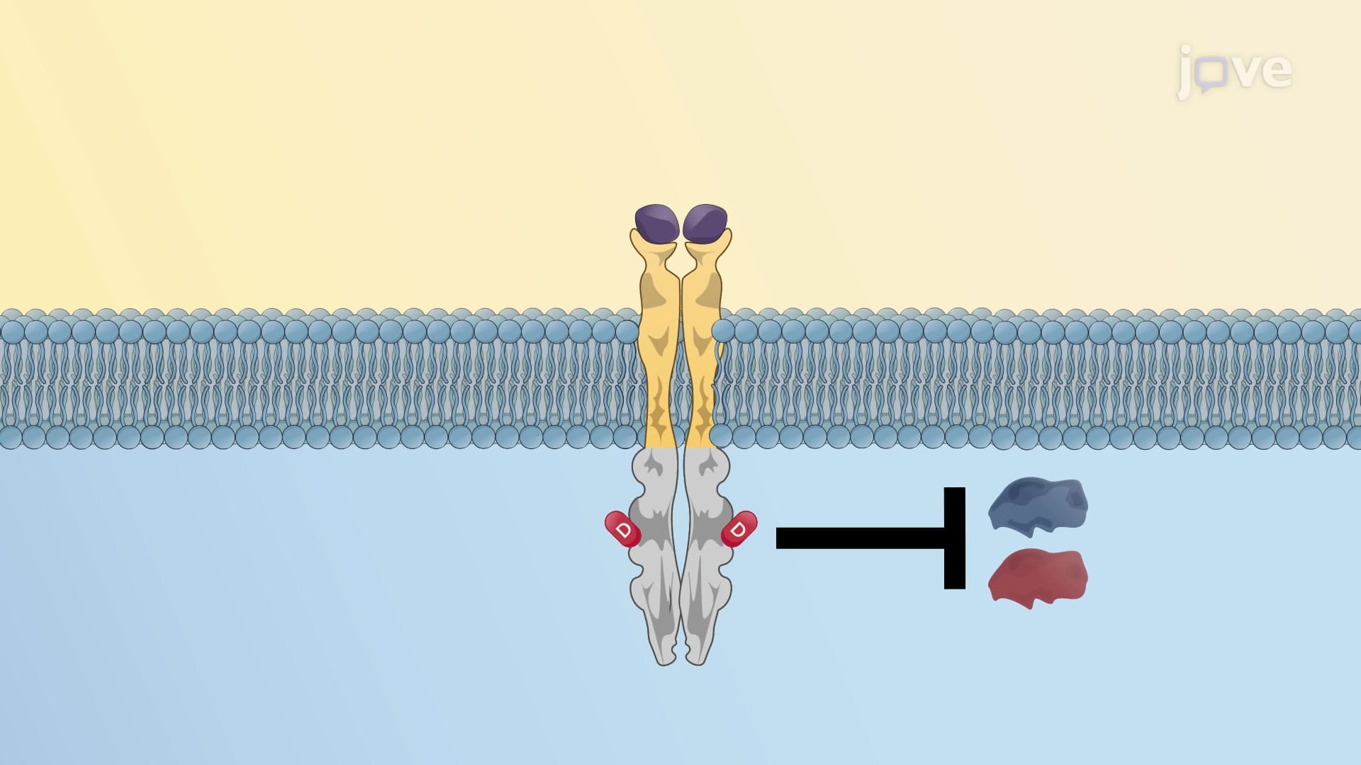
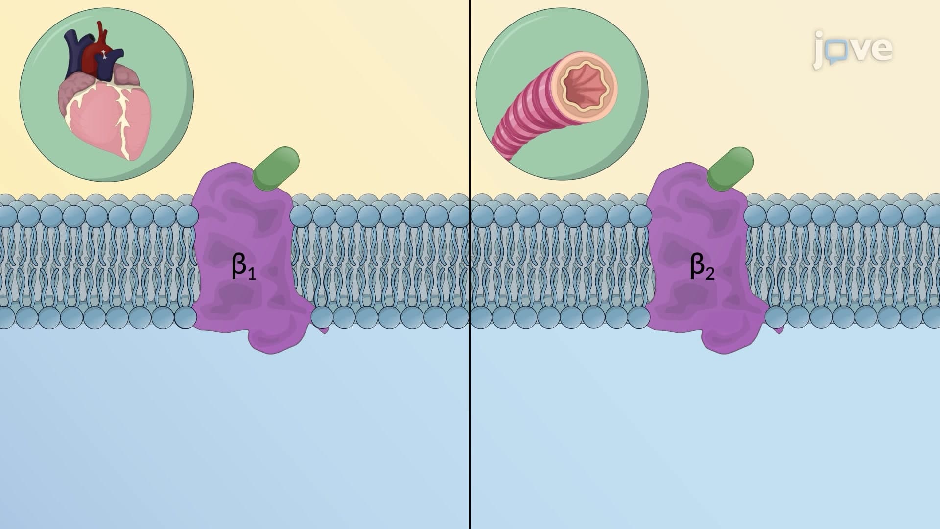
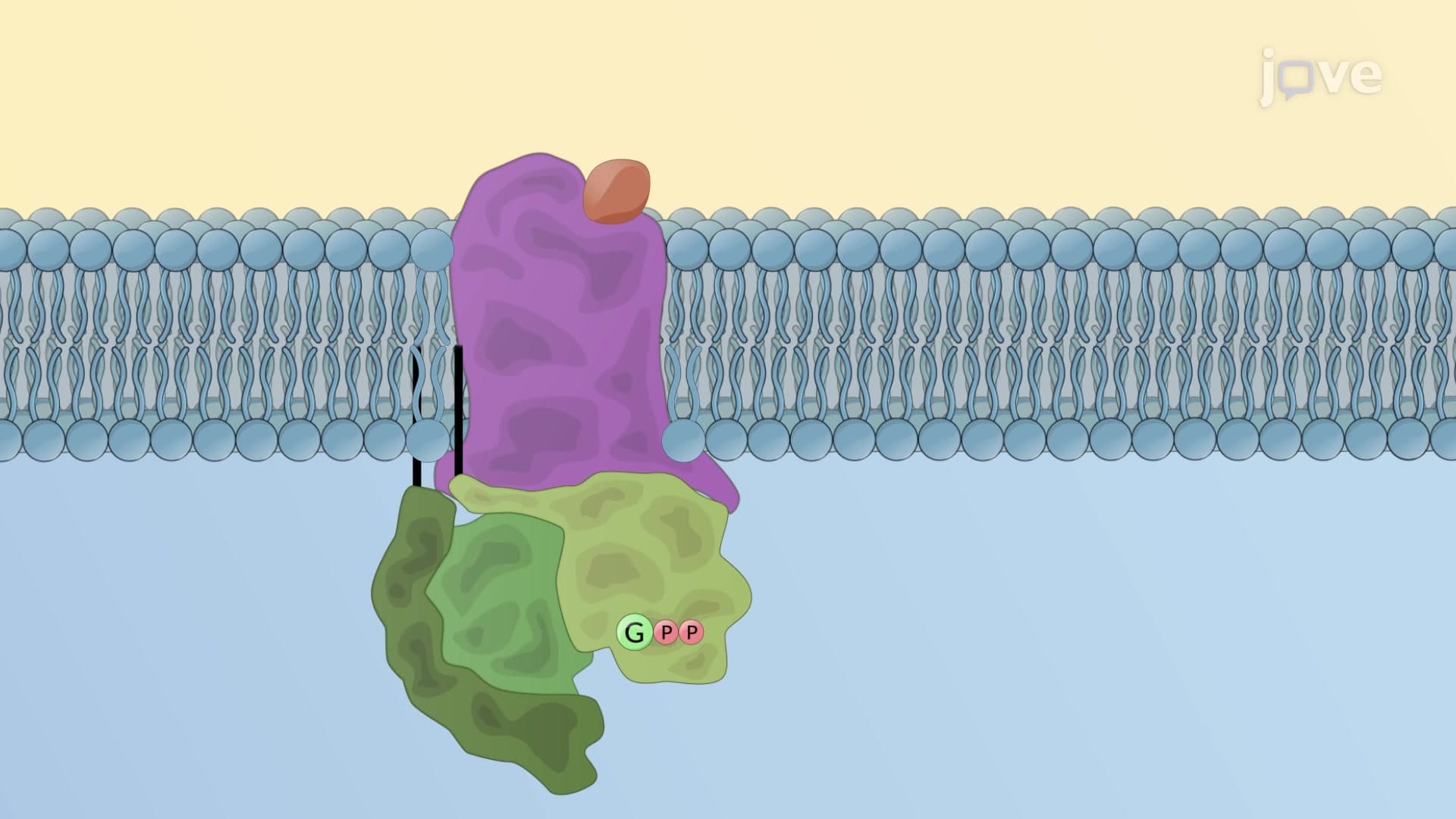
GPCRs share a distinctive seven-transmembrane domain structure that spans the cell membrane like a molecular serpent. The extracellular loops form precise binding pockets that recognize specific ligands with remarkable selectivity. Consider how the beta-adrenergic receptors in cardiac muscle specifically bind epinephrine released during stress, while ignoring thousands of other circulating molecules. This selectivity stems from the unique three-dimensional shape created by the extracellular domains, which act like molecular locks awaiting the correct chemical key.
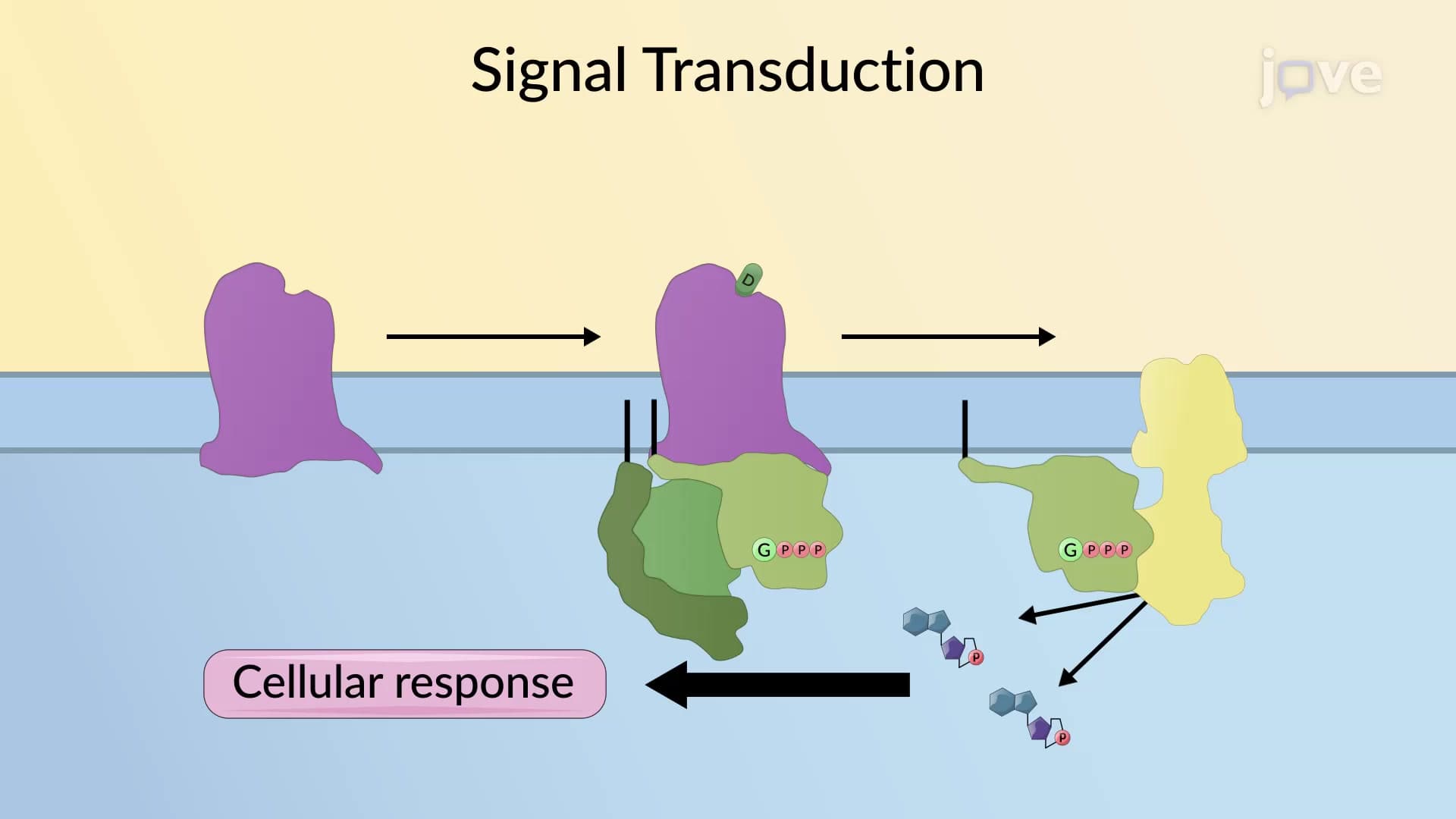
The cytosolic loops, particularly the third intracellular loop, serve as docking stations for heterotrimeric G proteins. These G proteins consist of alpha, beta, and gamma subunits that remain inactive when bound to GDP (guanosine diphosphate). The precision of this molecular architecture explains why pharmaceutical companies invest billions in developing drugs that target specific GPCR subtypes.
When a ligand binds to its GPCR, it triggers a cascade of conformational changes that represent one of biology's most elegant molecular machines. The activated receptor undergoes structural rearrangements that increase its affinity for the G protein complex. This receptor-G protein interaction catalyzes the exchange of GDP for GTP (guanosine triphosphate) on the alpha subunit, effectively "charging" the G protein with energy.
The GTP-bound alpha subunit then dissociates from both the receptor and the beta-gamma dimer, becoming a molecular messenger that activates downstream effector proteins. These effectors include enzymes like adenylyl cyclase, phospholipase C, and ion channels that amplify the original signal thousands of times over. This amplification explains how a single hormone molecule can trigger massive cellular responses.
Understanding GPCR transduction mechanisms has revolutionized medicine. Beta-blockers used in treating hypertension work by blocking beta-adrenergic GPCRs in the heart, preventing excessive stimulation by stress hormones. Opioid painkillers activate mu-opioid receptors in the brain and spinal cord, demonstrating how GPCR targeting can provide therapeutic relief while also explaining addiction potential.
Students preparing for the MCAT or AP Biology exams should focus on connecting GPCR structure to function, particularly how conformational changes enable signal transduction. College biochemistry courses often test understanding of the GDP-GTP exchange mechanism and downstream signaling cascades, making this concept essential for academic success in life sciences.
Related Micro-courses