Loading video…


 24 Concepts
 33.1K views

Video Summary: Voltammetric Techniques Linear Scan E Guide
Ever wonder how forensic labs detect toxic mercury levels in fish samples from the Great Lakes? A linear scan voltammetry guide reveals how scientists use controlled electrical sweeps to identify and quantify specific compounds in complex mixtures. This Voltammetric Techniques Linear Scan E Guide demonstrates how polarography and hydrodynamic voltammetry create characteristic current-potential plots that act like molecular fingerprints. From analyzing heavy metals in drinking water to detecting drug metabolites in clinical samples, these techniques power countless analytical procedures across US laboratories. Watch the full video on JoVE Coach to master this concept with expert-led visuals and step-by-step explanations.
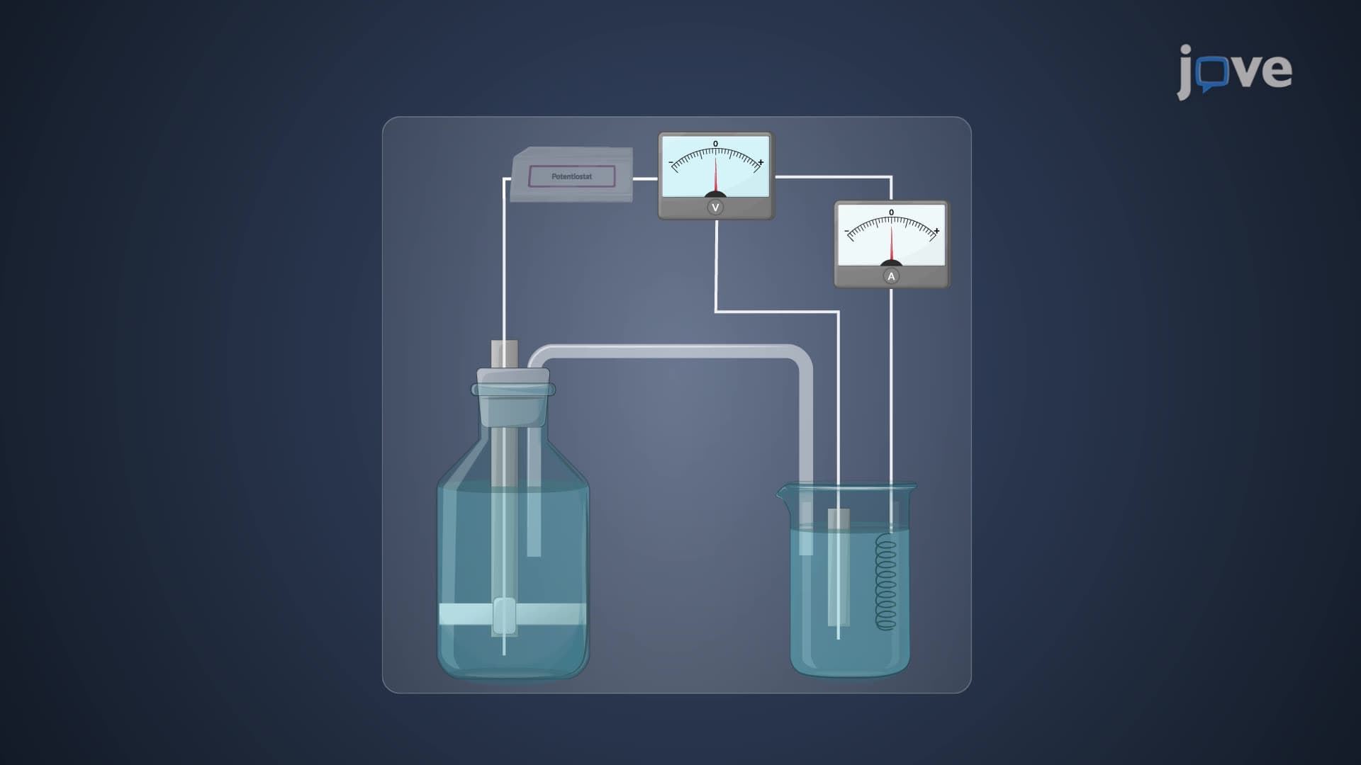
Linear scan voltammetry explained begins with a simple yet powerful concept: applying a steadily changing voltage to an electrode while monitoring the resulting current. Think of it like gradually increasing the pressure on a gas pedal while watching your speedometer, the response tells you about your engine's performance. In voltammetry, the current response reveals information about the chemical species present in solution.
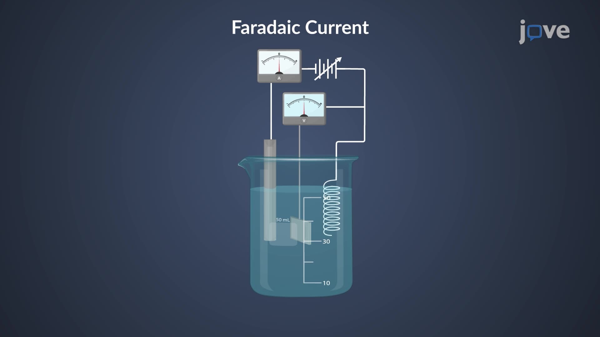
The technique operates on Faraday's laws of electrolysis, where current directly correlates to the rate of electron transfer reactions at the electrode surface. As potential increases linearly with time, different compounds undergo oxidation or reduction at characteristic voltages, creating peaks in the current-voltage plot that serve as molecular signatures.
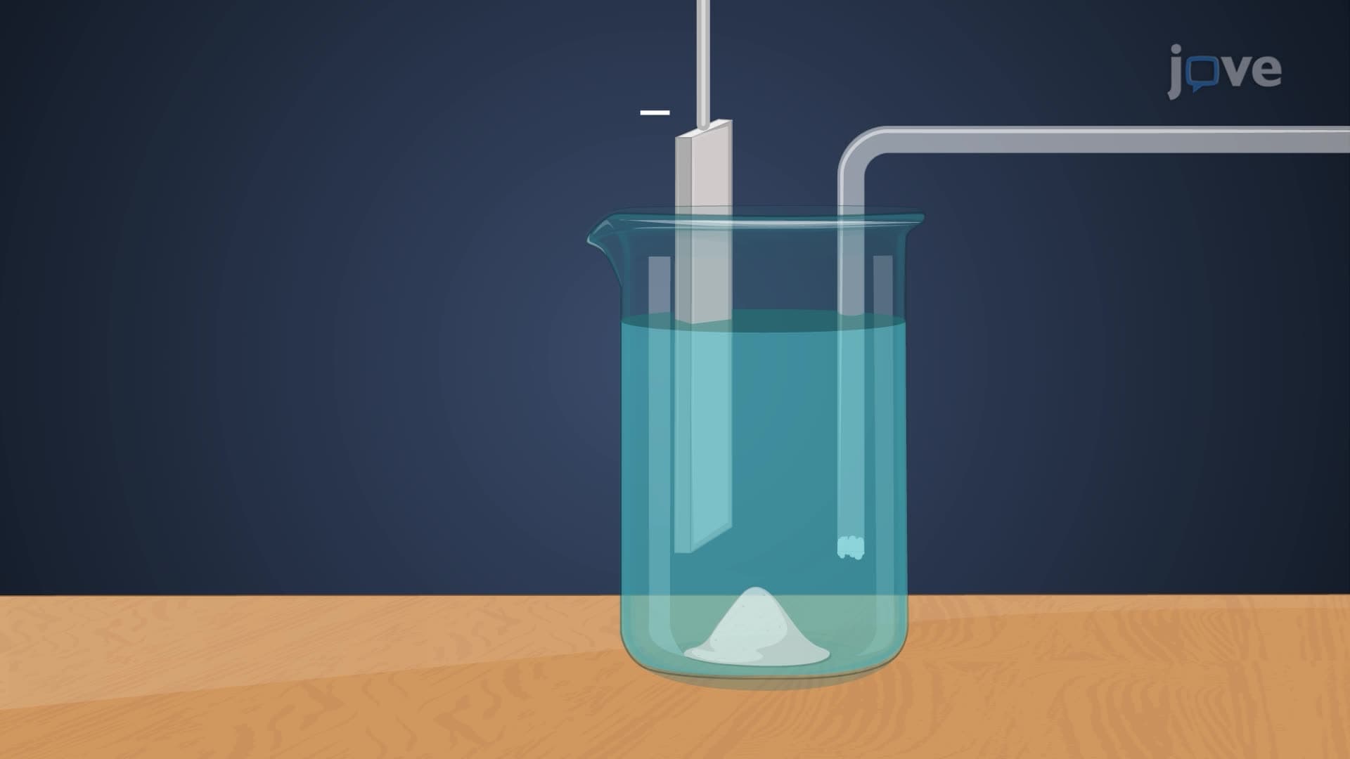
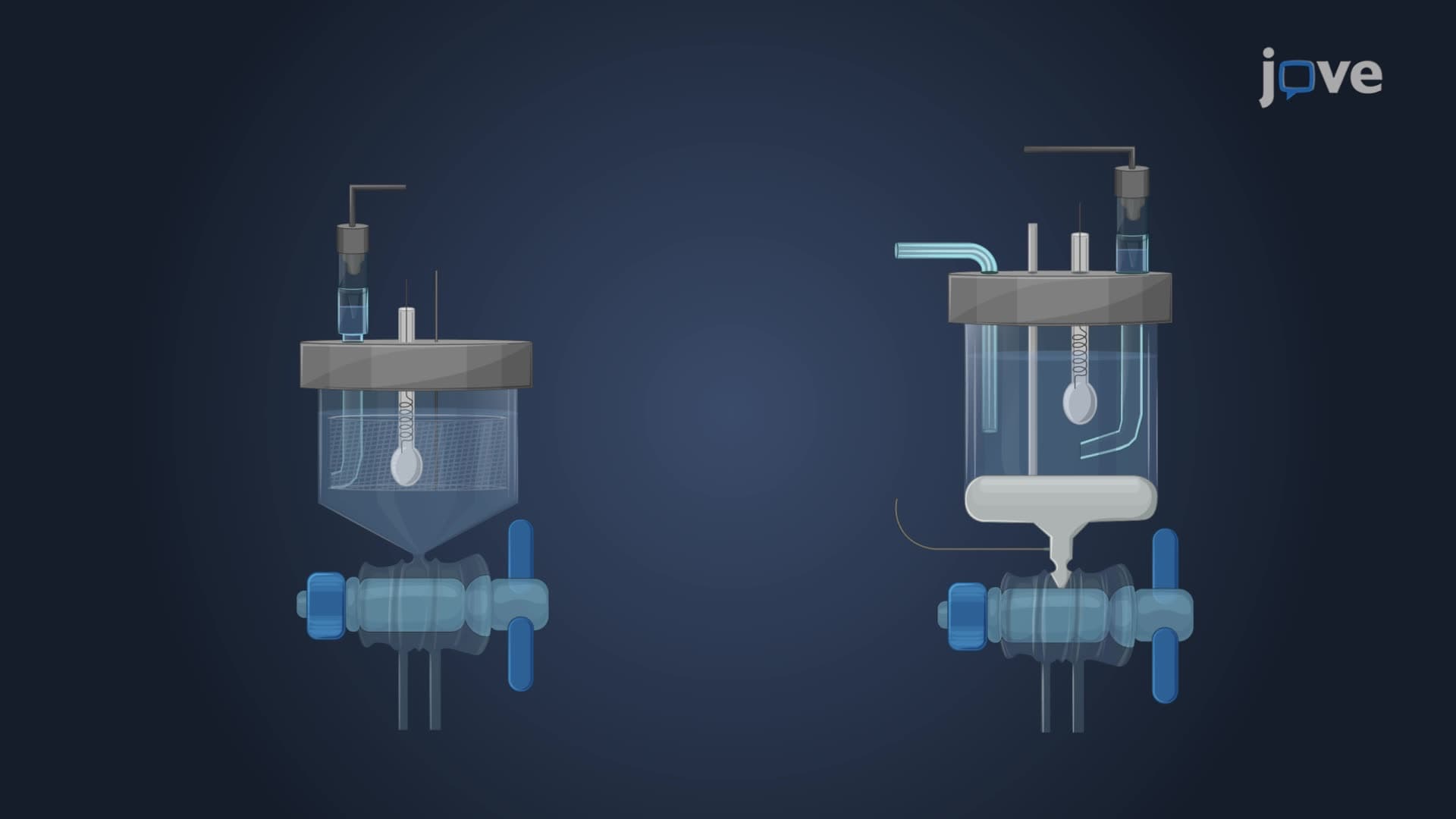
Polarography, the classical form of linear sweep voltammetry, employs a dropping mercury electrode (DME) that creates a fresh, reproducible surface for each measurement. This constantly renewed interface eliminates surface contamination issues that plague other electrode materials. The EPA frequently uses polarographic methods for heavy metal analysis in environmental samples, particularly for lead, cadmium, and zinc determination in water supplies.
The scan rate linear voltammetry parameter critically affects peak shape and resolution. Faster scans (100-1000 mV/s) enhance sensitivity but may compromise resolution between closely spaced peaks. Slower scans (1-50 mV/s) improve peak separation but increase analysis time, a trade-off frequently tested in AP Chemistry and college-level analytical chemistry courses.
The peak potential linear scan occurs when the surface concentration of electroactive species approaches zero, creating maximum current flow. This peak potential serves as a qualitative identifier, while peak height provides quantitative information. For reversible systems, peak potential shifts predictably with concentration according to the Nernst equation.
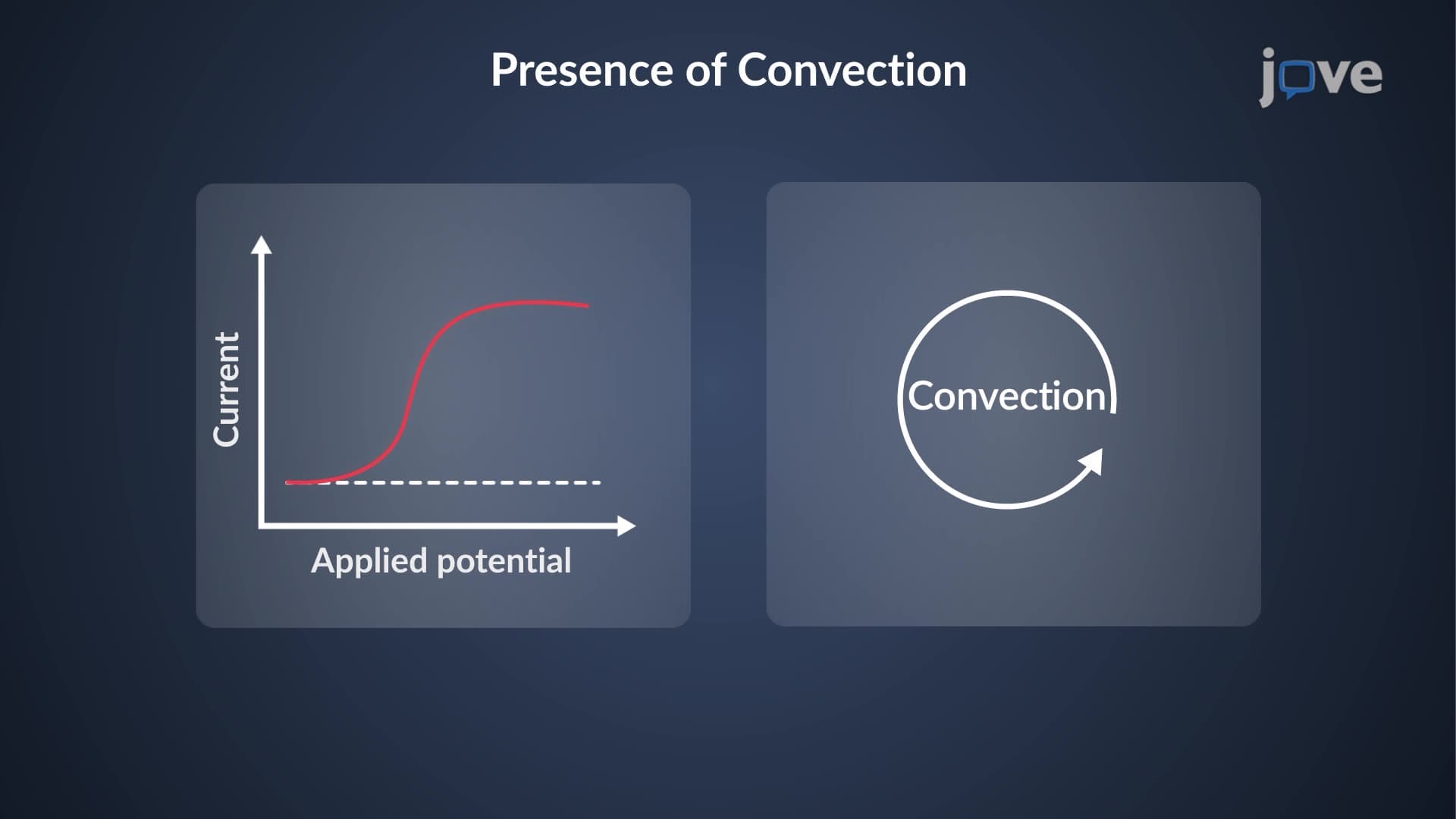
Diffusion limited current LSV dominates the response when mass transport becomes the rate-limiting step. This occurs when all electroactive species reaching the electrode surface immediately react, creating a concentration gradient that drives further diffusion from bulk solution. Understanding this principle helps explain why voltammetric techniques work best for species present in micromolar to millimolar concentrations.
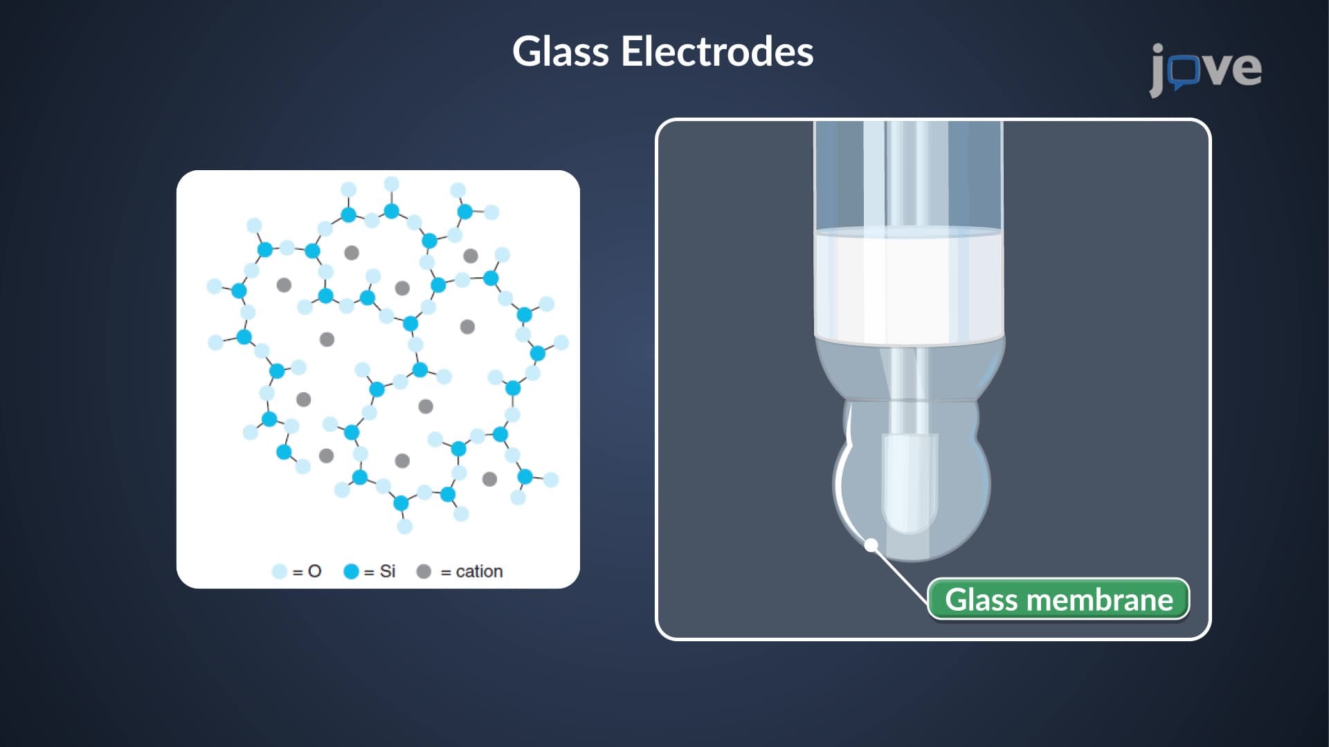
How linear scan voltammetry works in contemporary laboratories extends far beyond mercury electrodes. Solid electrodes like glassy carbon, gold, and platinum enable analysis of organic compounds, pharmaceuticals, and biological molecules. The FDA uses voltammetric methods for drug purity testing, while clinical laboratories employ them for glucose monitoring and neurotransmitter detection.
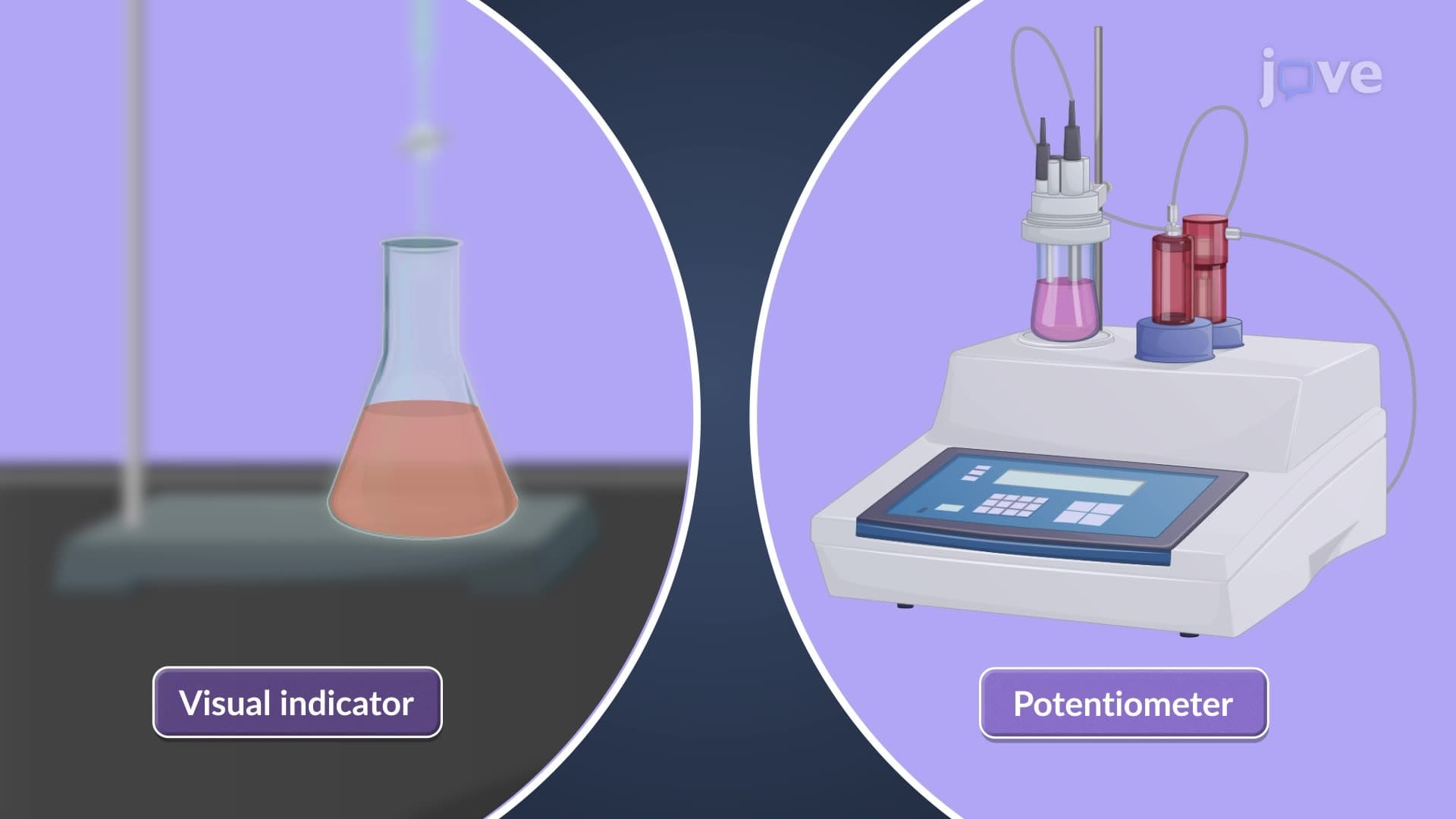
Modern instrumentation automatically compensates for background currents and applies sophisticated data processing to enhance signal-to-noise ratios. This advancement makes voltammetric techniques accessible for routine analysis in undergraduate teaching laboratories across US universities, from basic electrochemistry demonstrations to advanced research projects investigating new electrode materials and electrocatalysts.
Related Micro-courses