Loading subject page…


27,607 views

Video Summary: What Is Bones Upper Limb Ulna
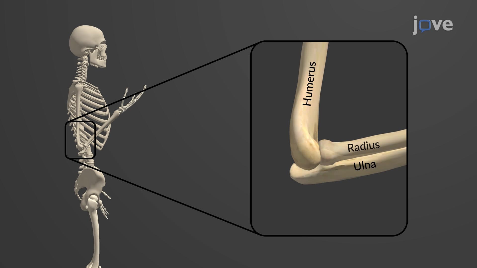
When you bump your "funny bone," you're actually hitting your ulna bone anatomy at the elbow! The ulna bone is the larger of two forearm bones that creates the stable framework allowing you to throw a baseball or type on your laptop. This medial forearm bone features specialized projections like the olecranon and coronoid processes that form your elbow joint with the humerus. Watch the full video on JoVE Coach to master this concept with expert-led visuals and step-by-step explanations.
The ulna bone anatomy represents one of the most structurally important bones in the human arm. As the longer and more medially positioned of the two forearm bones, the ulna provides the primary structural foundation for elbow stability and forearm function. Unlike its partner bone the radius, which rotates during forearm movement, the ulna remains relatively stationary, serving as the stable anchor point for arm mechanics.
The proximal end of the ulna features two critical bony projections that define elbow function. The olecranon process forms the pointed tip of your elbow-the part that hits the table when you lean on it. The coronoid process projects forward, creating a hook-like structure. Between these two processes lies the trochlear notch, a C-shaped depression that perfectly accommodates the trochlea of the humerus. This precise fit creates the hinge joint mechanism that allows your elbow to bend and straighten with remarkable stability.
During flexion, as when doing bicep curls in a gym, the coronoid process slides into the coronoid fossa of the humerus. During extension, the olecranon fits snugly into the olecranon fossa, preventing hyperextension that could damage surrounding structures. This anatomical design explains why your elbow can't bend backward naturally-a protective mechanism that prevents injury during activities like weightlifting or playing tennis.
The ulnar shaft features several important anatomical landmarks. The radial notch, located on the lateral aspect of the proximal ulna, accommodates the head of the radius bone, forming the proximal radioulnar joint. This joint allows the radius to rotate around the ulna during pronation and supination-the movements that turn your palm up or down.
At the distal end, the ulna terminates in the ulnar head, a rounded structure that contributes to wrist mechanics. The ulnar styloid process extends from this head, serving as an attachment point for ligaments that stabilize the wrist. A triangular fibrocartilage complex separates the ulnar head from the carpal bones, providing cushioning and stability during wrist movement.
Understanding ulna bone anatomy proves essential for students preparing for the MCAT, where questions often focus on joint mechanics and bone relationships. In AP Biology courses, the ulna serves as an excellent example of how bone structure directly relates to function. Physical therapy students studying for NPTE examinations must understand ulnar anatomy to treat conditions like tennis elbow or ulnar nerve entrapment. Medical students encounter ulnar fractures frequently in orthopedic rotations, making this knowledge clinically relevant for future physicians.
Related Micro-courses