Loading video…


 6 Concepts
 27.6K views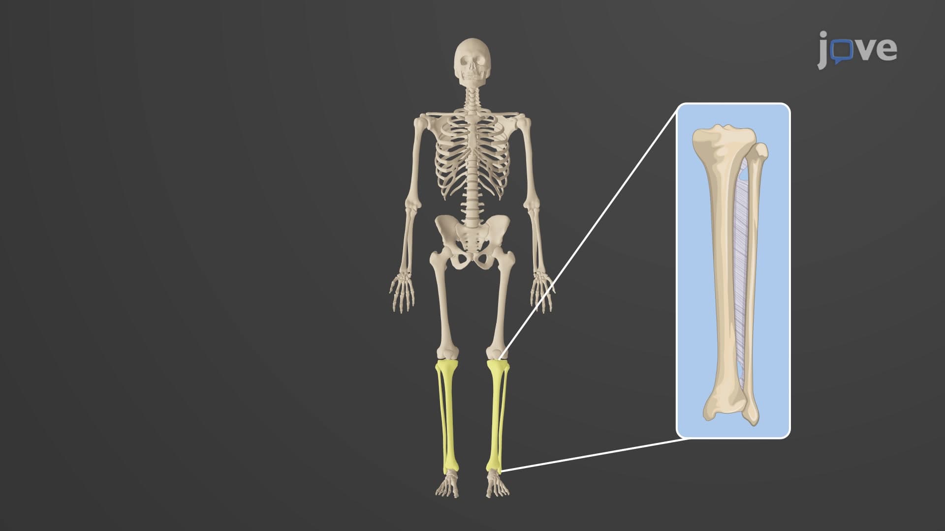

Video Summary: Bones Lower Limb Tibia and Fibula Explained
Ever wondered why soccer players often suffer "shin splints" or why ankle injuries are so common in basketball? The tibia fibula anatomy lower limb reveals how these two crucial bones work together to support your entire body weight while enabling complex movements. When NBA players like Stephen Curry recover from ankle injuries, understanding the intricate relationship between the tibia and fibula becomes essential for proper rehabilitation. Watch the full video on JoVE Coach to master this concept with expert-led visuals and step-by-step explanations.
The lower leg bones anatomy forms a sophisticated mechanical system that balances strength with mobility. The tibia, commonly called the "shinbone," ranks as the second-largest bone in the human body and bears approximately 80-90% of the body's weight during standing and walking. In contrast, the slender fibula primarily serves as a muscle attachment site and ankle stabilizer, handling minimal weight-bearing responsibilities.
The tibial anatomy reveals three distinct regions optimized for different functions. The proximal end features the tibial plateau medial condyle and lateral condyle, which create a stable platform for femoral articulation at the knee joint. This design proves crucial for athletes-consider how NFL linemen must absorb tremendous impact forces during blocking maneuvers. The tibial tuberosity crest, located on the anterior surface, serves as the attachment point for the patellar tendon, making it essential for powerful knee extension movements seen in jumping and sprinting.
The tibial shaft demonstrates remarkable engineering, with its triangular cross-section providing maximum strength while minimizing weight. Medical students studying for the MCAT often focus on how this shape resists bending forces during activities like marathon running. The distal end features the medial malleolus tibia, the prominent bump on the inner ankle that provides medial stability and articulates with the talus bone.
Despite its narrow diameter, the leg bone tibia fibula partnership demonstrates perfect functional complementarity. The fibula head lateral malleolus system creates a socket-like structure around the talus, preventing excessive side-to-side ankle movement. This proves critical in sports requiring rapid direction changes-basketball players depend on this stability when executing crossover dribbles or defensive slides.
Understanding what are the tibia and fibula bones in anatomy becomes essential for healthcare professionals treating lower extremity injuries. Emergency room physicians at major US trauma centers, such as those at Johns Hopkins or Mayo Clinic, frequently encounter tibial fractures from motor vehicle accidents or sports injuries. The fibula's role in ankle stability makes lateral malleolus fractures particularly significant for patient mobility and recovery outcomes.
Related Micro-courses