Loading video…


 6 Concepts
 27.6K views

Video Summary: What Is Bones Upper Limb Radius
Ever wonder why you can flip your palm up and down while keeping your elbow still? The radius bone anatomy reveals the secret behind this remarkable forearm flexibility. This lateral forearm bone works like a sophisticated pivot system, with its unique nail-shaped head rotating within the elbow joint. Medical students at Johns Hopkins University spend considerable time studying the radius because fractures here-especially near the wrist-are among the most common injuries in emergency rooms across America. Watch the full video on JoVE Coach to master this concept with expert-led visuals and step-by-step explanations.
The radius stands as one of anatomy's most functionally elegant bones, serving as the lateral powerhouse of the forearm. Unlike its medial partner the ulna, the radius bears the primary load during wrist movements and plays the starring role in forearm rotation. Pre-med students often struggle initially with radius identification, but understanding its distinctive "nail-shaped" head makes recognition straightforward on both cadaveric specimens and radiographic images.
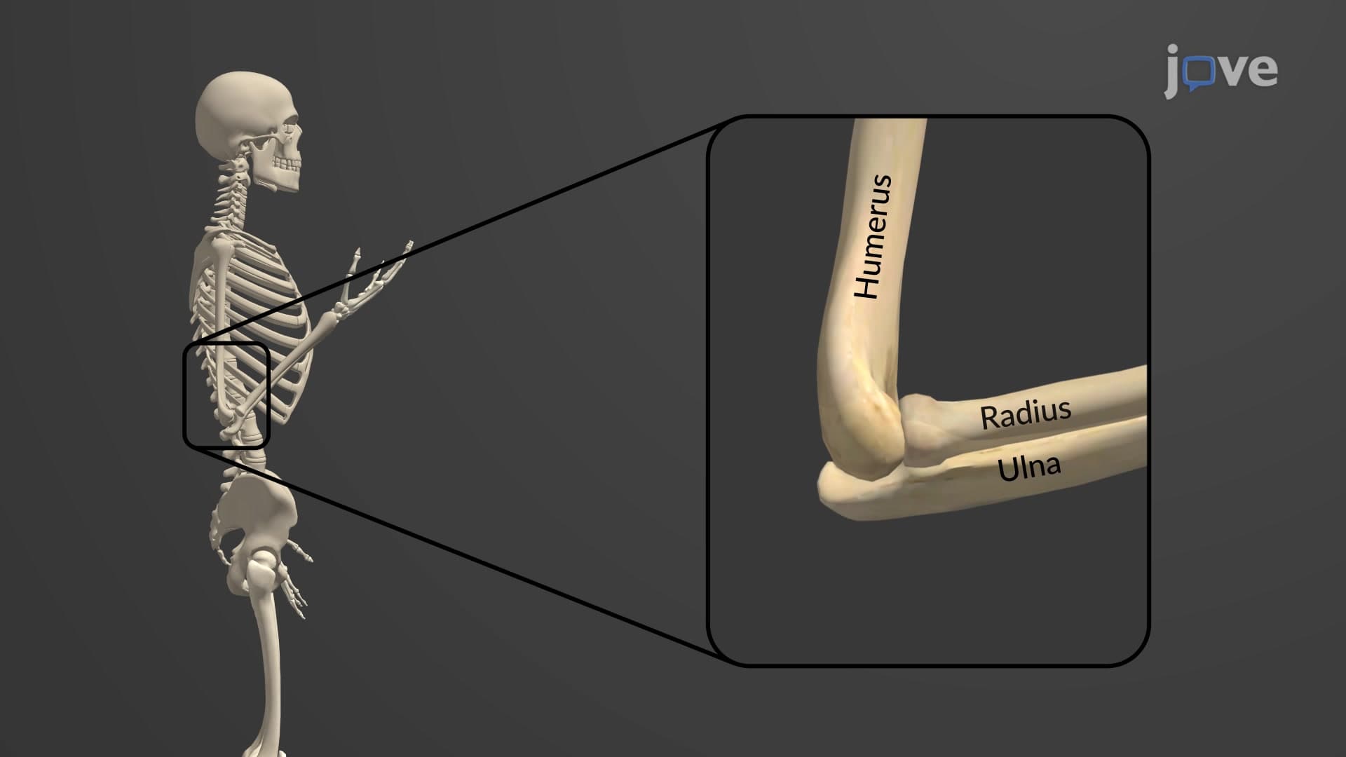
The radial head neck tuberosity complex represents mechanical engineering at its finest. The circular radial head articulates with the humeral capitulum, creating the radiocapitellar joint that enables smooth elbow flexion. During pronation and supination-the movements that rotate your palm-this head spins within the radial notch of the ulna like a wheel on an axle. The radial tuberosity, located just below the neck, serves as the insertion point for the biceps brachii muscle, explaining why bicep curls involve both elbow flexion and slight forearm rotation.
The distal radius anatomy becomes critically important in clinical settings, particularly in orthopedic emergency departments. The radial styloid process extends further distally than its ulnar counterpart, creating the natural anatomical "snuffbox" visible when you extend your thumb. This prominence makes distal radius fractures-often called Colles fractures-extremely common, especially among elderly patients who fall on outstretched hands. Emergency medicine residents learn to identify these fractures quickly, as they account for approximately 15% of all bone fractures treated in US hospitals.
The radius ulnar notch connection appears frequently on MCAT passages dealing with biomechanics, while AP Biology exams often test understanding of bone tissue structure using the radius as an example. Medical students encounter detailed radius anatomy in their musculoskeletal system blocks, with particular emphasis on the lateral forearm radius positioning during physical examination techniques. Understanding these anatomical relationships proves essential for future healthcare providers, as radius-related injuries span from pediatric "nursemaid's elbow" to geriatric osteoporotic fractures.
Related Micro-courses