Loading video…


 6 Concepts
 27.6K views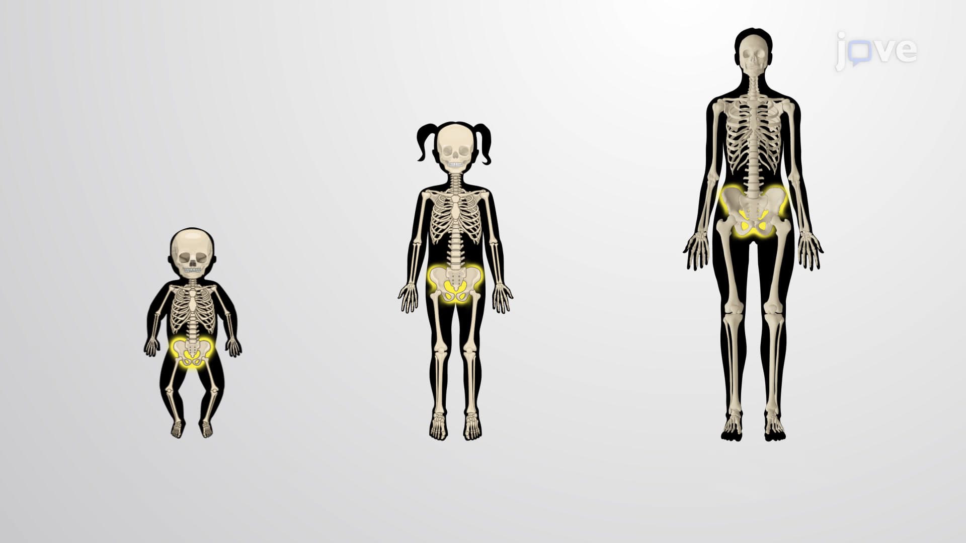

Video Summary: Changes in the Appendicular Skeleton Explained
Did you know that your arms and legs were once tiny cartilage buds smaller than a pencil eraser? Appendicular skeleton changes biology reveals how these limb buds transform into the complex bone structures that allow a basketball player at Duke University to dunk or a gymnast to perform on uneven bars. Understanding Changes in The Appendicular Skeleton Explained helps us grasp why teenage growth spurts happen and why elderly adults face increased fracture risks. Watch the full video on JoVE Coach to master this concept with expert-led visuals and step-by-step explanations.
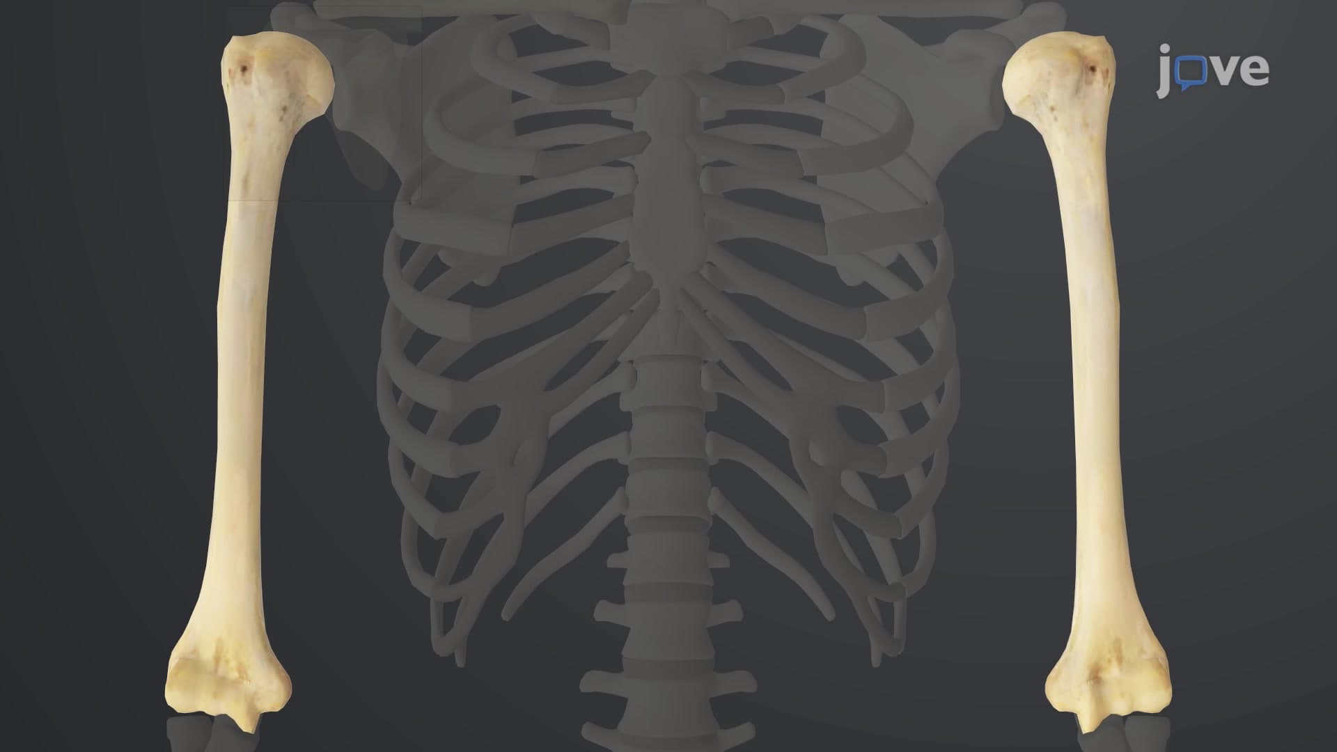
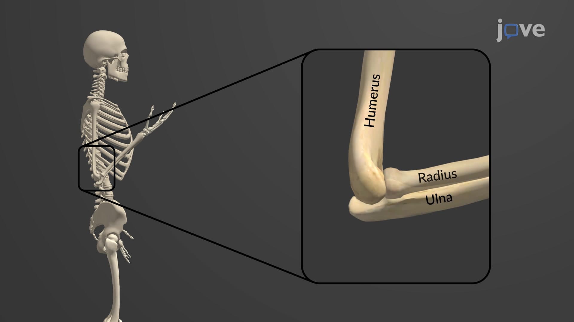
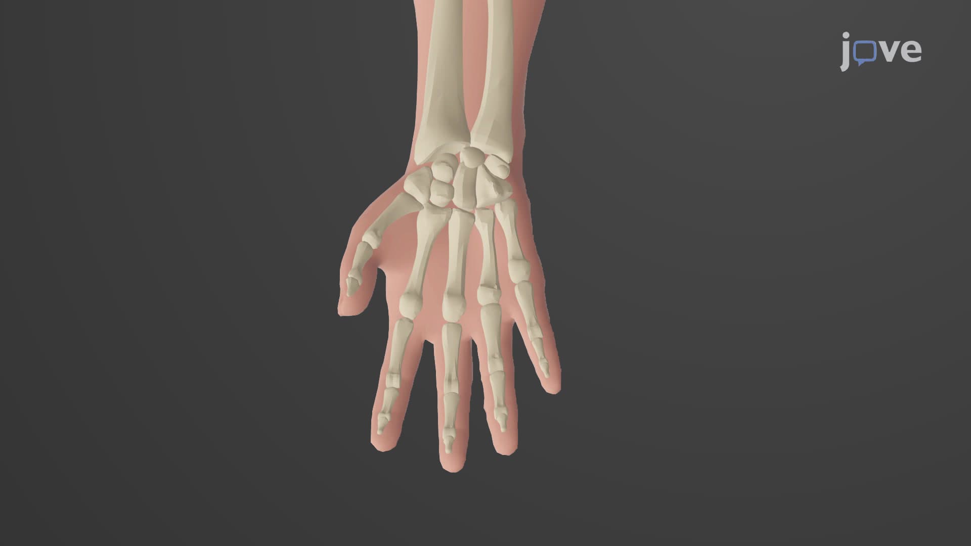
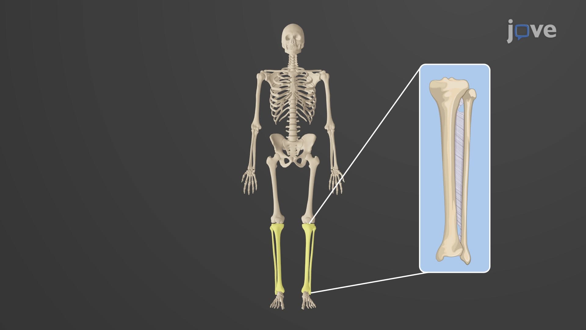
The appendicular skeleton changes biology encompasses a fascinating journey from embryonic development through aging. Unlike the axial skeleton (skull, spine, ribs), the appendicular skeleton-comprising arms, legs, pelvis, and shoulder girdle-undergoes distinct developmental patterns that directly impact human mobility and function.
During the first eight weeks of human development, appendicular skeleton development begins as small tissue projections called limb buds. These structures initially consist entirely of hyaline cartilage, serving as templates for future bones. The transformation process varies significantly between different bones. Most appendicular bones develop through endochondral ossification, where cartilage gradually gets replaced by bone tissue in a highly organized manner.
However, the clavicles (collarbones) represent a unique exception in appendicular skeleton variation explained. These bones develop through intramembranous ossification, forming directly from mesenchymal tissue without a cartilage intermediate. This difference explains why clavicular fractures heal differently than other appendicular bones-a concept frequently tested in medical school anatomy courses and MCAT physiology sections.
What changes occur in the appendicular skeleton varies dramatically with age and biological sex. During childhood, lower limbs experience rapid growth, achieving proportional balance with the trunk by age ten. This growth surge explains why children often appear "top-heavy" in early years.
Sex differences appendicular skeleton become particularly pronounced during puberty. Female pelvic bones undergo significant broadening to accommodate potential childbearing, with changes continuing until approximately age 25. These modifications involve both increased bone width and altered pelvic angles-concepts crucial for understanding obstetric anatomy in nursing programs and medical school curricula.
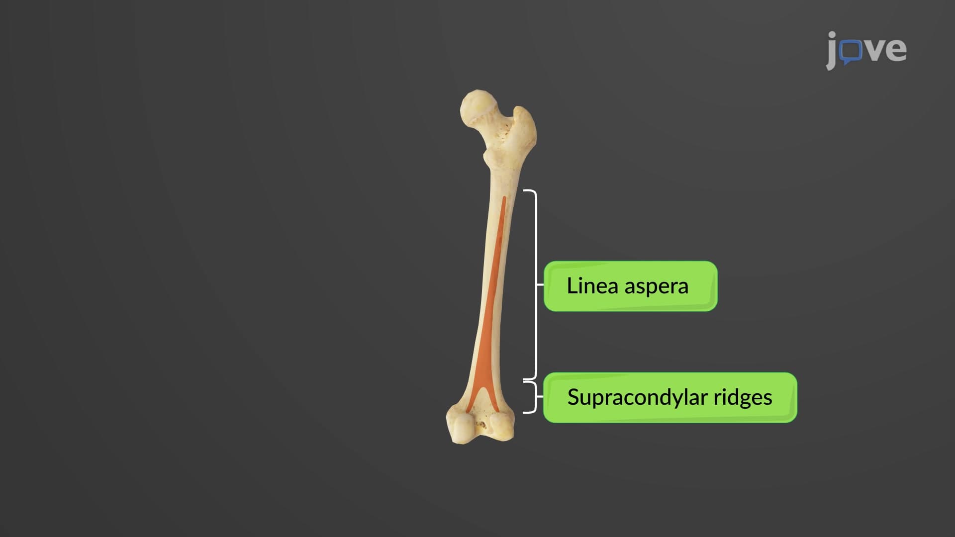
After age 25, healthy individuals maintain bone homeostasis through balanced resorption and deposition. However, aging appendicular changes accelerate after middle age when bone breakdown exceeds formation. This imbalance particularly affects weight-bearing bones like the femur, making the femoral neck a common fracture site in elderly patients-a critical consideration for physical therapy students and healthcare providers.
The limb bone variation observed in aging populations reflects decreased osteoblast activity and increased osteoclast function, processes frequently examined in biochemistry courses and tested on standardized exams like the USMLE Step 1.
Related Micro-courses