Loading subject page…


138,522 views

Video Summary: What Is Atomic Force Microscopy
Did you know scientists can now "touch" individual atoms to create incredibly detailed images? Atomic force microscopy represents one of the most powerful imaging techniques available today, capable of magnifying samples up to 1,000,000 times-far beyond traditional optical microscopes. Researchers at Stanford University use AFM to study everything from cancer cells to computer chip surfaces with nanometer precision. What is Atomic Force Microscopy becomes clear when you see how this technology uses a tiny probe to map surface features atom by atom, creating three-dimensional images that reveal the hidden world of the very small. Watch the full video on JoVE Coach to master this concept with expert-led visuals and step-by-step explanations.
Atomic force microscopy represents a revolutionary advancement in imaging technology that has transformed our ability to visualize matter at the nanoscale. Unlike conventional microscopy techniques that rely on light or electron beams, AFM operates on an entirely different principle-mechanical interaction between a sharp probe and the sample surface. This atomic force microscopy definition encompasses both the physical mechanism and the unprecedented resolution capabilities that make it invaluable for modern scientific research.
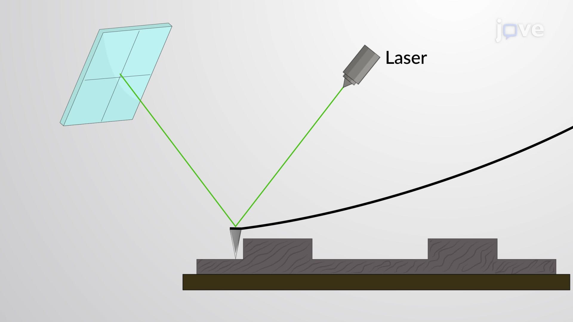
The core principle behind AFM involves a nanoscale probe mounted on a flexible cantilever that physically scans across the sample surface. As the probe encounters surface features-whether hills, valleys, or individual molecules-the cantilever bends in response to attractive or repulsive forces. A sophisticated laser detection system monitors these minute deflections, with the reflected laser beam hitting a position-sensitive photodetector that converts mechanical movement into electrical signals. This atomic force microscopy overview reveals how mechanical forces at the atomic level translate into detailed topographical maps.
The extraordinary magnification potential of AFM-up to 1,000,000 times-stems from the probe's ability to interact with individual atoms and molecules. Research facilities like those at MIT and the National Institute of Standards and Technology (NIST) routinely use AFM to characterize materials with sub-nanometer precision, enabling breakthrough discoveries in fields ranging from semiconductor manufacturing to drug development.
Understanding atomic force microscopy becomes particularly important for students preparing for advanced coursework and standardized exams. The AP Physics exam frequently includes questions about microscopy principles, while the MCAT tests understanding of imaging techniques used in biological research. College-level materials science and biology courses extensively cover AFM applications, from studying protein folding dynamics to characterizing nanoparticle surfaces.
In practical applications, AFM has revolutionized quality control in the semiconductor industry. Companies like Intel and IBM use AFM to inspect computer chips at the nanoscale, ensuring defect-free manufacturing. Biomedical researchers employ AFM to study living cells in real-time, observing processes like membrane dynamics and cellular division as they occur. This atomic force microscopy study guide perspective emphasizes how theoretical knowledge translates into technological innovation and scientific discovery.
Related Micro-courses