Loading video…


 17 Concepts
 138.5K views

Video Summary: What Is Immunofluorescence Microscopy
Ever wonder how researchers at Johns Hopkins can pinpoint a single cancerous cell among millions of healthy ones? Immunofluorescence microscopy transforms invisible cellular targets into glowing beacons of light, revolutionizing medical diagnosis and research. This powerful technique uses fluorescent antibodies to illuminate specific proteins or antigens within tissues and cells, making the invisible visible. From detecting Alzheimer's protein plaques in brain tissue to diagnosing lupus through blood tests, what is immunofluorescence microscopy becomes clear when you see its life-saving applications in American hospitals and research labs. Watch the full video on JoVE Coach to master this concept with expert-led visuals and step-by-step explanations.
Immunofluorescence microscopy represents a cornerstone technique in modern biological research and clinical diagnostics, combining the specificity of immunology with the visual power of fluorescence. This method leverages the natural ability of antibodies to recognize and bind specific molecular targets, enhanced by fluorescent molecules that make these interactions visible under specialized microscopes.
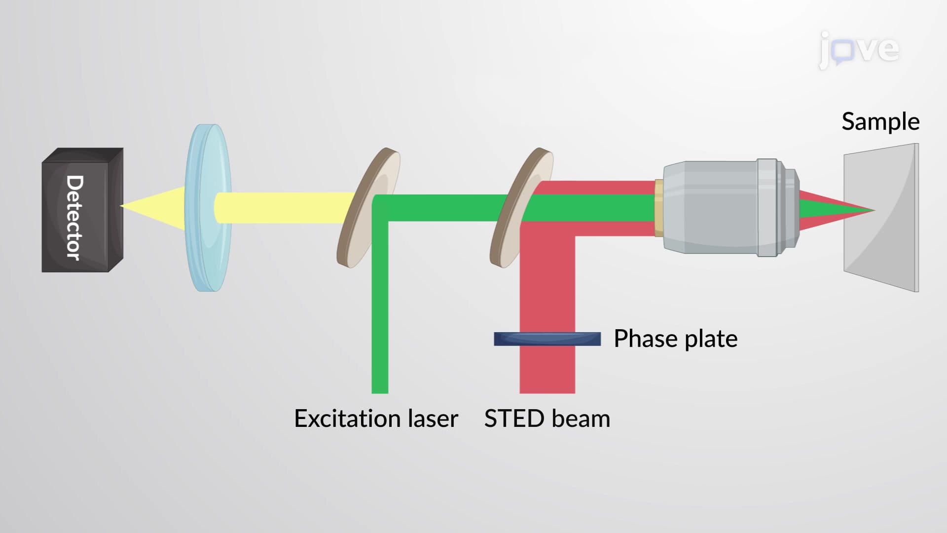
The underlying physics involves exciting fluorophore molecules with high-energy light, typically blue or ultraviolet, causing electrons to jump to higher energy states. When these excited electrons return to their ground state, they release energy as light of a longer wavelength-often appearing as green, red, or yellow fluorescence. This Stokes shift principle ensures that the emitted light differs from the excitation light, allowing clear visualization of labeled targets.
The technique branches into two distinct methodologies, each with specific advantages. Direct immunofluorescence employs primary antibodies already conjugated with fluorophores, creating a one-step binding process. This approach proves particularly valuable in clinical settings like dermatopathology labs at Mayo Clinic, where rapid diagnosis of skin conditions requires detecting specific protein deposits in tissue biopsies.
Indirect immunofluorescence utilizes a two-step process: unlabeled primary antibodies first bind the target, followed by fluorophore-conjugated secondary antibodies that recognize the primary antibodies. This amplification system produces stronger signals because multiple secondary antibodies can bind each primary antibody, significantly enhancing detection sensitivity-crucial for identifying low-abundance targets in research applications.
American healthcare systems extensively utilize immunofluorescence for autoimmune disease diagnosis. The antinuclear antibody (ANA) test, a standard screening tool for conditions like systemic lupus erythematosus, employs indirect immunofluorescence to detect patient antibodies that react against cellular components. Similarly, direct immunofluorescence helps pathologists at institutions like Cleveland Clinic identify abnormal protein aggregations in neurodegenerative diseases, including the amyloid plaques characteristic of Alzheimer's disease.
Students preparing for the MCAT will encounter immunofluorescence concepts in both biology and biochemistry sections, particularly regarding protein structure and cellular biology. AP Biology curricula increasingly emphasize this technique when discussing cell communication and immune system function. Understanding fluorescence principles also appears in college-level cell biology and biochemistry courses, where students learn to interpret fluorescence microscopy data and design experiments using appropriate antibody-fluorophore combinations.
Related Micro-courses