Loading video…


 17 Concepts
 138.5K views

Video Summary: What Is Immunogold Electron Microscopy
Ever wondered how scientists at Johns Hopkins can pinpoint the exact location of a single protein inside a cell? Immunogold electron microscopy revolutionizes cellular imaging by attaching tiny gold particles to antibodies, creating dark spots that reveal where specific molecules hide within cellular structures. This powerful technique combines the precision of immunolabeling with electron microscopy's incredible magnification, allowing researchers to map proteins with nanometer-level accuracy. Watch the full video on JoVE Coach to master this concept with expert-led visuals and step-by-step explanations.
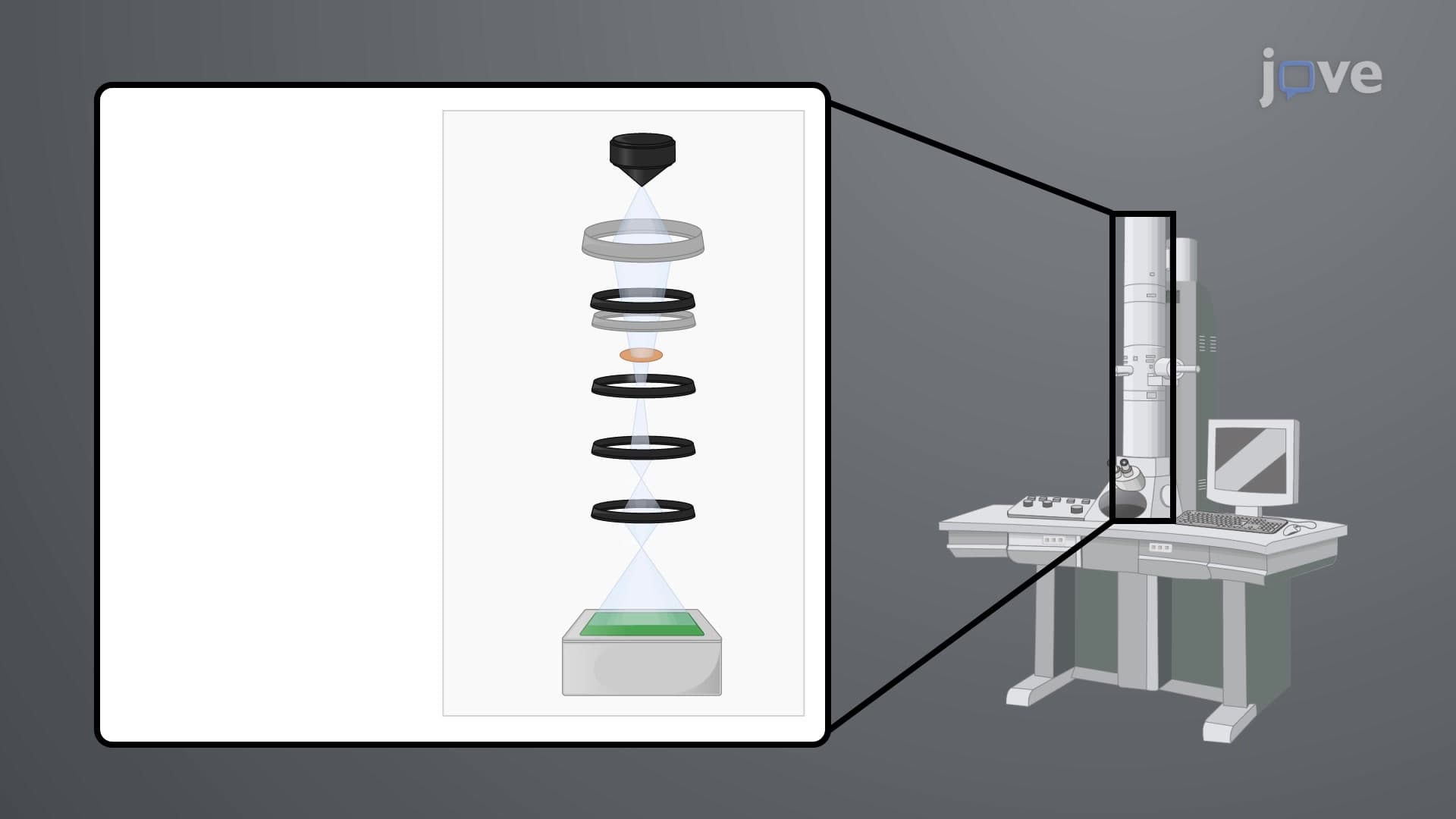
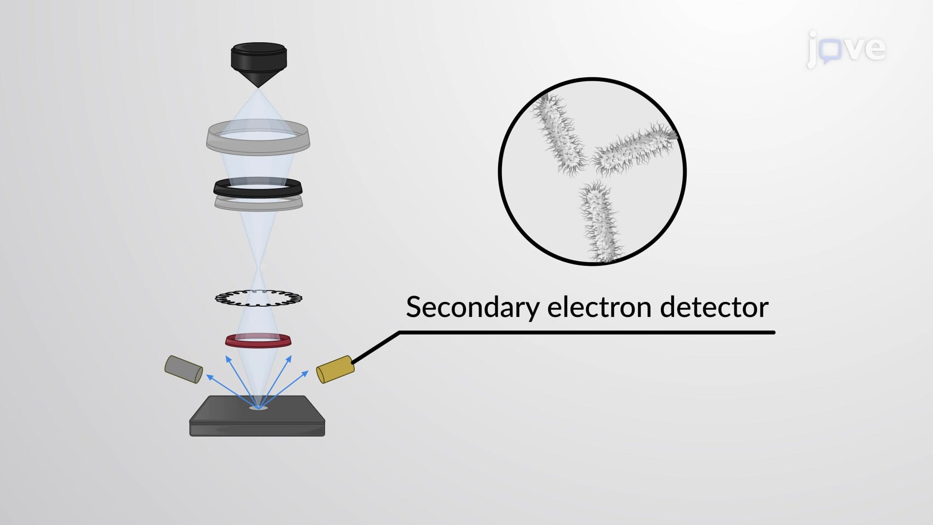
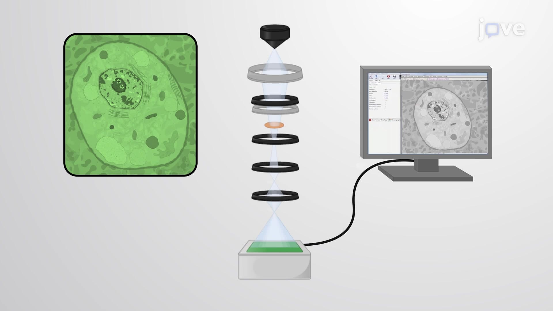
Immunogold electron microscopy represents a breakthrough fusion of immunological specificity and electron microscopy's ultra-high resolution. This technique leverages gold nanoparticles' unique electron-dense properties to create visible markers for target molecules within cellular structures. When electrons encounter gold particles during imaging, they scatter dramatically, producing distinct dark spots that stand out against lighter cellular components.
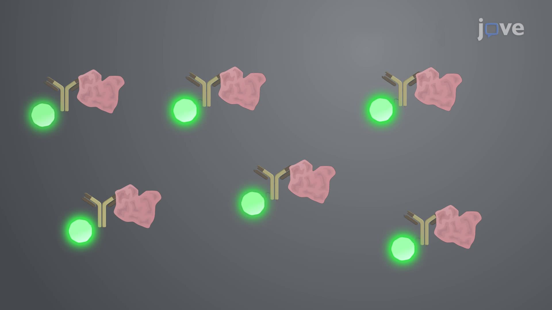
The foundation of this method lies in antibody-antigen interactions. Researchers can attach gold particles directly to primary antibodies (direct method) or use gold-labeled secondary antibodies that bind to unlabeled primary antibodies (indirect method). The indirect approach dominates research applications because secondary antibodies can bind multiple sites on primary antibodies, amplifying the signal and improving detection sensitivity-crucial when studying low-abundance proteins.
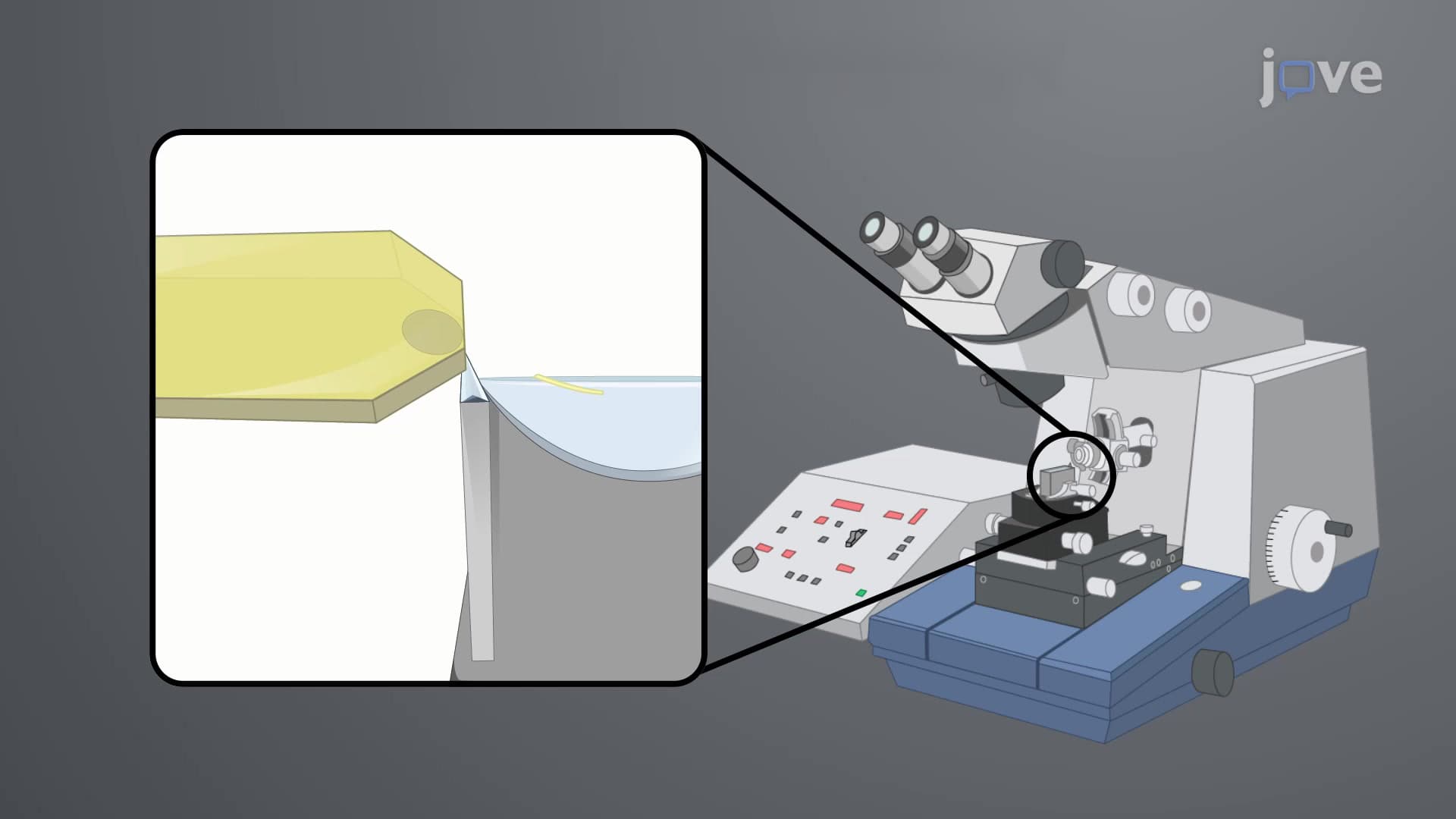
Three main preparation methods serve different research needs. Post-embedding immunolabeling involves embedding samples in resin, creating ultrathin sections, then applying gold-labeled antibodies. This approach works excellently for studying intracellular protein distributions and is commonly used in cancer research labs across institutions like MD Anderson Cancer Center.
Pre-embedding techniques reverse this order-samples receive antibody treatment first, followed by resin embedding and sectioning. This method excels when studying surface proteins or when antibody penetration might be limited post-embedding. Whole-mount preparations skip resin embedding entirely, allowing direct antibody application to samples on electron microscopy grids, ideal for studying isolated organelles or macromolecular complexes.
Immunogold electron microscopy plays vital roles in medical diagnostics and pharmaceutical development. Pathologists use it to identify viral particles in patient samples, while pharmaceutical companies employ it to track drug distribution within cells. At the CDC, researchers utilize this technique to study pathogen-host interactions, providing crucial data for vaccine development.
This concept frequently appears in advanced biology courses and standardized exams. MCAT students encounter immunogold microscopy in cell biology sections, while AP Biology students may see it in molecular biology contexts. Understanding the principle of signal amplification through indirect labeling helps students grasp broader immunological concepts tested across multiple exam formats.
The technique's integration of multiple scientific disciplines-immunology, materials science, and microscopy-makes it an excellent example of interdisciplinary research approaches increasingly emphasized in modern curricula.
Related Micro-courses