Loading video…


 17 Concepts
 138.5K views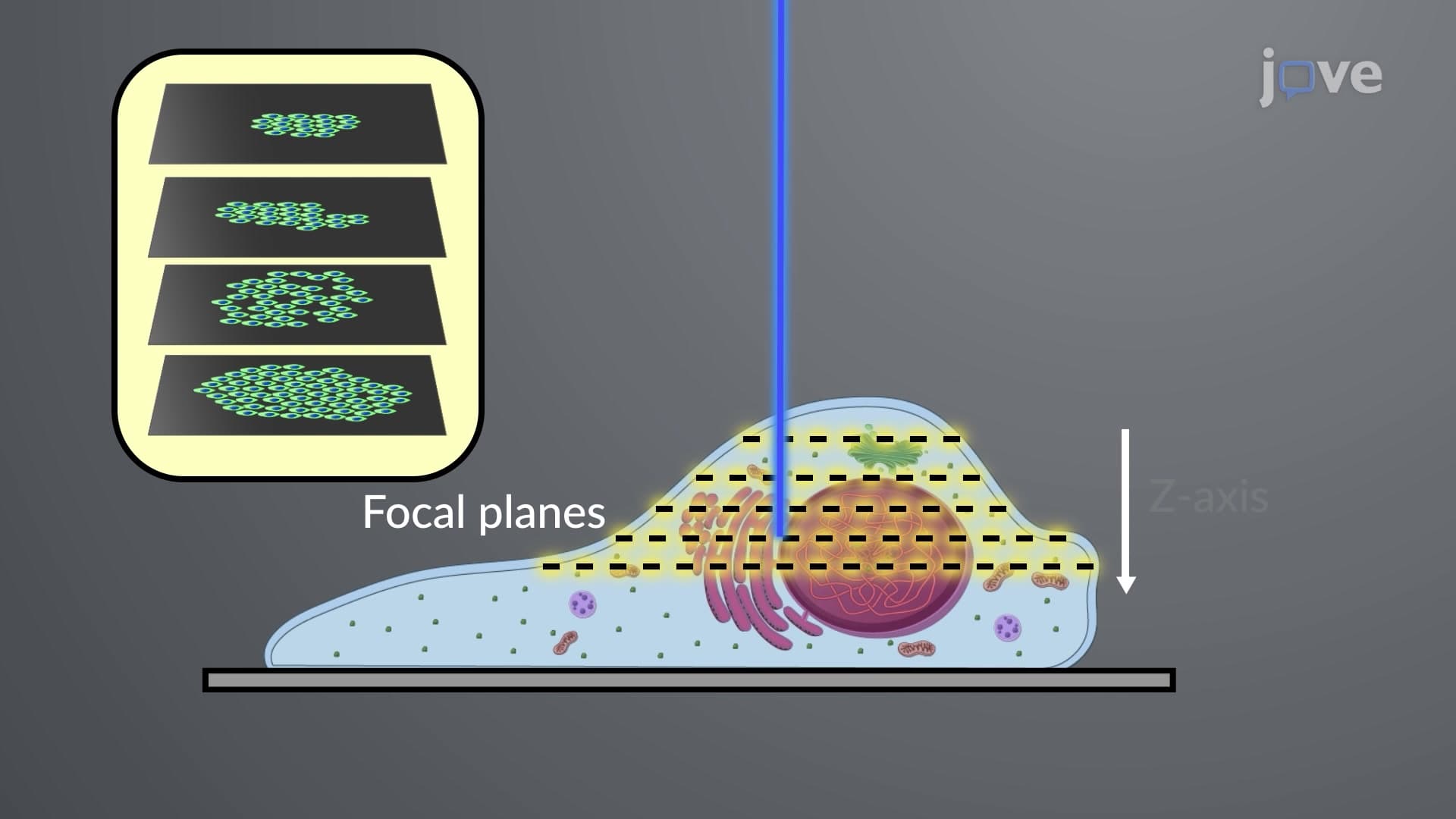

Video Summary: What Is Confocal Fluorescence Microscopy
Ever wondered how scientists at the CDC create those stunning 3D images of viruses that look like they're floating in space? Confocal fluorescence microscopy revolutionizes biological imaging by using laser beams and precise pinholes to capture razor-sharp optical slices of specimens. This advanced technique allows researchers at institutions like Johns Hopkins to build detailed three-dimensional reconstructions from multiple focal planes, eliminating the blurriness that plagues traditional fluorescence microscopy. Watch the full video on JoVE Coach to master this concept with expert-led visuals and step-by-step explanations.
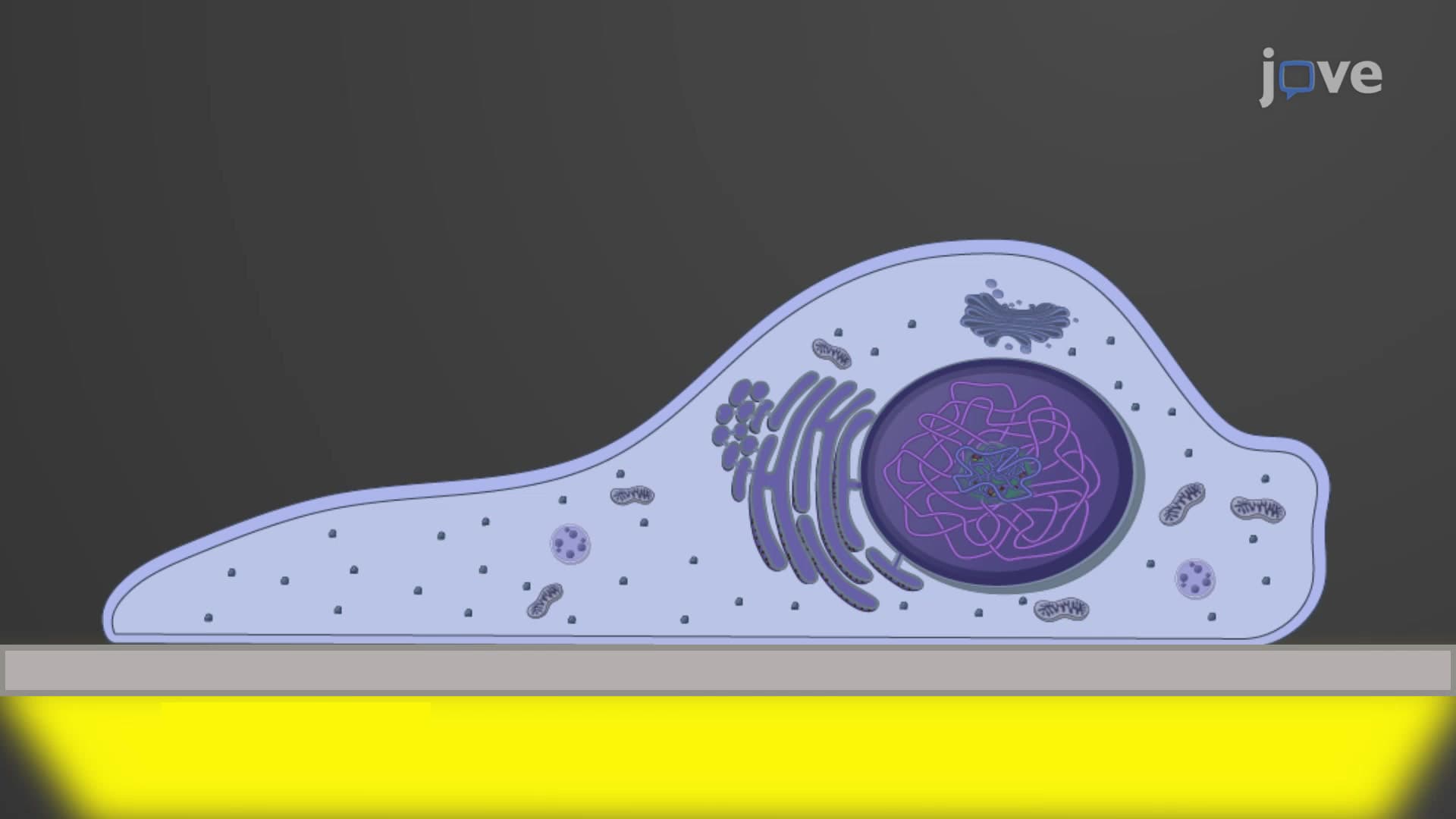
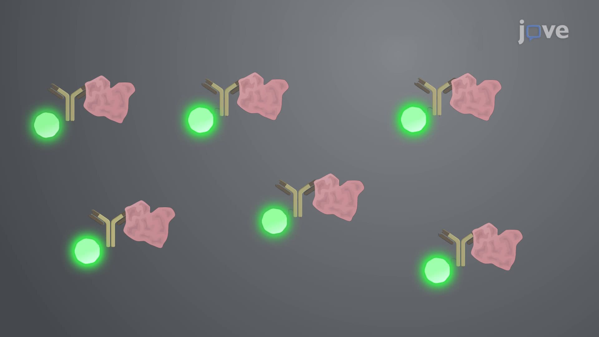
Confocal fluorescence microscopy represents a quantum leap in biological imaging technology, fundamentally changing how scientists visualize cellular structures and molecular processes. Unlike traditional fluorescence microscopes that illuminate entire specimens simultaneously, confocal systems use focused laser beams to excite fluorophores at precisely defined points within the sample. This point-by-point scanning approach, combined with sophisticated pinhole apertures, creates images with unprecedented clarity and depth resolution.
The genius of confocal fluorescence microscopy lies in its dual-pinhole system. The illumination pinhole focuses laser light to a diffraction-limited spot, while the detection pinhole acts as a spatial filter, rejecting out-of-focus light from above and below the focal plane. This optical sectioning capability allows researchers to peer into thick specimens layer by layer, much like a medical CT scanner creates cross-sectional images of the human body. The result? Crystal-clear images free from the haze and blur that compromise traditional fluorescence microscopy.
Students preparing for the MCAT or AP Biology exams should understand that this technology operates on the principle of confocality-where illumination and detection focal points coincide precisely. This geometric relationship ensures that only light from the desired focal plane contributes to the final image, dramatically improving contrast and resolution.
Major US research institutions leverage confocal fluorescence microscopy for groundbreaking discoveries. At the National Institutes of Health, scientists use confocal imaging to track protein interactions in living cells, while researchers at Stanford University employ this technology to study neural connections in brain tissue. In clinical settings, confocal microscopy aids dermatologists in diagnosing skin conditions and helps ophthalmologists examine retinal structures without invasive procedures.
The power of confocal fluorescence microscopy extends beyond single-plane imaging through Z-stack acquisition. By systematically capturing optical sections at different depths, researchers can reconstruct three-dimensional models of cellular structures with remarkable detail. This capability proves invaluable for understanding complex biological architectures, from the intricate branching patterns of neurons to the dynamic organization of cellular organelles. College students studying cell biology will encounter numerous examples where Z-stack confocal data reveals spatial relationships invisible in conventional 2D images.
Related Micro-courses