Loading video…


 17 Concepts
 138.5K views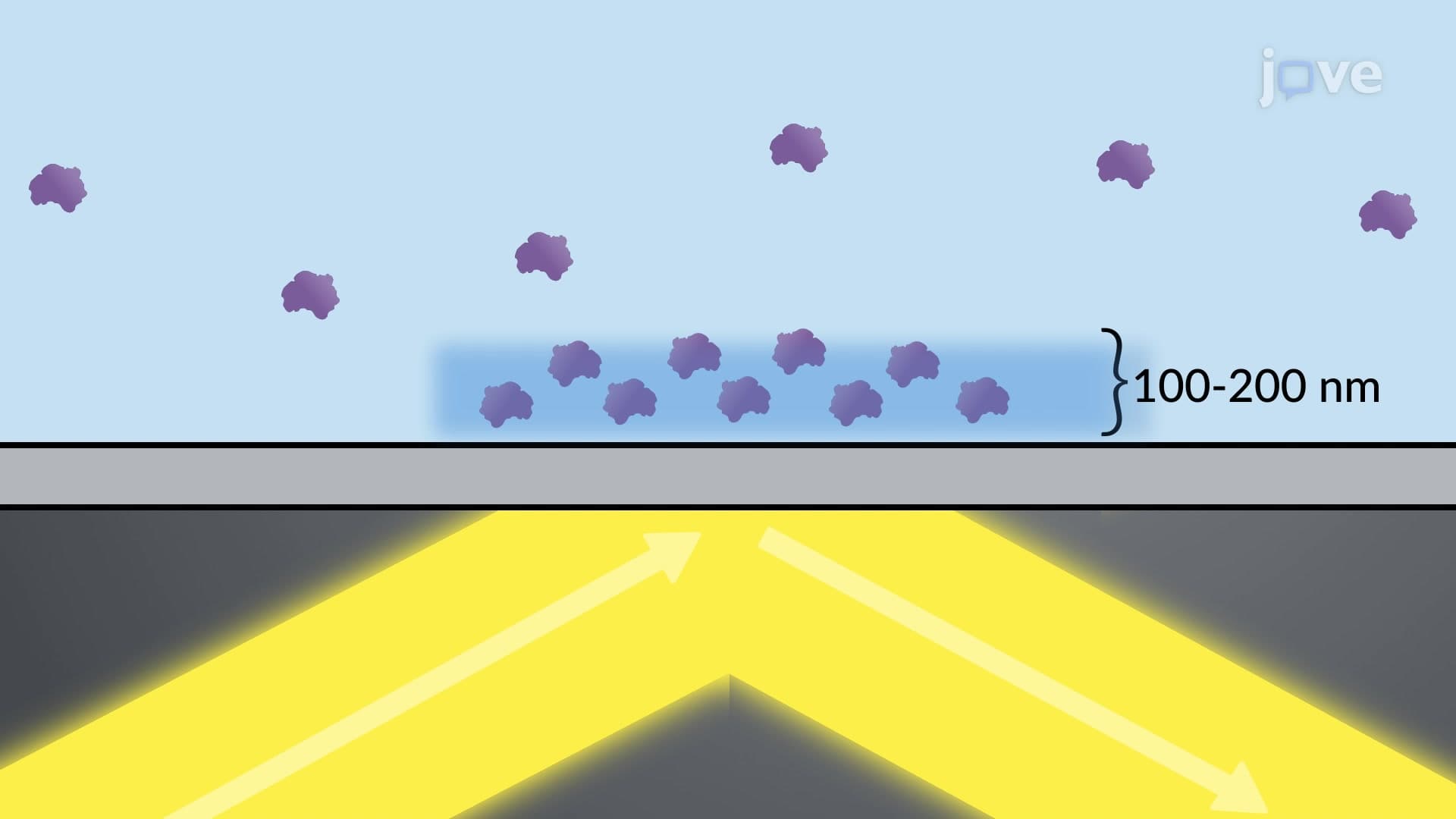

Video Summary: Total Internal Reflection Fluorescence Microscopy Explained
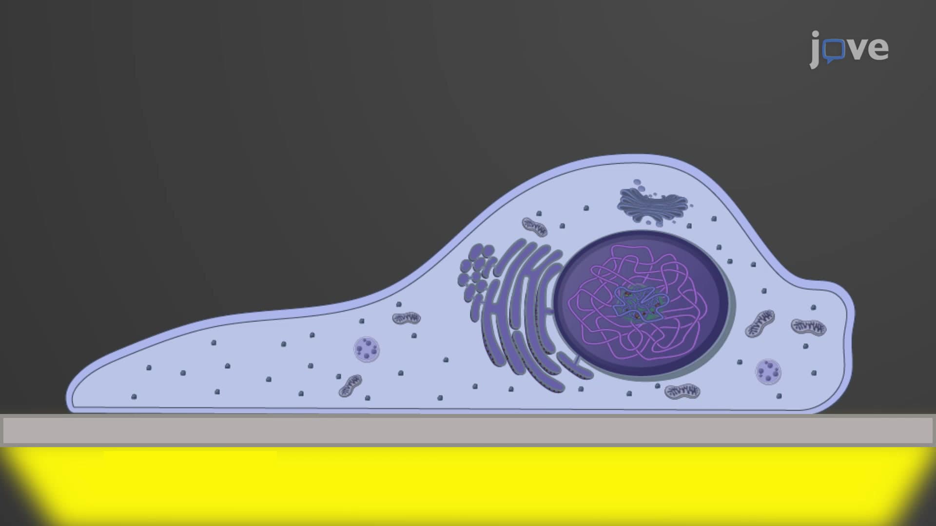
Ever wonder how scientists at Harvard Medical School can watch individual protein molecules moving along cellular highways in real-time? Total internal reflection fluorescence microscopy (TIRF) makes this possible by creating an ultra-thin light field that illuminates only molecules within 200 nanometers of a cell's surface. This revolutionary technique eliminates background noise, producing crystal-clear images of cellular processes like motor proteins transporting cargo along microtubules. Watch the full video on JoVE Coach to master this concept with expert-led visuals and step-by-step explanations.
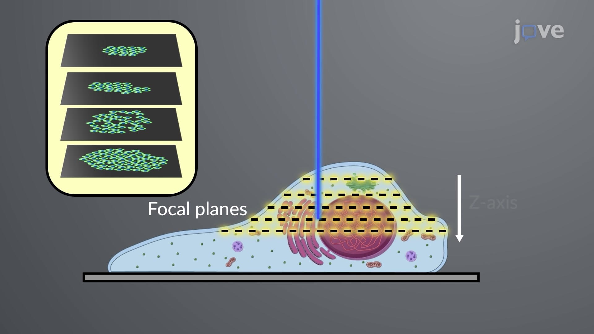
Total internal reflection fluorescence microscopy represents a sophisticated imaging technique that exploits fundamental optical physics to achieve unprecedented clarity in biological observation. Unlike conventional fluorescence microscopy, which excites fluorophores throughout the entire sample depth, TIRF creates a highly selective excitation zone by harnessing the phenomenon of total internal reflection.
The technique relies on the critical angle principle, where light traveling from a high refractive index medium (glass coverslip, n ≈ 1.52) encounters a lower refractive index medium (aqueous biological sample, n ≈ 1.33). When the incident angle exceeds the critical angle-approximately 61 degrees for this glass-water interface-total internal reflection occurs, preventing light transmission into the sample.
During total internal reflection, an electromagnetic phenomenon creates an evanescent wave that penetrates only 100-200 nanometers into the sample. This exponentially decaying field selectively excites fluorophores near the interface while leaving deeper molecules unaffected. For context, this penetration depth is roughly equivalent to the thickness of a cellular plasma membrane plus a small portion of the cytoplasm.
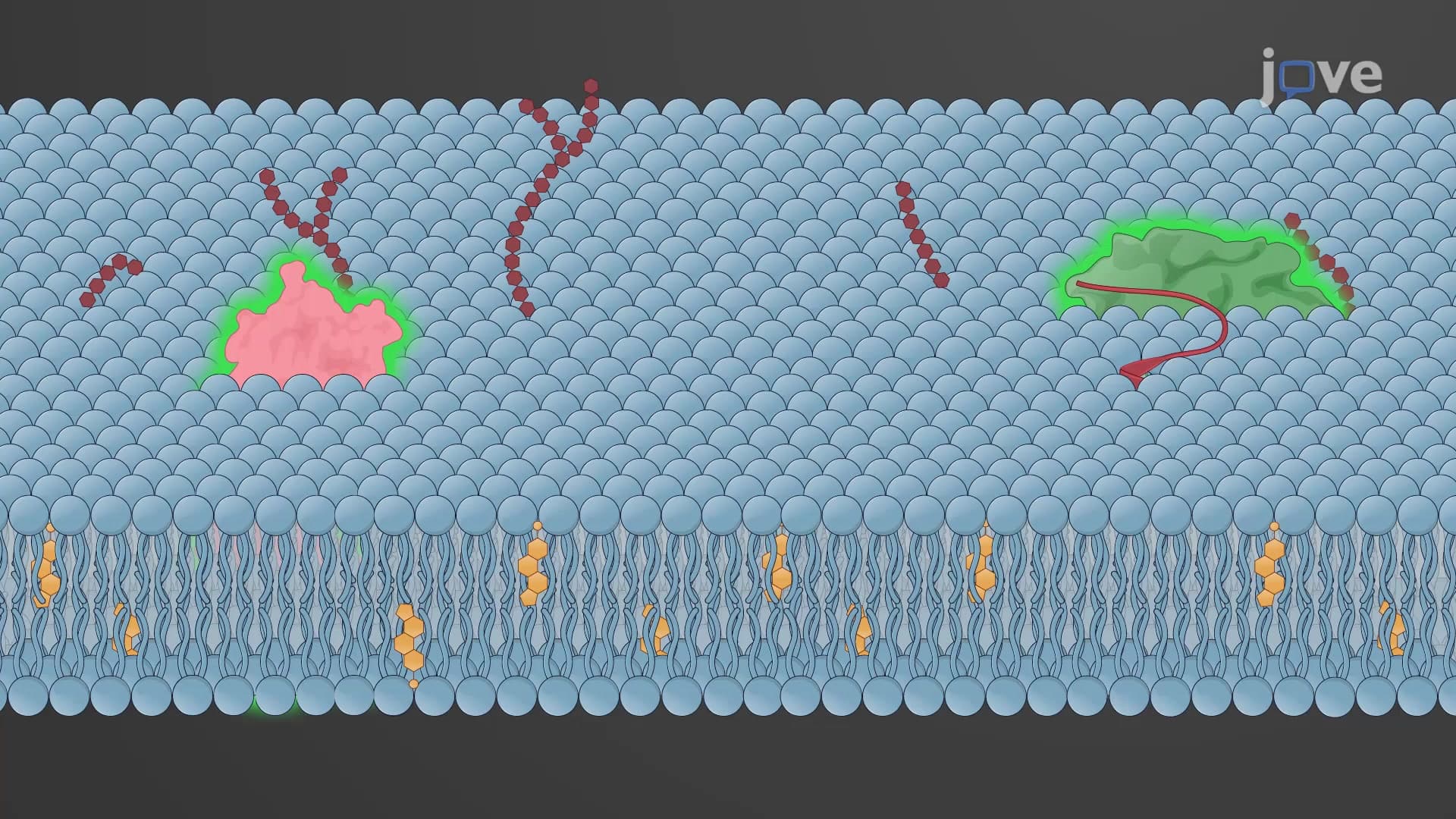
Research institutions like Stanford University and MIT extensively employ TIRF microscopy to study membrane dynamics, endocytosis, and exocytosis. The technique proves invaluable for observing single-molecule events, such as vesicle fusion during neurotransmitter release or the stepwise movement of molecular motors like kinesin and dynein along cytoskeletal tracks.
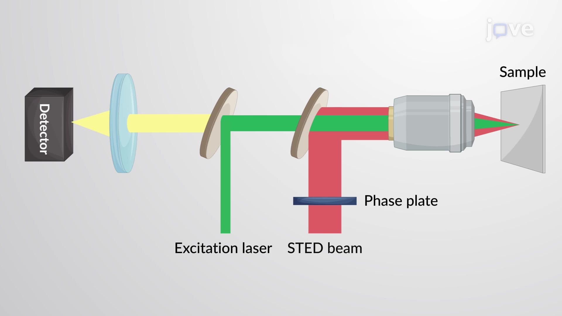
Modern TIRF systems integrate laser excitation sources with high-numerical-aperture objectives to achieve the necessary incident angles. Students preparing for AP Biology or college-level cell biology courses encounter TIRF applications when studying membrane trafficking, signal transduction, and cytoskeletal dynamics.
The technique's selectivity eliminates the need for extensive image processing, making it particularly valuable for studying rapid biological processes in real-time. This capability has revolutionized understanding of cellular mechanisms, from how cancer cells metastasize to how neurons communicate at synapses.
For students pursuing STEM careers, understanding TIRF principles provides insight into how advanced microscopy techniques overcome fundamental limitations of light-based imaging, demonstrating the practical application of physics concepts in biological research.
Related Micro-courses