Loading subject page…


561,001 views

Video Summary: Enzymes Structure Function and Catalytic Mechanisms
Did you know that without enzymes, a single hamburger would take over 80 years to digest? Enzymes: structure, function, and catalytic mechanisms reveal how these protein catalysts accelerate biochemical reactions by millions of times. From breaking down food in your stomach to enabling DNA replication in your cells, enzymes like pepsin demonstrate the lock-and-key binding specificity that makes life possible. Watch the full video on JoVE Coach to master this concept with expert-led visuals and step-by-step explanations.
Enzymes represent one of biology's most elegant solutions to the challenge of making chemical reactions occur at biologically useful rates. These remarkable catalysts achieve reaction rate increases of 10^6 to 10^17 times faster than uncatalyzed reactions, making them essential for life processes from cellular respiration to DNA replication.
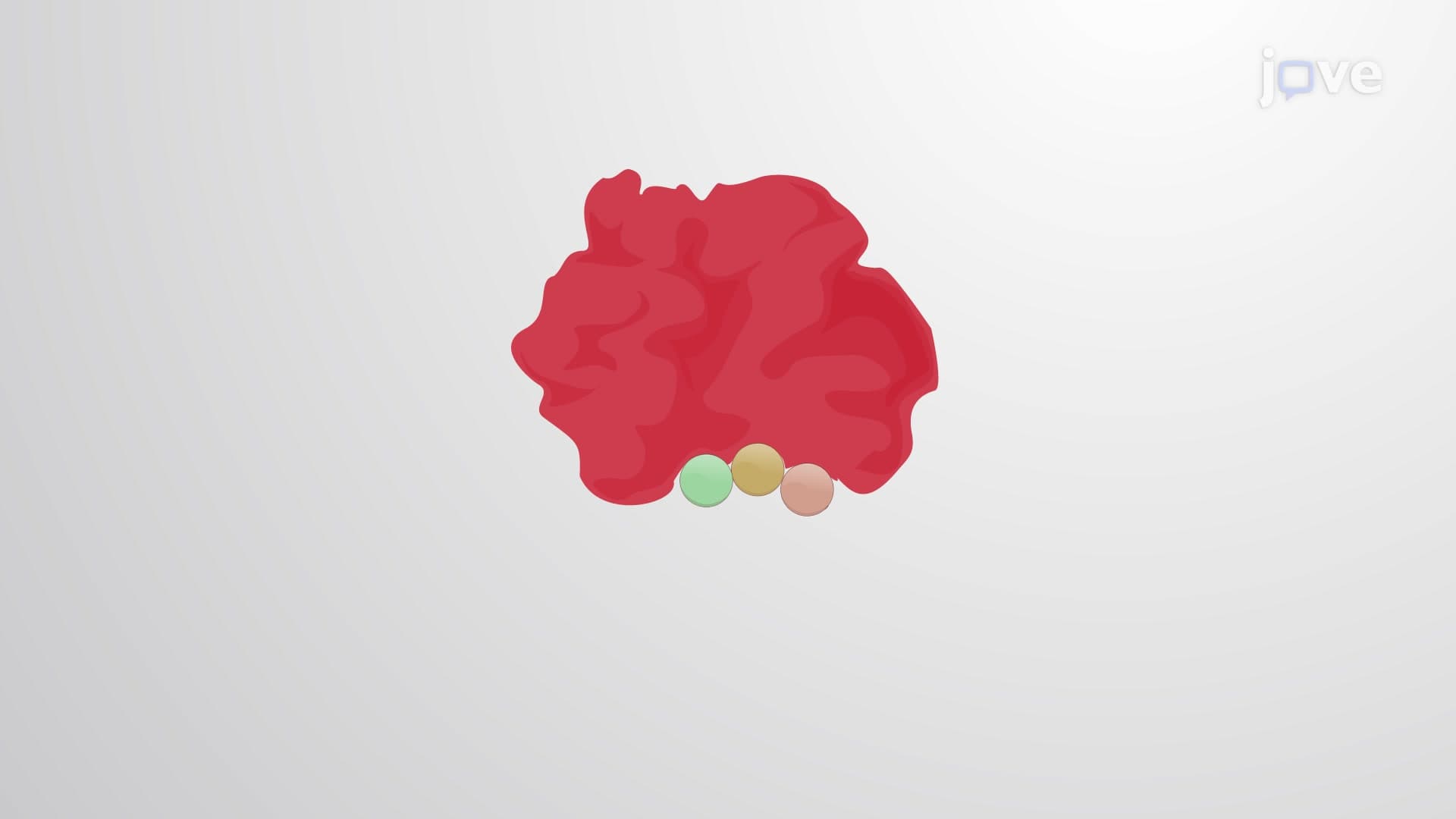
The three-dimensional structure of enzymes directly determines their catalytic capabilities. Most enzymes are globular proteins with distinct regions: the active site where catalysis occurs, allosteric sites that regulate activity, and structural domains that maintain stability. The active site's precise geometry, created by specific amino acid arrangements, determines substrate specificity. For example, hexokinase, the first enzyme in glucose metabolism, has an active site that perfectly accommodates glucose while excluding similar sugars like galactose.
This structural specificity appears frequently on the MCAT and AP Biology exams, where students must analyze how amino acid changes affect enzyme function. Understanding protein folding patterns helps predict how temperature, pH, and denaturants impact enzymatic activity-concepts crucial for pre-med students preparing for the USMLE.
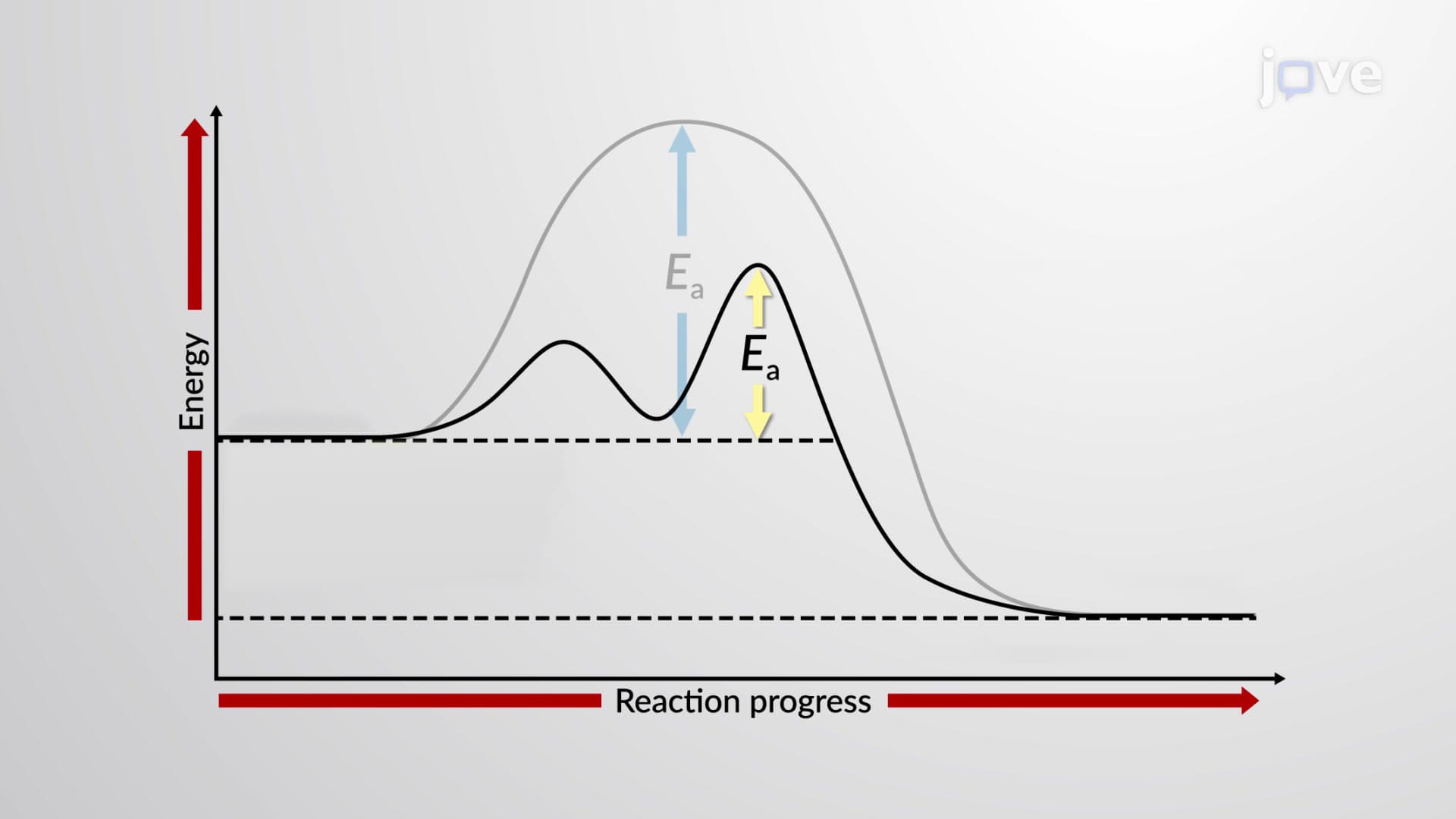
Enzymes lower activation energy through several sophisticated mechanisms. They stabilize transition states, strain substrate bonds to facilitate breaking, and position reactive groups optimally for bond formation. The induced-fit model explains how enzymes undergo conformational changes upon substrate binding, creating an optimal catalytic environment. This dynamic interaction contrasts with the simpler lock-and-key model taught in introductory courses.
Consider carbonic anhydrase, found in red blood cells, which catalyzes CO2 hydration 10^7 times faster than the uncatalyzed reaction. This enzyme demonstrates how precise positioning of catalytic residues (histidine and zinc ion) creates an optimal microenvironment for the reaction.
Enzyme inhibition mechanisms have profound medical applications. Competitive inhibitors like statins block HMG-CoA reductase to lower cholesterol synthesis, while noncompetitive inhibitors such as aspirin permanently modify cyclooxygenase enzymes to reduce inflammation. Understanding these mechanisms proves essential for pharmacy students taking the NAPLEX and nursing students preparing for the NCLEX, where drug mechanism questions frequently appear.
Allosteric regulation adds another layer of complexity, allowing enzymes like phosphofructokinase to respond to cellular energy status through feedback inhibition-a concept that bridges biochemistry and physiology in advanced coursework.
Related Micro-courses