Loading subject page…

- Anatomy and Physiology
- The Appendicular Skeleton
- Bones of the Lower Limb: Femur and Patella
Micro-course: The Appendicular Skeleton
27,600 views
Learn key concepts and earn your certificate.

Bones of the Lower Limb: Femur and Patella

00:00
01:16
Video Summary: Bones Lower Limb Femur and Patella Explained
Ever wonder why football players can generate such powerful kicks that send a ball soaring 60 yards downfield? The secret lies in the femur patella anatomy lower limb structure, where the femur-your body's longest and strongest bone-works with the patella to create incredible force during athletic movements. When NFL kickers like Justin Tucker attempt record-breaking field goals, they rely on the precise biomechanics of these crucial lower limb bones. Understanding Bones Lower Limb Femur And Patella Explained reveals how your skeletal system transforms muscle contractions into explosive athletic performance. Watch the full video on JoVE Coach to master this concept with expert-led visuals and step-by-step explanations.
- Understand the anatomical structure and function of bones in the lower limb, specifically focusing on Bones Lower Limb Femur And Patella Explained concepts
- Identify key anatomical landmarks including the femoral head, neck, greater trochanter, and femoral condyles on the thigh bone
- Analyze how the patella functions as a sesamoid bone within the quadriceps tendon system
- Compare the structural differences between the femur's proximal and distal ends and their functional significance
- Evaluate the biomechanical relationship between femur and patella during knee extension movements
- Apply knowledge of femoral anatomy to understand muscle attachment sites and their clinical importance
- Recognize the developmental timeline of patella ossification from birth through puberty
- Explore how anatomical variations in femur and patella structure can impact athletic performance and injury patterns
Understanding Bones Lower Limb Femur And Patella Explained: Foundation of Human Locomotion
The lower limb's skeletal architecture represents one of human anatomy's most remarkable engineering solutions. The femur patella anatomy lower limb system forms the structural foundation that enables everything from walking to Olympic-level athletic performance. These bones don't just provide support-they create a sophisticated lever system that amplifies muscle force and enables the complex movements that define human mobility.
The Femur: Engineering Marvel of the Human Body
The thigh bone kneecap anatomy begins with understanding why the femur holds the distinction as our body's largest bone. Measuring approximately 18 inches in an average adult, the femur length largest bone designation isn't just about size-it's about function. The femur must withstand forces up to 12 times body weight during activities like jumping, making its robust construction essential for survival.
The femoral head neck greater trochanter complex at the proximal end creates the hip joint's ball-and-socket mechanism. The spherical femoral head fits snugly into the acetabulum of the hip bone, while the neck provides the crucial angle that allows for the wide range of hip motion. The greater and lesser trochanters serve as attachment points for powerful muscles like the gluteus medius and iliopsoas, which are critical for maintaining balance and generating locomotor force.
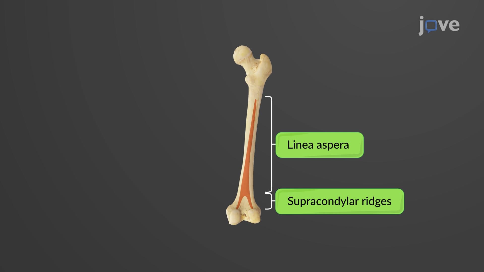
Along the femur's posterior shaft, the linea aspera provides attachment for the adductor muscles and hamstrings. This roughened ridge demonstrates how bone structure directly reflects functional demands-areas of greater muscle attachment develop more pronounced surface features. At the distal end, the femoral condyle epicondyle structures create the knee joint's complex articulation, with the medial and lateral condyles providing weight-bearing surfaces while the intercondylar fossa accommodates the crucial anterior cruciate ligament.
The Patella: Small Bone, Major Function
The patella sesamoid kneecap represents a unique category of bone-sesamoid bones that develop within tendons. The patella quadriceps attachment system creates a mechanical advantage that increases the quadriceps' leverage by approximately 30%, transforming this small triangular bone into a force multiplier that's essential for activities requiring knee extension.
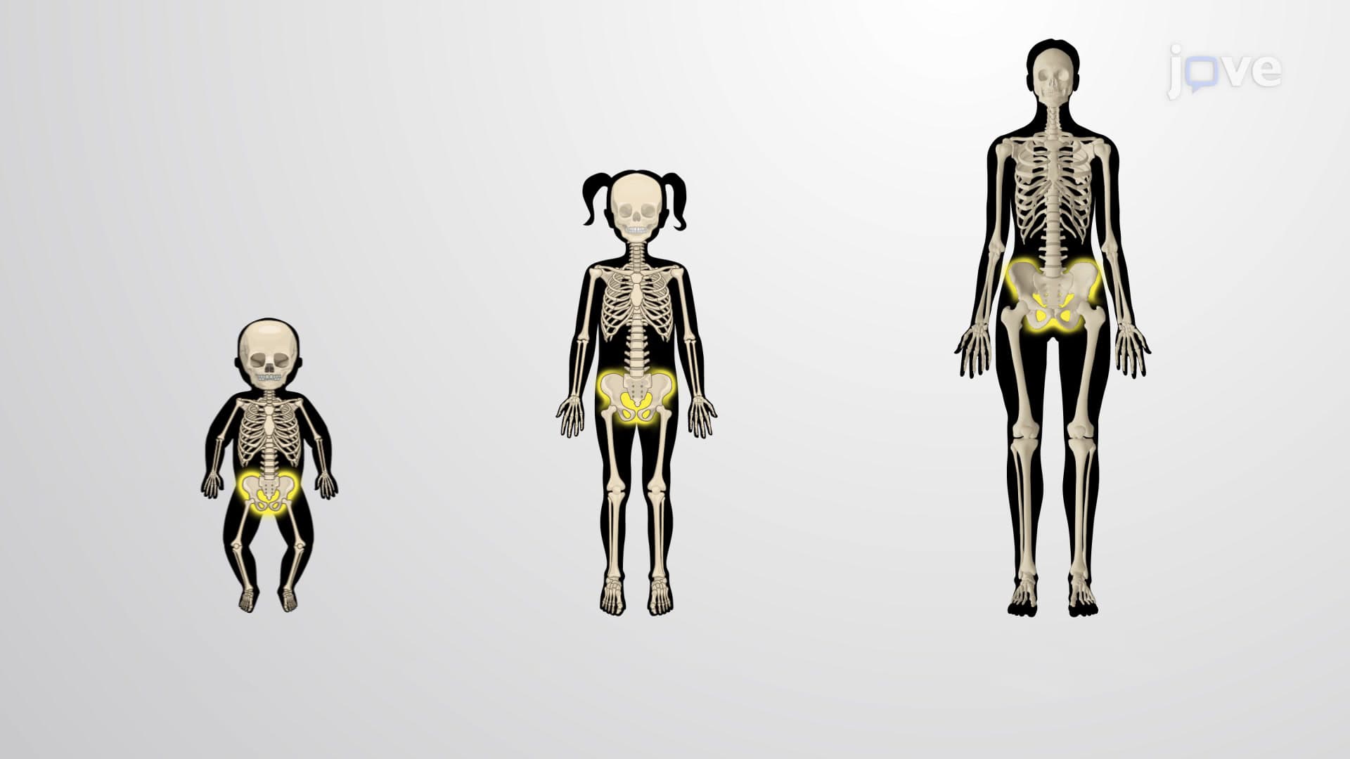
Understanding what are the femur and patella bones in lower limb anatomy becomes particularly relevant for students preparing for AP Biology or college-level anatomy courses. The patella's development timeline-cartilaginous at birth, beginning ossification at ages 2-3, and completing the process at puberty-frequently appears on exams like the MCAT and USMLE Step 1. This developmental pattern reflects the bone's functional importance, as it only begins significant ossification when walking and running place substantial mechanical demands on the knee extension system.
The patella's gliding mechanism within the femur's patellar groove demonstrates perfect form-function relationships. This groove guides the patella's movement during knee flexion and extension, preventing lateral dislocation while maintaining optimal muscle function. When this system fails, as in patellar tracking disorders common among runners and basketball players, the resulting dysfunction illustrates the critical importance of proper anatomical alignment.
Frequently Asked Questions
The femur and patella are essential bones in your thigh and knee region that work together to enable walking, running, and jumping. The femur is your body's longest and strongest bone, extending from hip to knee, while the patella (kneecap) is a small triangular bone that increases the efficiency of your quadriceps muscles. Together, they form a mechanical system that can withstand forces many times your body weight during athletic activities.
MCAT questions frequently test femur and patella anatomy in both biological sciences and physics contexts. You'll encounter questions about bone development timelines, muscle attachment sites, and biomechanical principles like leverage and force multiplication. The patella's role as a sesamoid bone and its ossification pattern from ages 2-3 through puberty are particularly high-yield topics for exam success.
Focus on the femoral head, neck, greater and lesser trochanters, linea aspera, supracondylar ridges, and medial/lateral condyles. These landmarks appear consistently on practical exams because they represent major muscle attachment sites. The intercondylar fossa is also crucial as it houses important knee ligaments like the ACL and PCL.
Professional athletes like NFL kickers rely on the femur-patella system to generate explosive leg movements. The patella increases quadriceps leverage by about 30%, allowing players to achieve greater knee extension force. This biomechanical advantage is why proper patella tracking is crucial for athletic performance-when the kneecap doesn't glide properly in the femur's groove, power output decreases significantly.
Not at all-lower limb anatomy builds logically on basic principles you already understand. Think of the femur as a mechanical lever and the patella as a pulley system that improves muscle efficiency. Starting with these simple concepts and adding anatomical details gradually makes even complex structures like the intercondylar fossa manageable for high school and college students.
Create concept maps linking structure to function, focusing on how bone features relate to muscle attachments and movements. Practice identifying anatomical landmarks using online resources and relate each structure to its biomechanical purpose. The AP Biology exam often tests developmental biology, so memorize the patella's ossification timeline and understand why sesamoid bones develop within tendons.
Femur anatomy integrates with muscle physiology, joint mechanics, developmental biology, and even evolutionary biology. The bone's structure reflects bipedal locomotion adaptations, while its development illustrates principles of bone formation and remodeling. Understanding femur-patella relationships prepares you for advanced topics like orthopedic medicine, sports science, and biomechanical engineering.
Learning bones first provides the structural framework for understanding muscle attachments and movements. The femur's trochanters, linea aspera, and condyles only make sense when you understand which muscles attach there and what movements they produce. This sequential approach-bones, then muscles, then movements-builds comprehensive understanding more effectively than studying systems in isolation.
Related Micro-courses