Loading video…


 22 Concepts
 178.8K views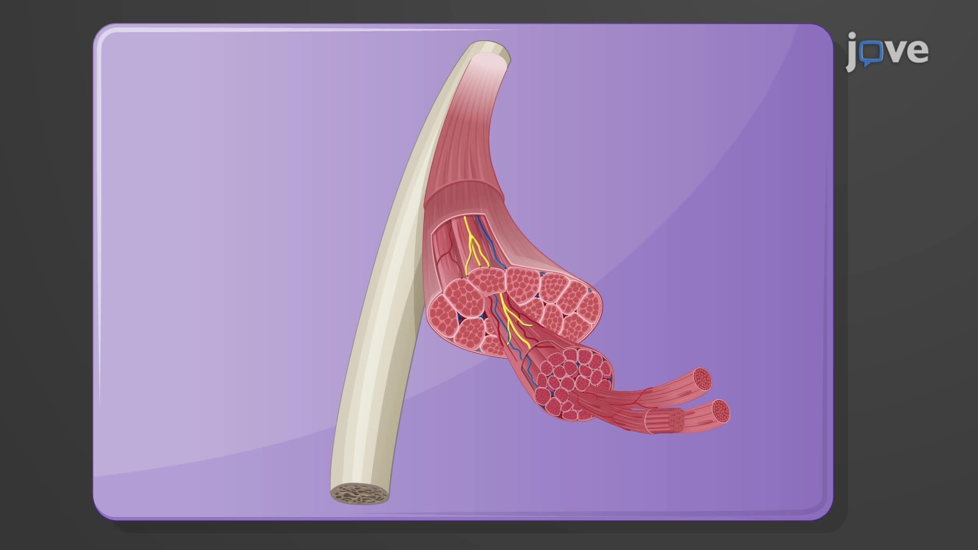

Video Summary: Gross Anatomy of Skeletal Muscles Explained
Ever wonder how a pitcher like Jacob deGrom generates the explosive power to throw a 100-mph fastball? The secret lies in skeletal muscle gross anatomy, where layered connective tissue sheaths transform microscopic muscle fiber contractions into powerful, coordinated movements. Understanding the gross anatomy of skeletal muscles reveals how epimysium, perimysium, and endomysium work together to create the structural framework that enables voluntary movement throughout your body. Watch the full video on JoVE Coach to master this concept with expert-led visuals and step-by-step explanations.
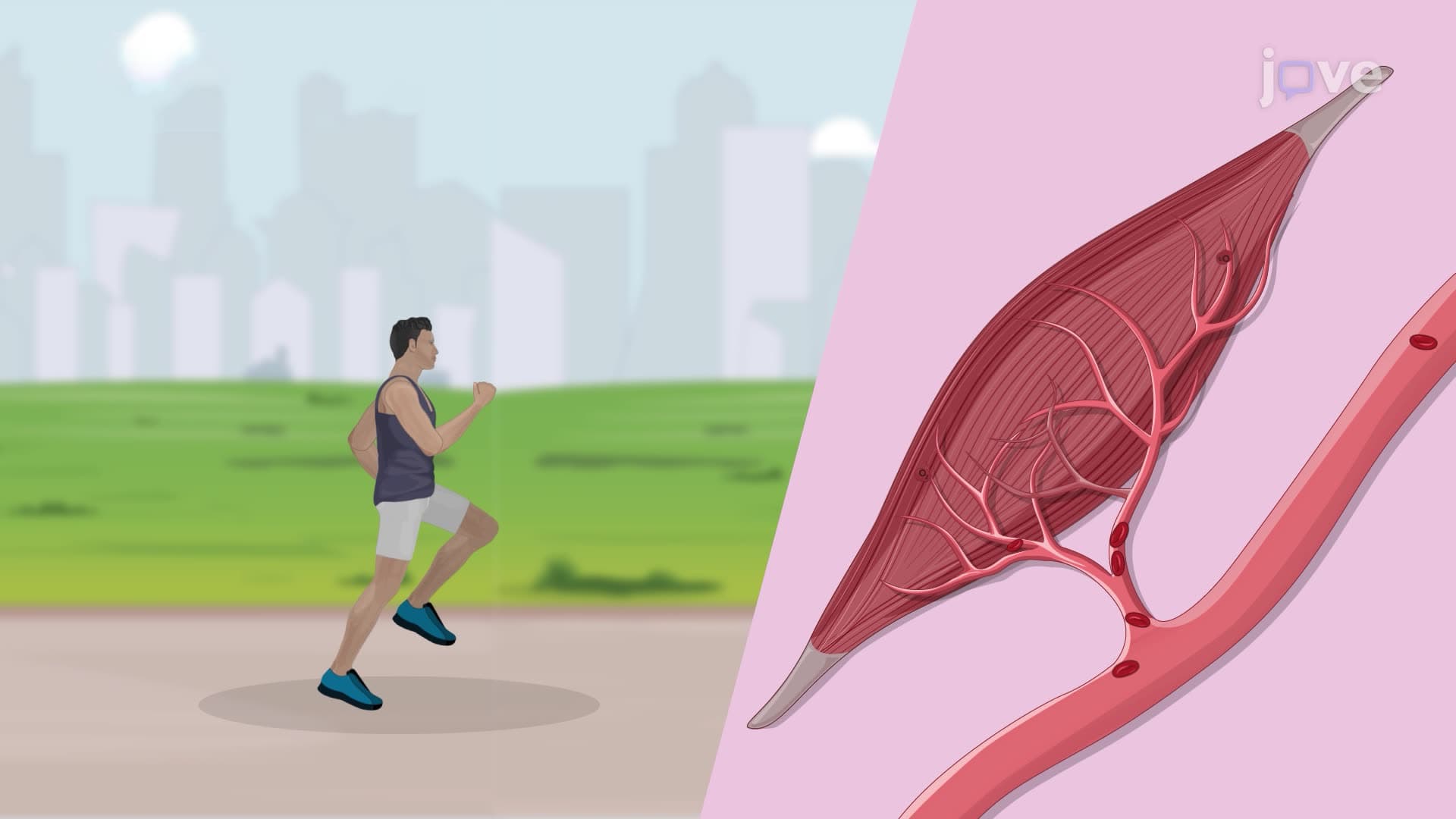
The gross anatomy of skeletal muscles represents one of anatomy's most elegant organizational systems. Unlike cardiac or smooth muscle, skeletal muscle displays a highly organized hierarchical structure visible to the naked eye. This macroscopic organization directly impacts how muscles generate force, coordinate movement, and maintain structural integrity during the demanding contractions required for activities ranging from delicate piano playing to powerlifting competitions.
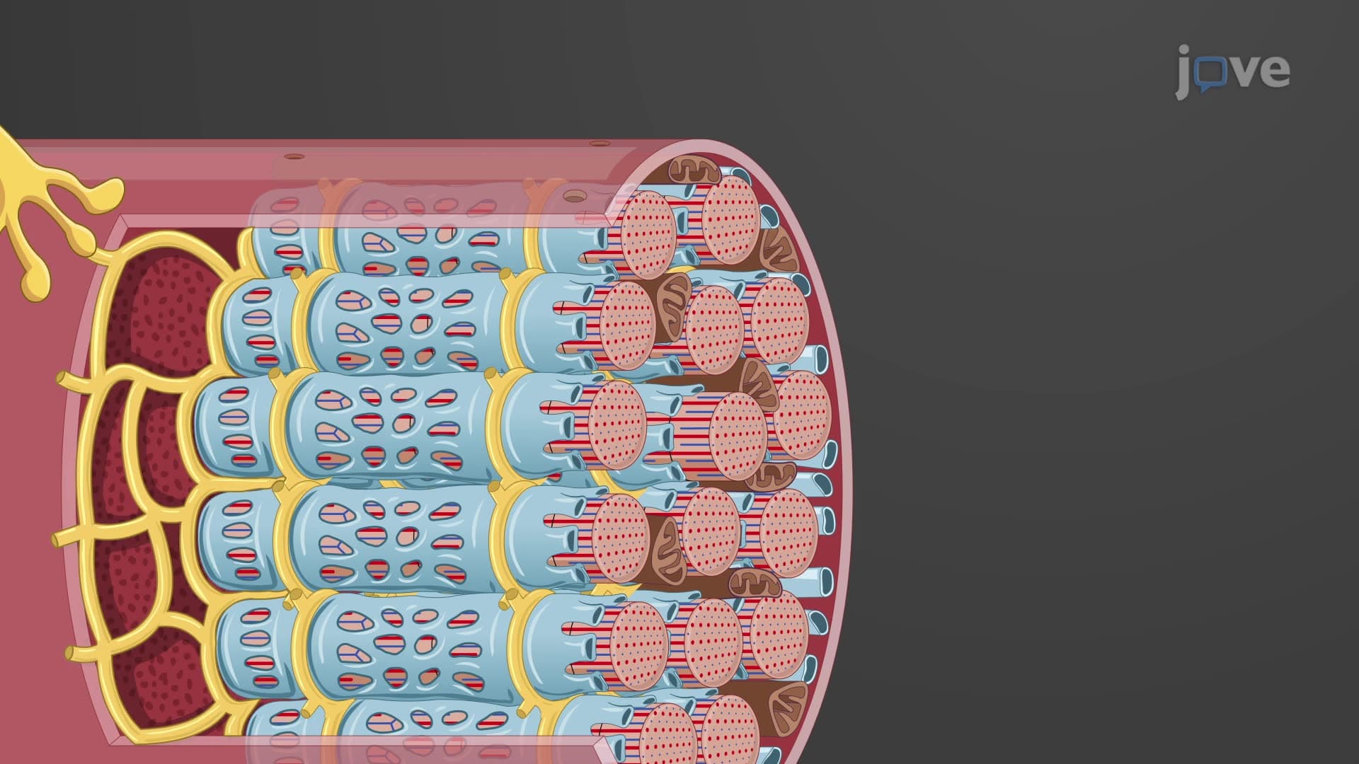
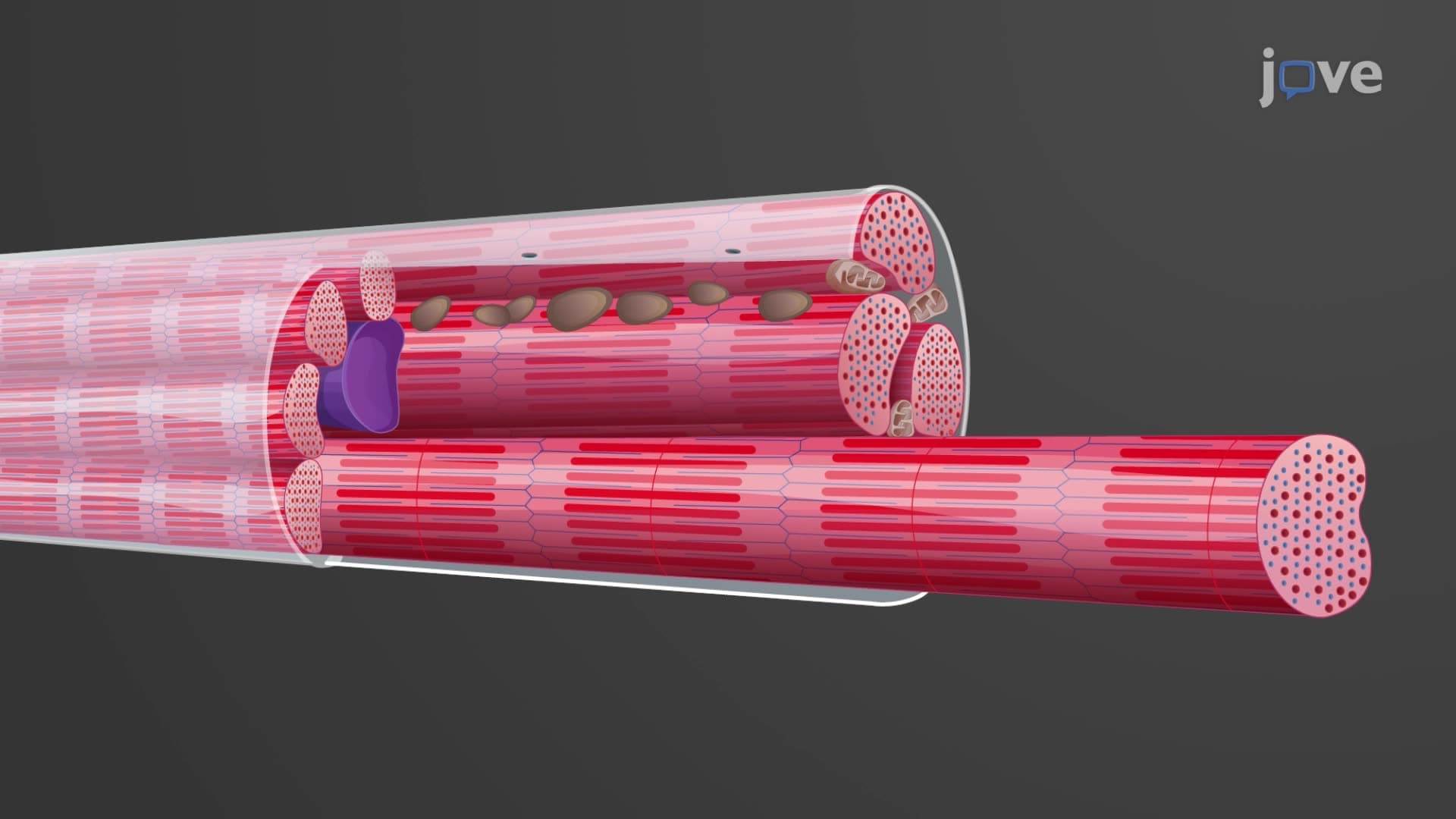
At the heart of muscle gross anatomy lies a sophisticated three-layer connective tissue system. The outermost epimysium encases the entire muscle like a protective jacket, similar to how the fascia surrounding your biceps brachii maintains its distinct shape during arm curls. This dense irregular connective tissue provides structural support while allowing the muscle to change shape during contraction.
Moving inward, the perimysium divides muscle tissue into fascicles-bundles of muscle fibers that function as coordinated units. Think of fascicles as the "grain" you see in a well-prepared steak; these visible bundles represent hundreds of muscle fibers working together. Finally, the endomysium wraps each individual muscle fiber, creating a microenvironment that supports cellular function while maintaining fiber alignment.
Fascicle arrangement directly impacts muscle function and appears frequently on AP Biology and college anatomy exams. Parallel fascicles, like those in the sartorius muscle, optimize for range of motion. Pennate arrangements, seen in the deltoid, maximize force production by packing more fibers into a given space. This architectural diversity explains why different muscles excel at different tasks-sprinters develop powerful pennate gastrocnemius muscles for explosive push-off, while marathon runners develop endurance-optimized parallel fiber arrangements.
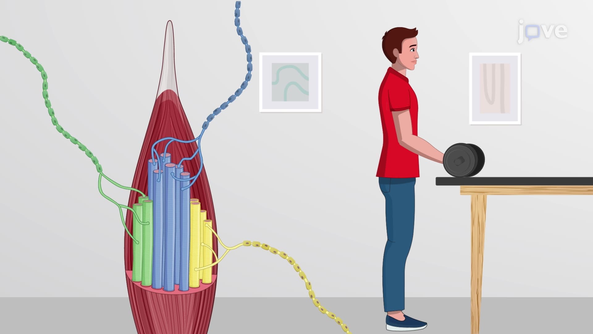
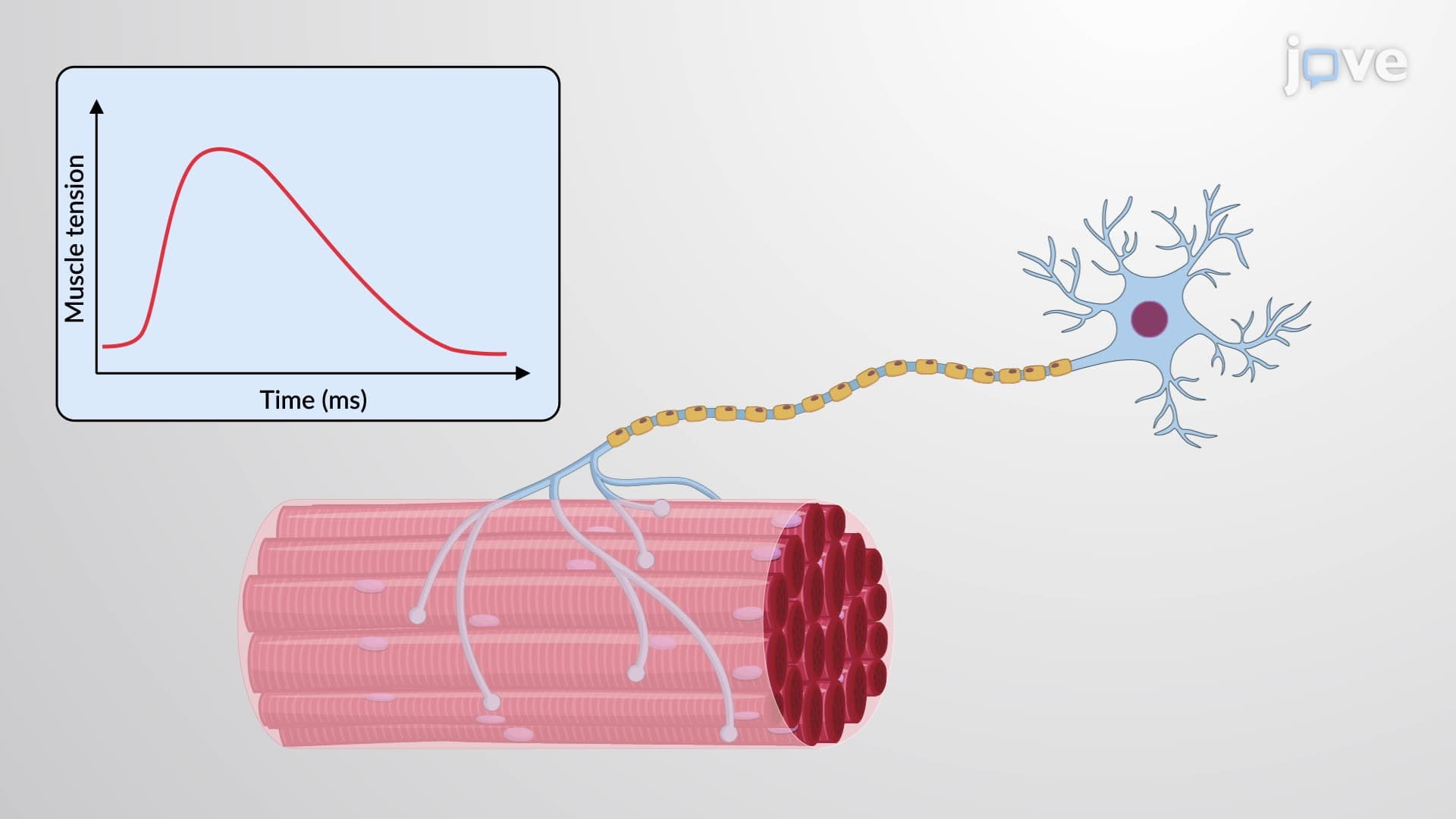
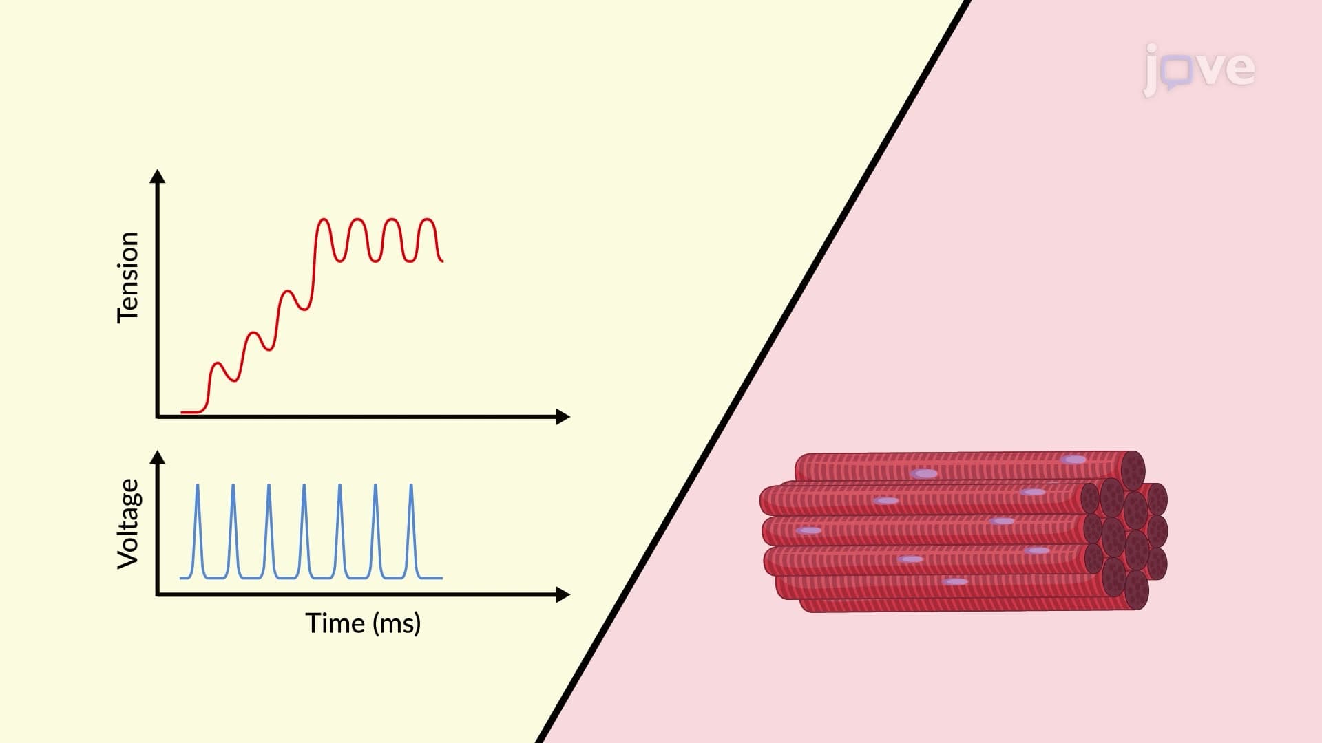
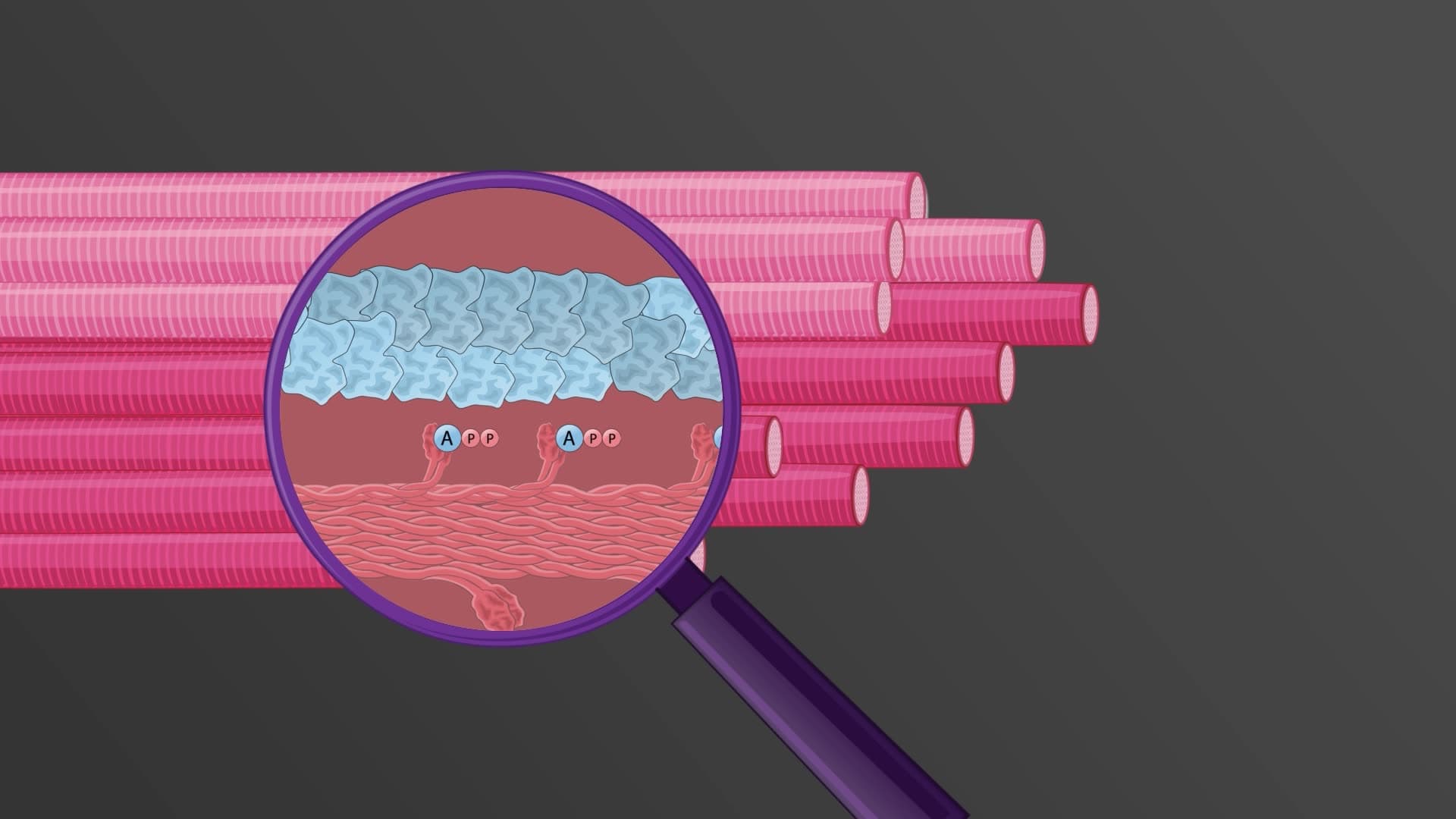
The genius of skeletal muscle gross anatomy becomes apparent during contraction. When individual muscle fibers contract, they pull on their surrounding endomysium. This force transmits through the perimysium to the fascicle level, then through the epimysium to the entire muscle. The connective tissue sheaths act as a mechanical transmission system, converting millions of microscopic contractions into unified, powerful movements capable of moving bones and joints.
This force transmission system extends beyond the muscle belly through tendons and aponeuroses. The Achilles tendon, connecting the gastrocnemius and soleus to the calcaneus, exemplifies how connective tissue continuity enables muscles to move bones located several inches away from the muscle belly itself.
Related Micro-courses