Loading subject page…


178,811 views

Video Summary: Microscopic Anatomy of Skeletal Muscles Explained
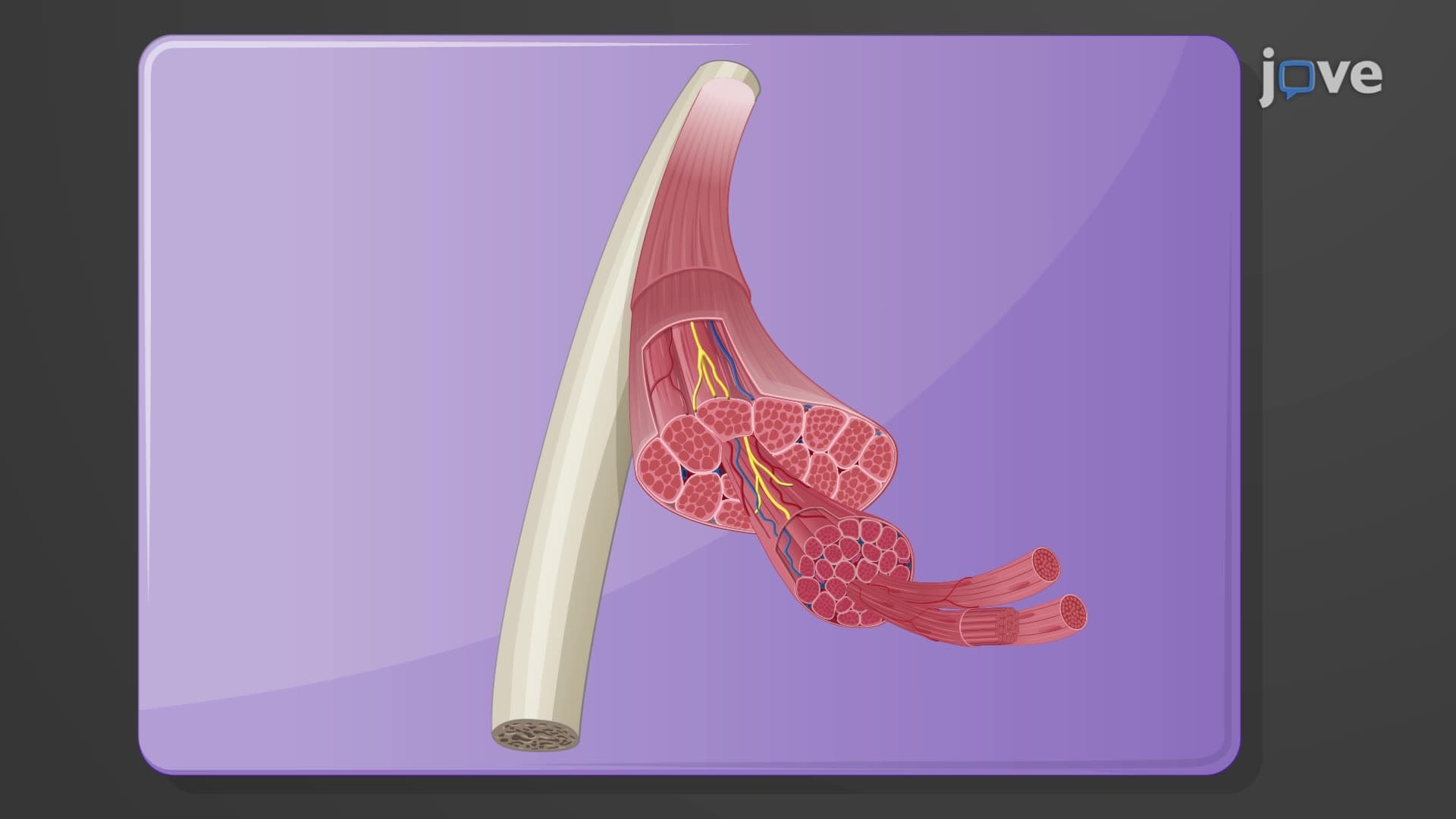
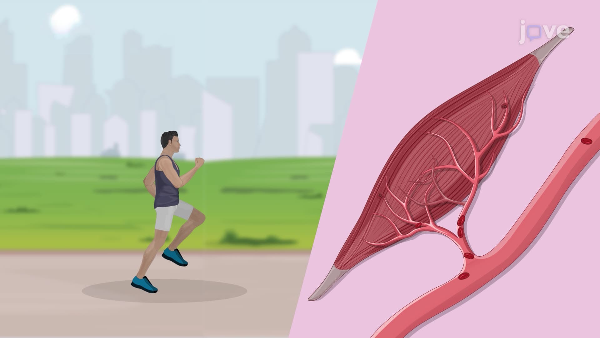
Ever wonder how your bicep contains millions of microscopic powerhouses that contract in perfect synchrony when you flex? Skeletal muscle microscopic anatomy reveals an intricate cellular architecture where multinucleated muscle fibers work like biological machines. Consider how NFL athletes generate explosive power-their muscle cells contain specialized structures like sarcolemma, T-tubules, and myofibrils that enable rapid, coordinated contractions. Watch the full video on JoVE Coach to master this concept with expert-led visuals and step-by-step explanations.
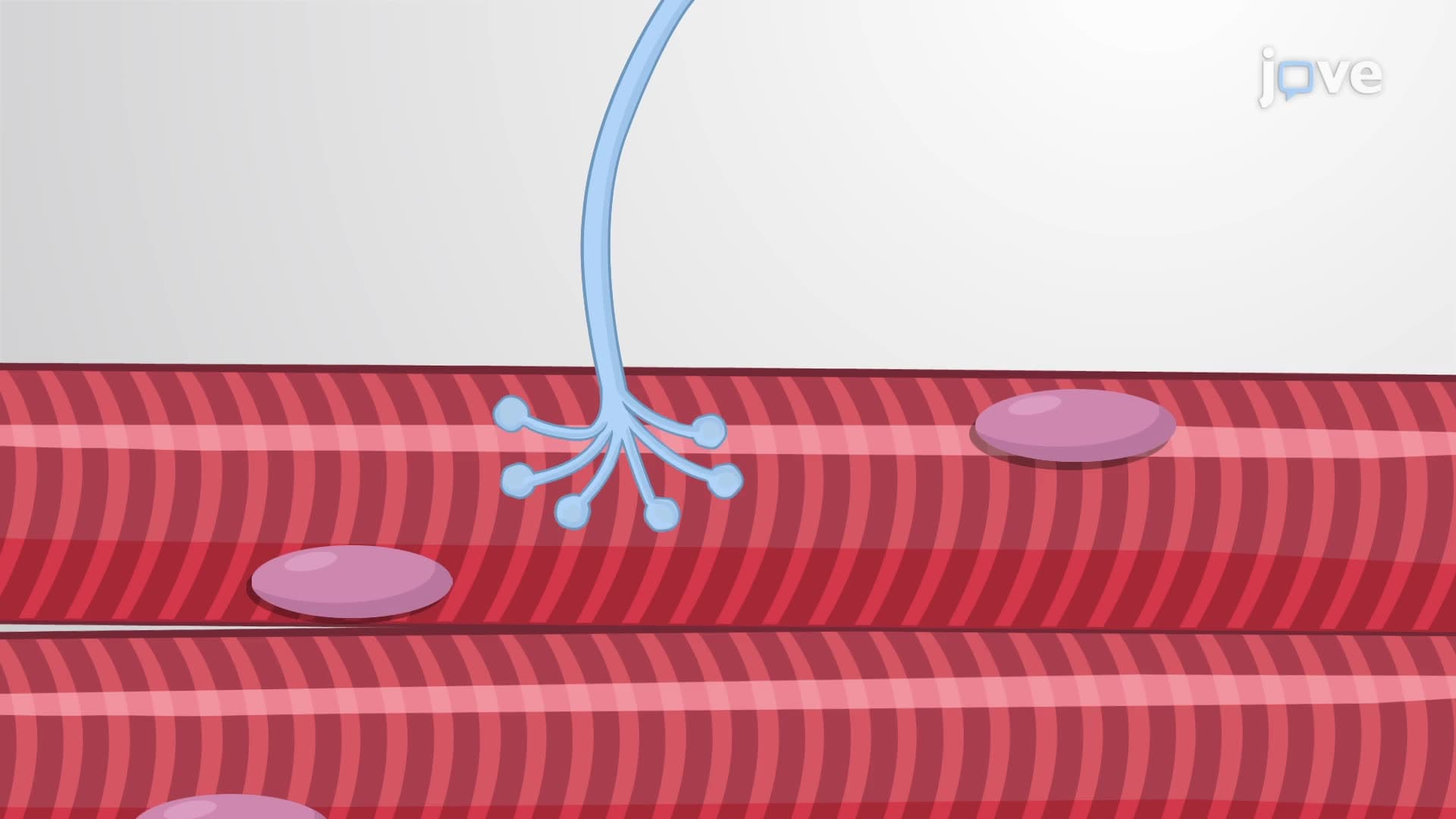
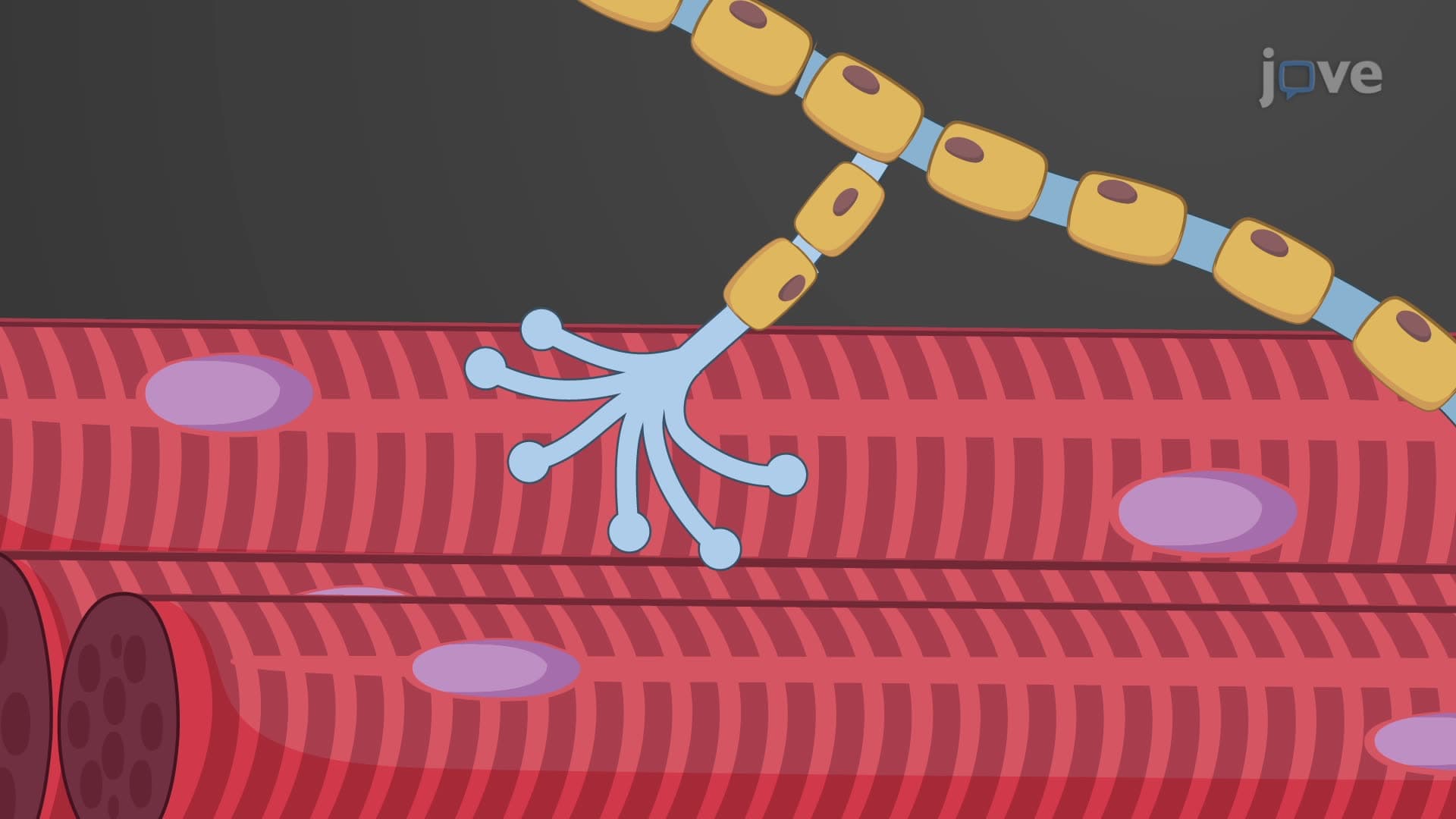
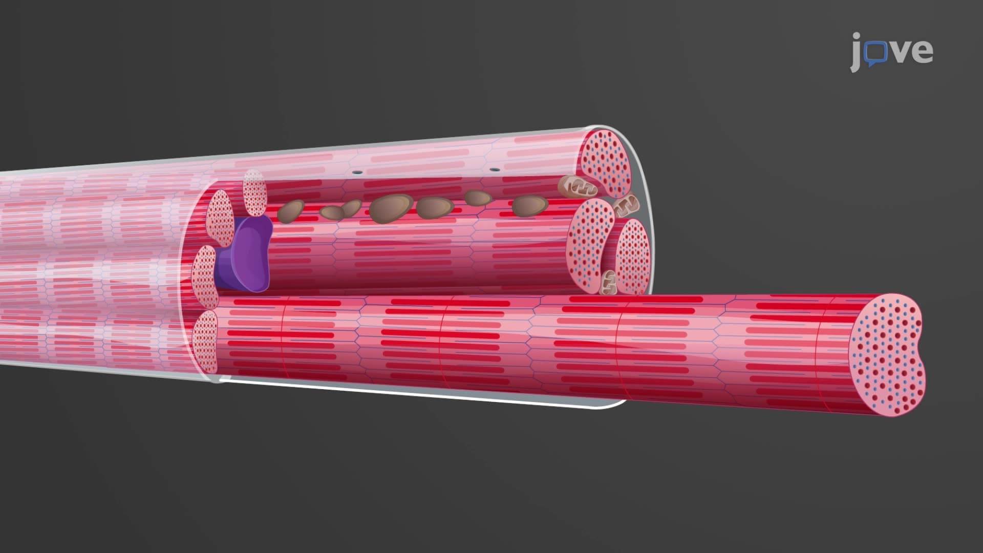
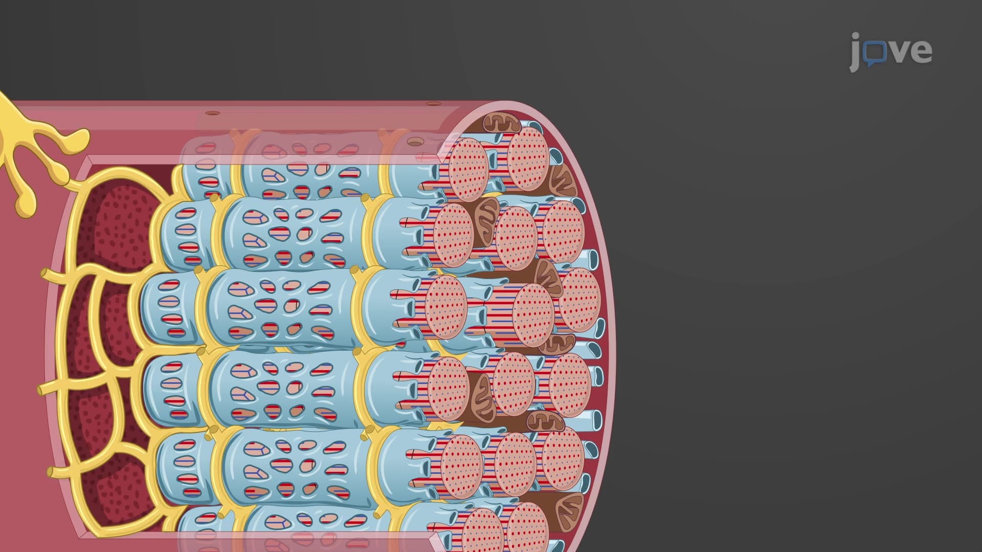
The microscopic anatomy of skeletal muscles represents one of biology's most sophisticated cellular designs. Unlike typical cells with single nuclei, skeletal muscle fibers are multinucleated giants-some extending several centimeters in length. This unique architecture allows for the massive protein synthesis needed to build and maintain contractile machinery.
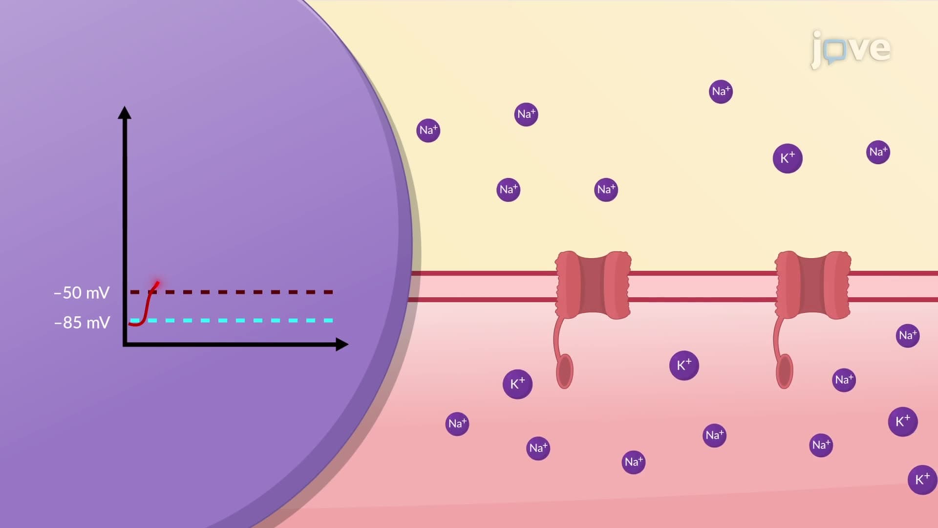
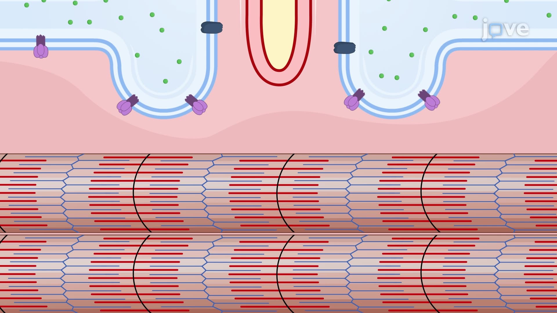
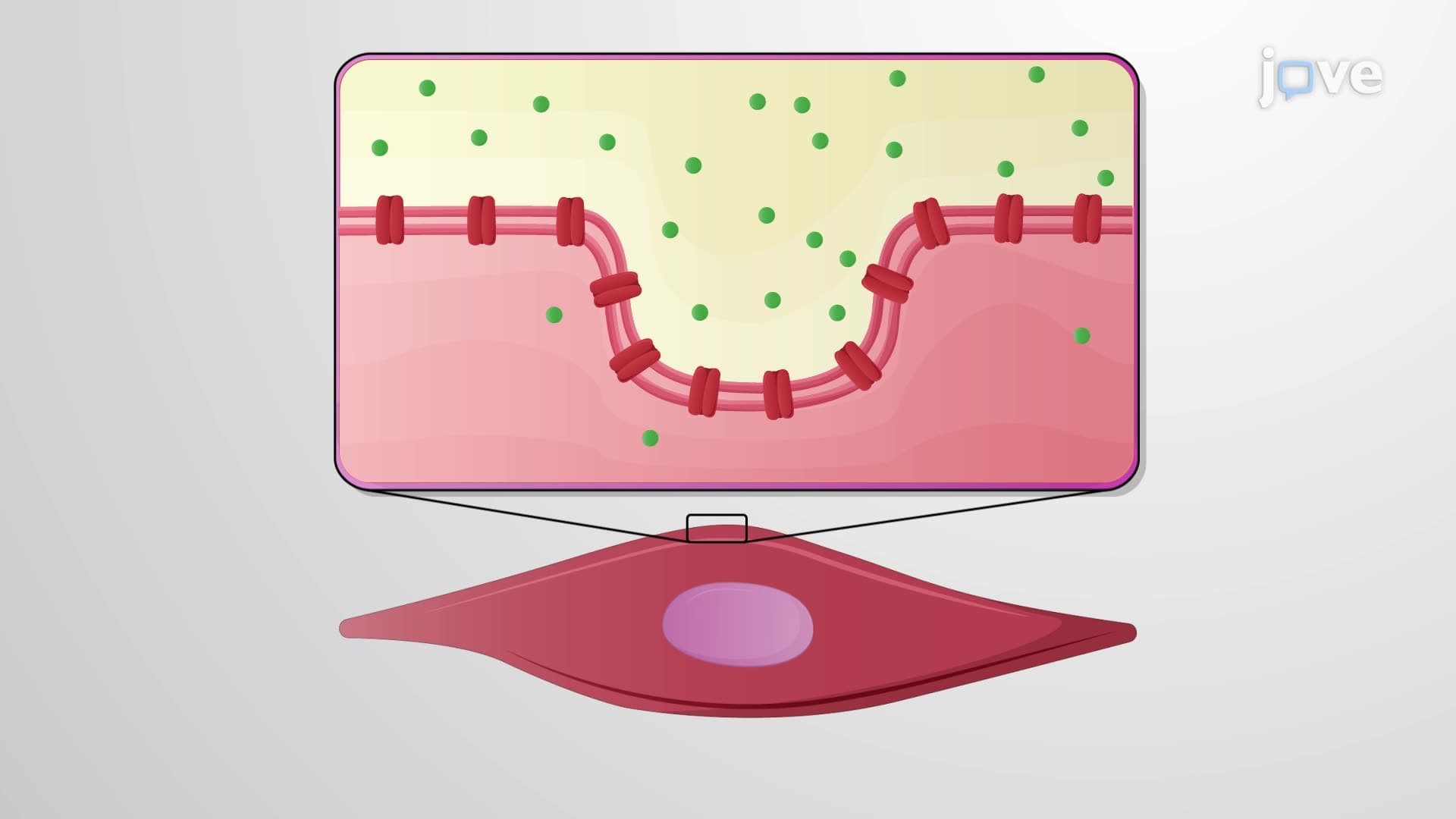
The muscle fiber microscopic biology begins with the sarcolemma, a specialized plasma membrane that acts like biological wiring. T-tubules, formed by sarcolemma invaginations, create an internal network ensuring electrical signals reach every corner of these massive cells simultaneously. This system is crucial for understanding muscle physiology questions on the MCAT, where students must explain how action potentials trigger uniform contractions across entire muscle fibers.
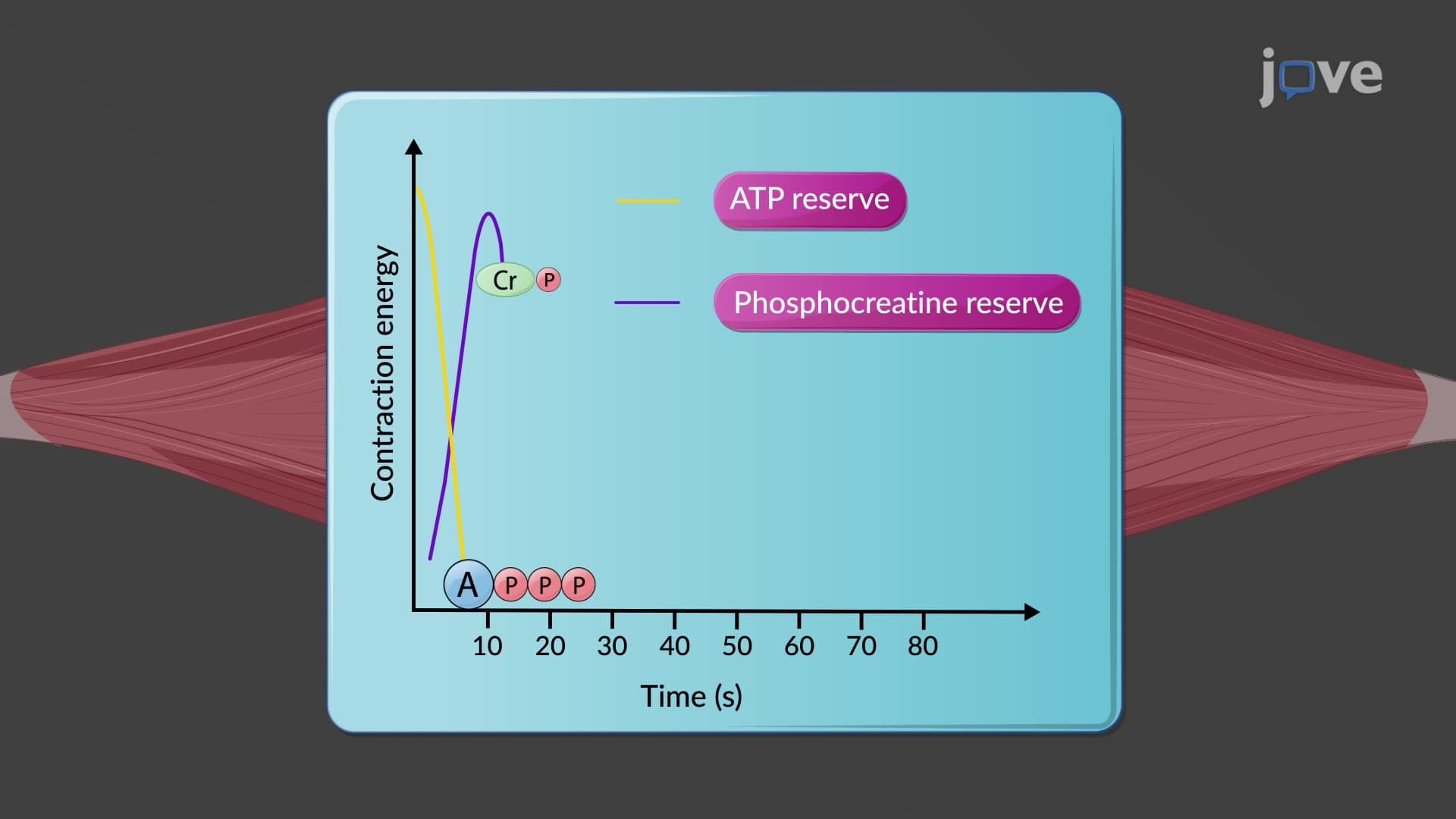
Within the sarcolemma T-tubule sarcoplasm environment, muscle cells pack extraordinary metabolic machinery. Abundant mitochondria provide ATP for contraction, while myoglobin stores oxygen for sustained activity-explaining why endurance athletes like marathon runners have darker, more myoglobin-rich muscles. Glycosomes contain enzymes for rapid glucose breakdown, supporting the explosive energy needs seen in Olympic sprinters.
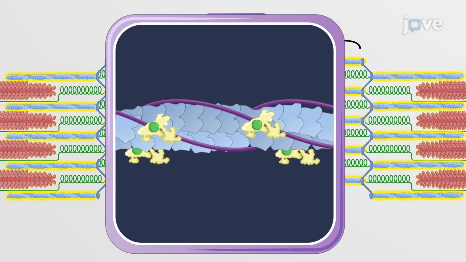
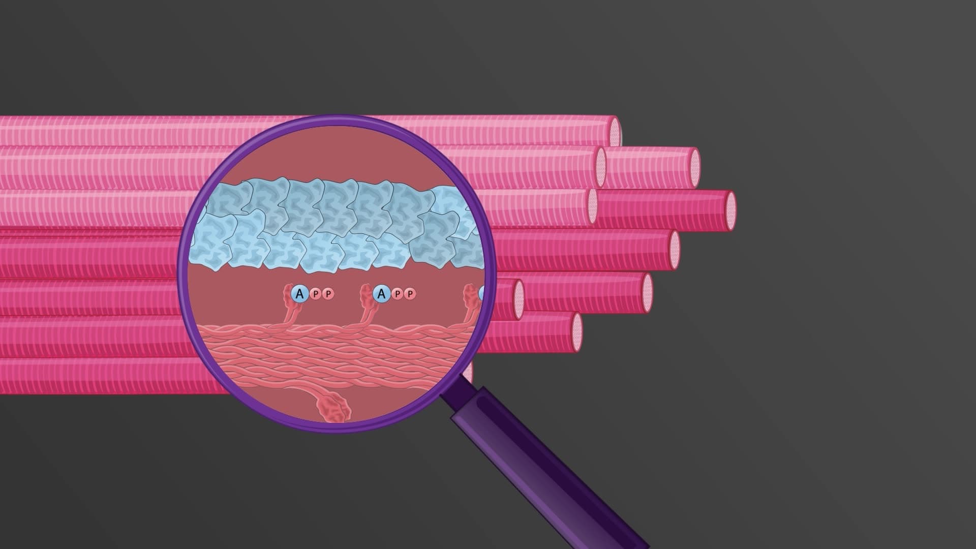
The muscle fiber myofibril structure represents the ultimate contractile design. Myofibrils contain repeating sarcomere units where actin myosin filament interactions generate force. The sarcoplasmic reticulum muscle system wraps around myofibrils like a calcium-storing net, releasing calcium ions to initiate contraction and resequestering them to stop it. This precise calcium control mechanism frequently appears in AP Biology exams and college physiology courses, where students analyze muscle contraction cycles and calcium's regulatory role.
Understanding muscle cell ultrastructure provides essential foundation knowledge for health science students pursuing careers in physical therapy, sports medicine, or clinical research, where muscle function analysis directly impacts patient care and athletic performance optimization.
Related Micro-courses