Loading subject page…


178,811 views

Video Summary: Structure and Organization of Smooth Muscles Explained
Did you know that the smooth muscles controlling your blood pressure right now lack the striped pattern found in your biceps? Smooth muscle structure organization involves two distinct arrangements: single-unit muscles that work together (like those in your intestines during digestion) and multi-unit muscles with individual cell control (such as those adjusting your pupil size in bright classroom lighting). Watch the full video on JoVE Coach to master this concept with expert-led visuals and step-by-step explanations.
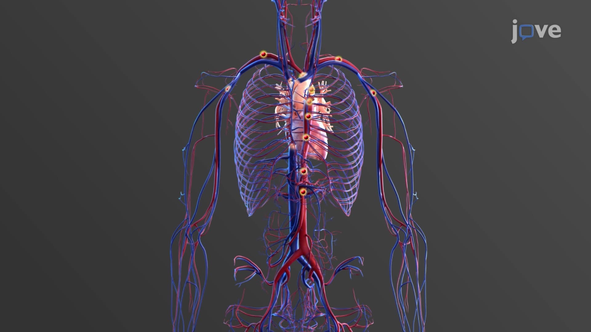
The Structure And Organization of Smooth Muscles Explained reveals a sophisticated biological system optimized for sustained, involuntary contractions. Unlike the highly organized skeletal muscles that move your arms during a basketball game, smooth muscles operate continuously without conscious control, regulating everything from blood flow during exercise to digestive processes after lunch.
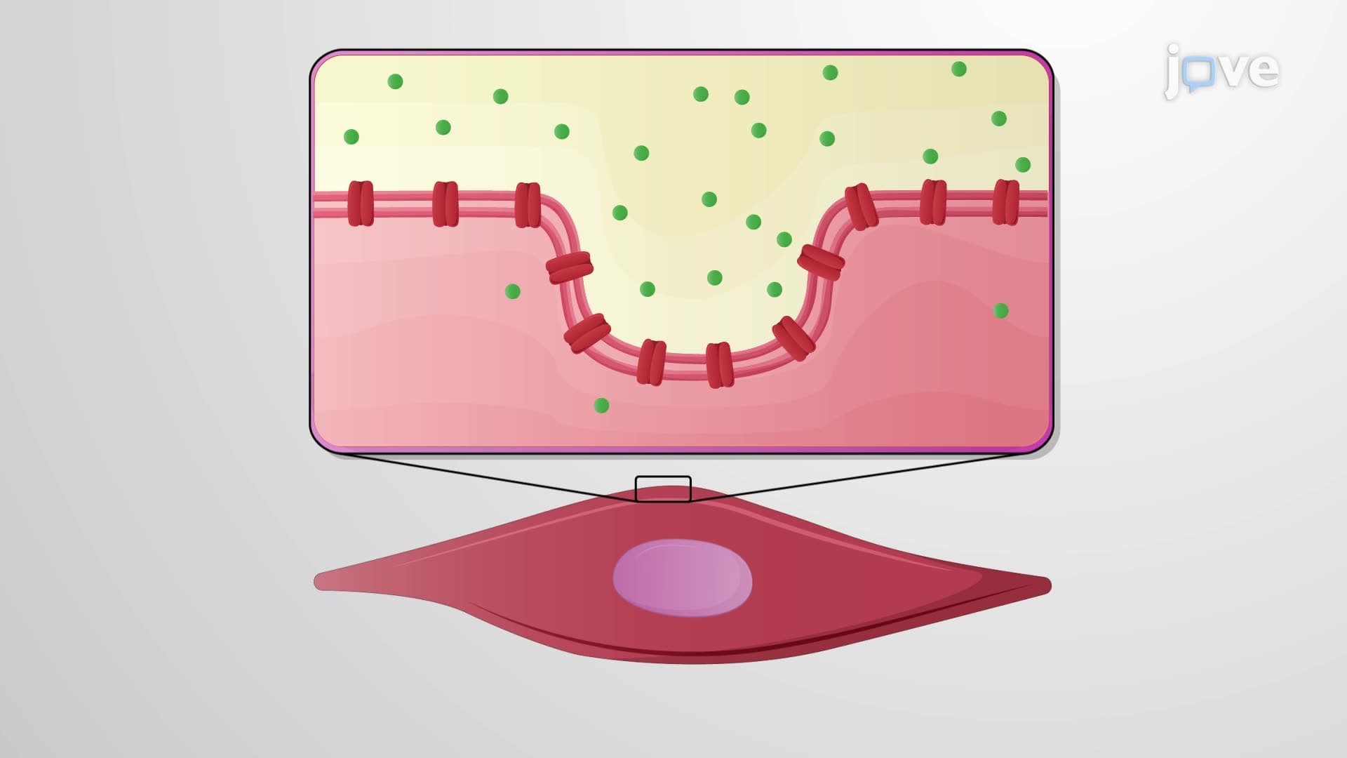
Smooth muscle cells exhibit distinctive spindle shaped smooth cell morphology, measuring 30-200 micrometers in length with diameters of 5-10 micrometers. Each cell contains a centrally located nucleus, distinguishing it from the multinucleated skeletal muscle fibers. This smooth muscle tissue anatomy reflects functional specialization-the elongated shape allows cells to generate force along their length while maintaining flexibility for organ wall movements.
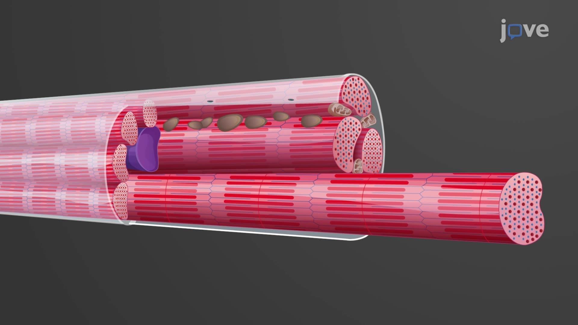
The absence of organized sarcomeres creates the characteristic non-striated appearance. Instead of the precise A-bands and I-bands seen in skeletal muscle, smooth muscle filament organization features irregularly arranged thick and thin filaments along the cell's long axis. This arrangement, while appearing less organized, actually provides greater versatility for the varied contractions required in organs like the stomach during digestion or blood vessels during exercise.
Dense body smooth muscle structures serve as crucial anchoring points, functionally replacing the Z-lines found in striated muscle. These protein-rich regions anchor thin filaments to the sarcolemma and connect to adjacent cells through intermediate filament networks. This creates a three-dimensional web that transmits contractile forces throughout the tissue-essential for coordinated contractions in organs like the uterus during childbirth or intestinal walls during peristalsis.
The single unit multi-unit classification system reflects functional differences in neural control. Visceral (single-unit) smooth muscles, found in digestive tract walls and blood vessels, contract as synchronized sheets through gap junctions-like a coordinated wave moving food through your esophagus. Multi-unit smooth muscles, controlling structures like iris muscles and hair follicles, allow precise individual cell control for fine adjustments.
This organizational understanding proves crucial for AP Biology students studying muscle physiology and pre-med students preparing for MCAT passages on cardiovascular or digestive system regulation.
Related Micro-courses