Loading subject page…


178,811 views

Video Summary: What Is the Neuromuscular Junction
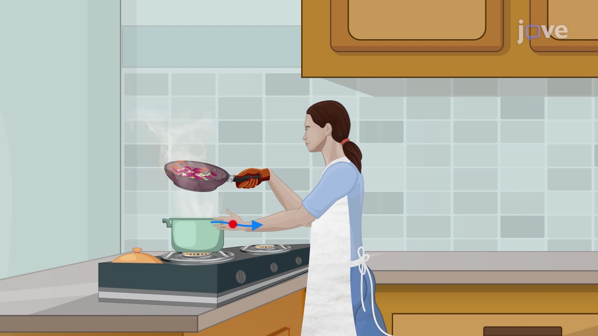
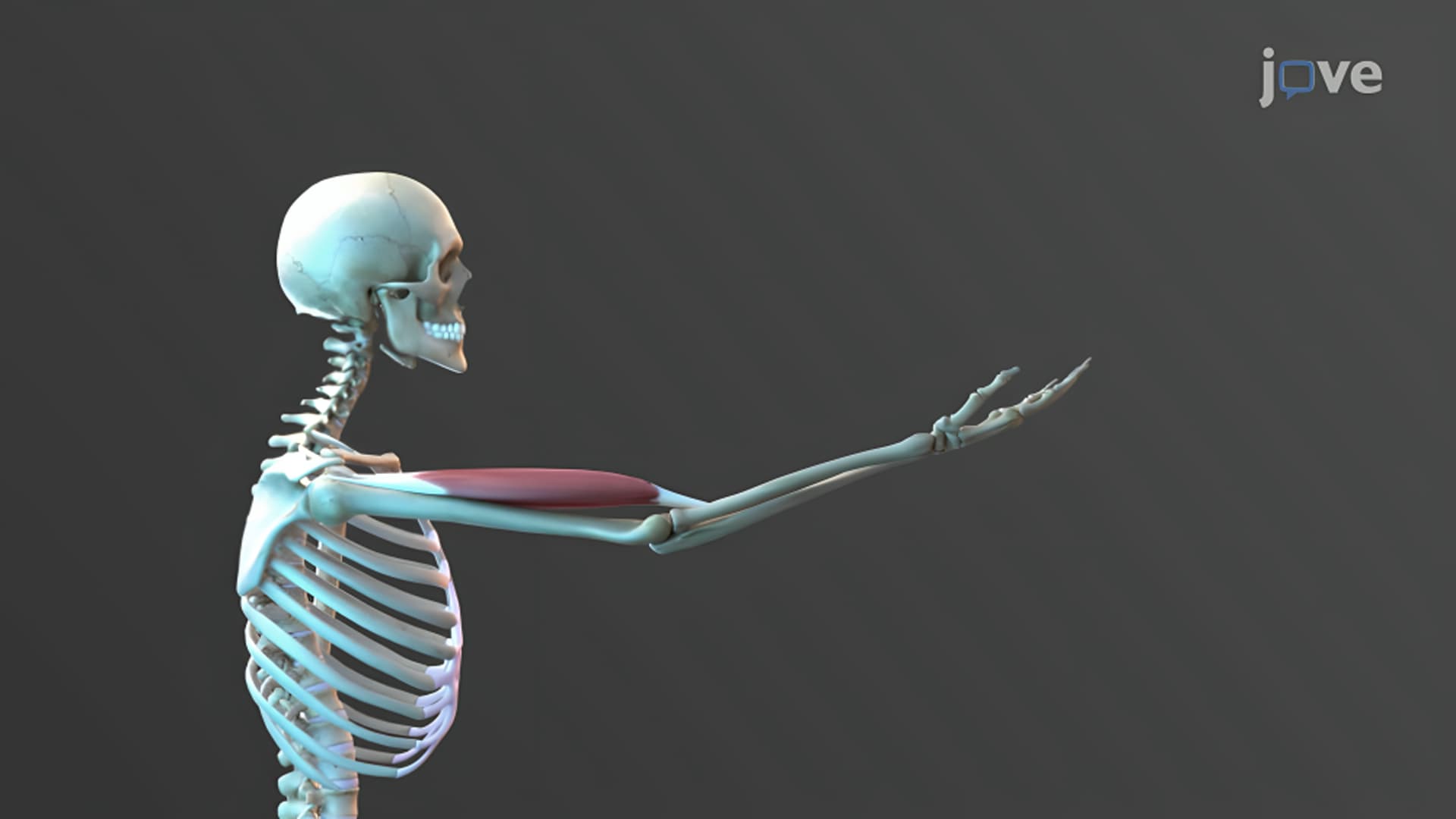
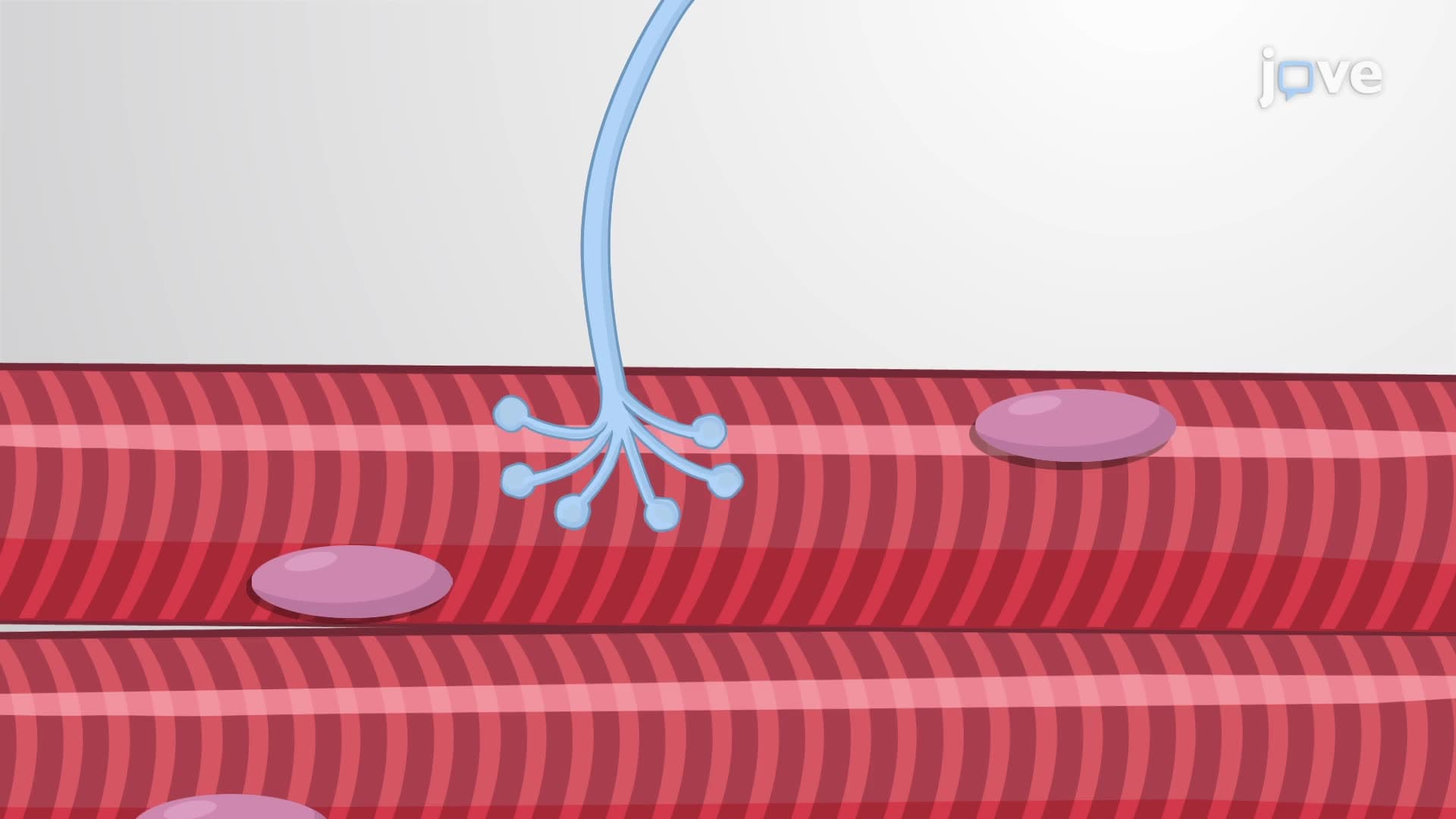
Ever wonder how your brain instantly commands your bicep to flex during a workout at your local gym? The neuromuscular junction anatomy reveals this remarkable process where motor neurons communicate directly with muscle fibers through a specialized connection called the neuromuscular junction. This critical interface allows electrical signals from your spinal cord to trigger muscle contractions that power everything from typing to sprinting. Watch the full video on JoVE Coach to master this concept with expert-led visuals and step-by-step explanations.
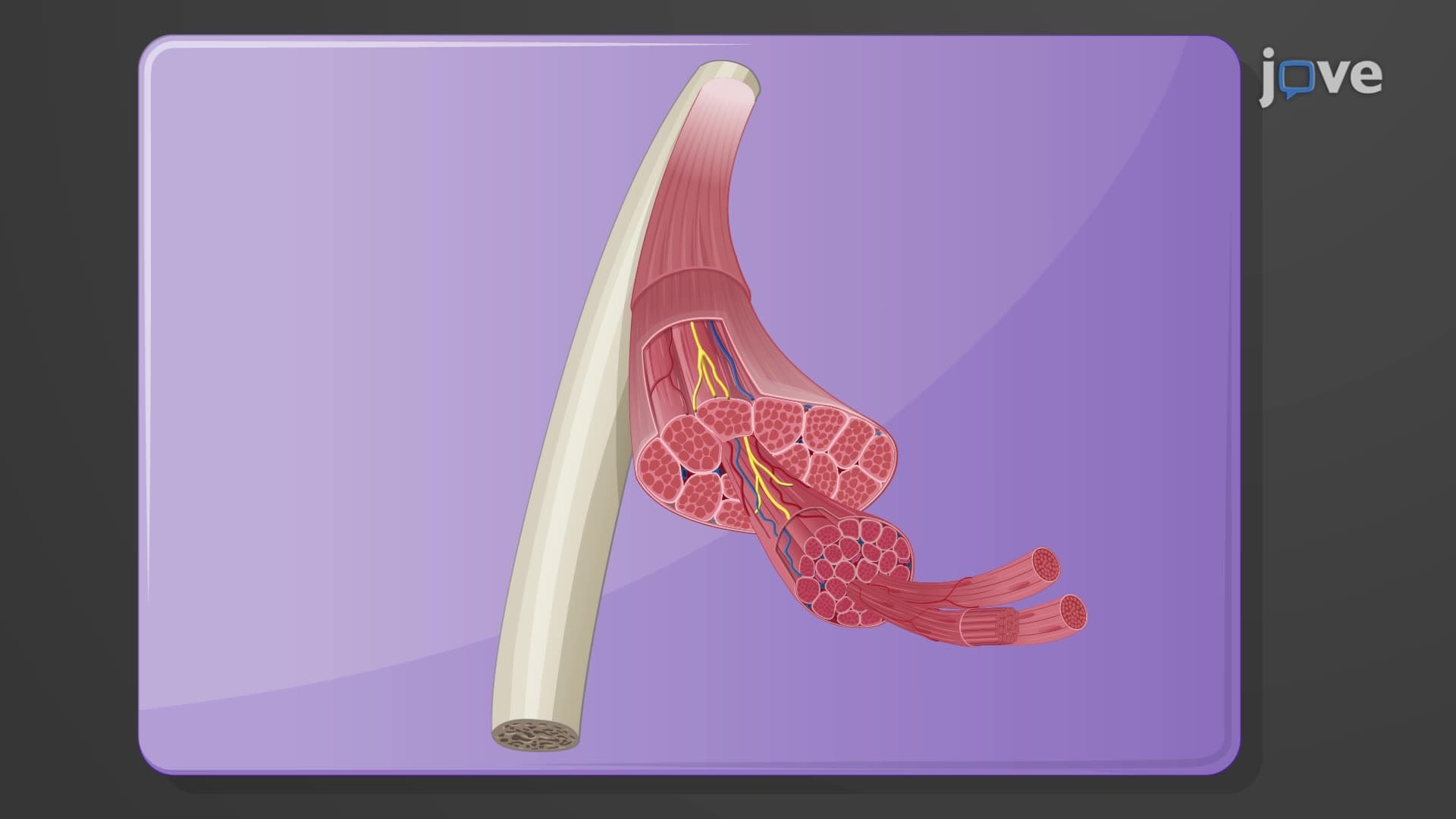
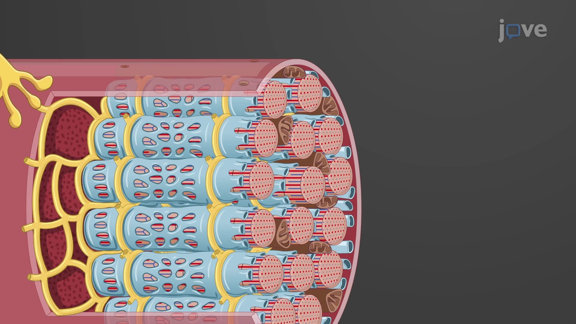
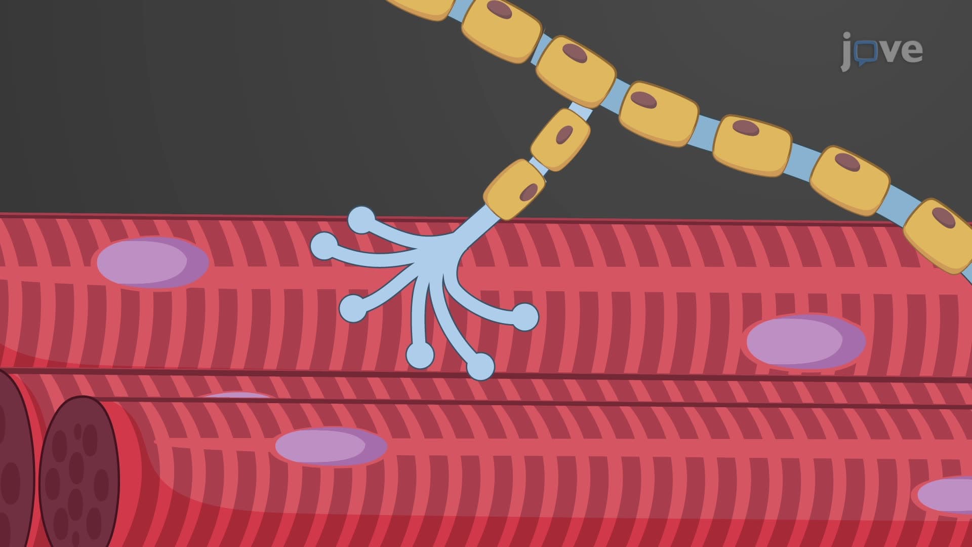
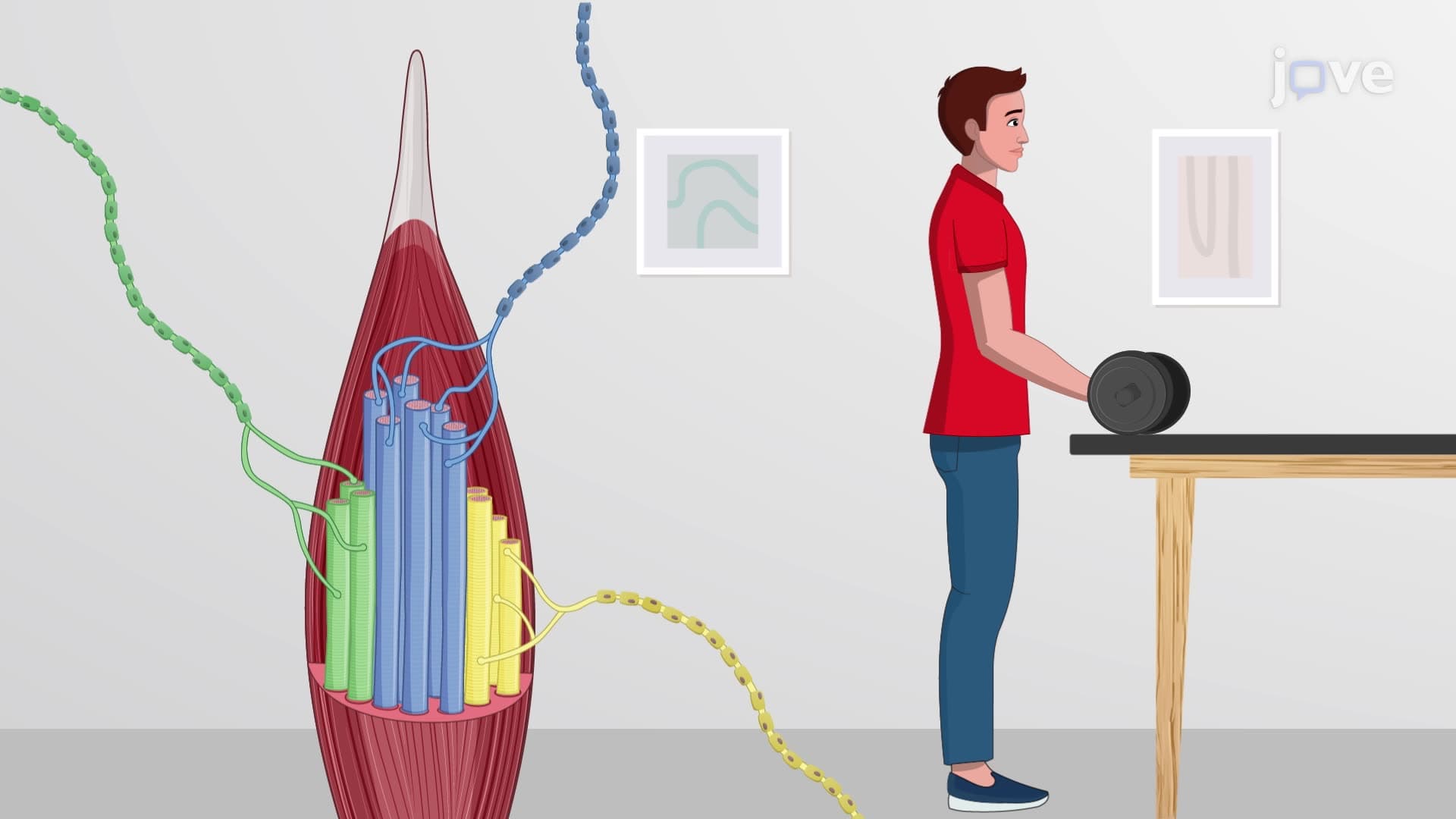
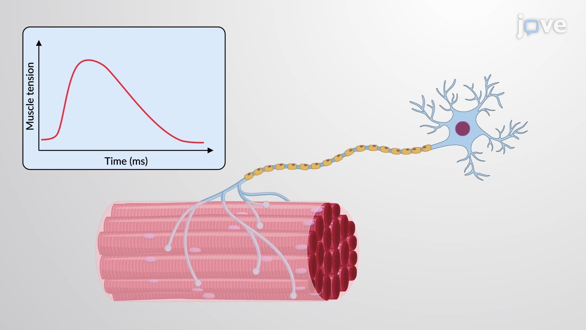
The neuromuscular junction anatomy represents one of the most precisely organized synapses in the human body. Unlike brain synapses that may involve multiple neurotransmitters, the NMJ exclusively uses acetylcholine for communication. Each motor neuron muscle fiber connection begins in the anterior horn of the spinal cord, where somatic motor neurons send their axons through peripheral nerves to reach target muscles. At Harvard Medical School and other top US medical programs, students learn that a single motor neuron can innervate anywhere from 10 to 2,000 muscle fibers, forming what physiologists call a "motor unit."
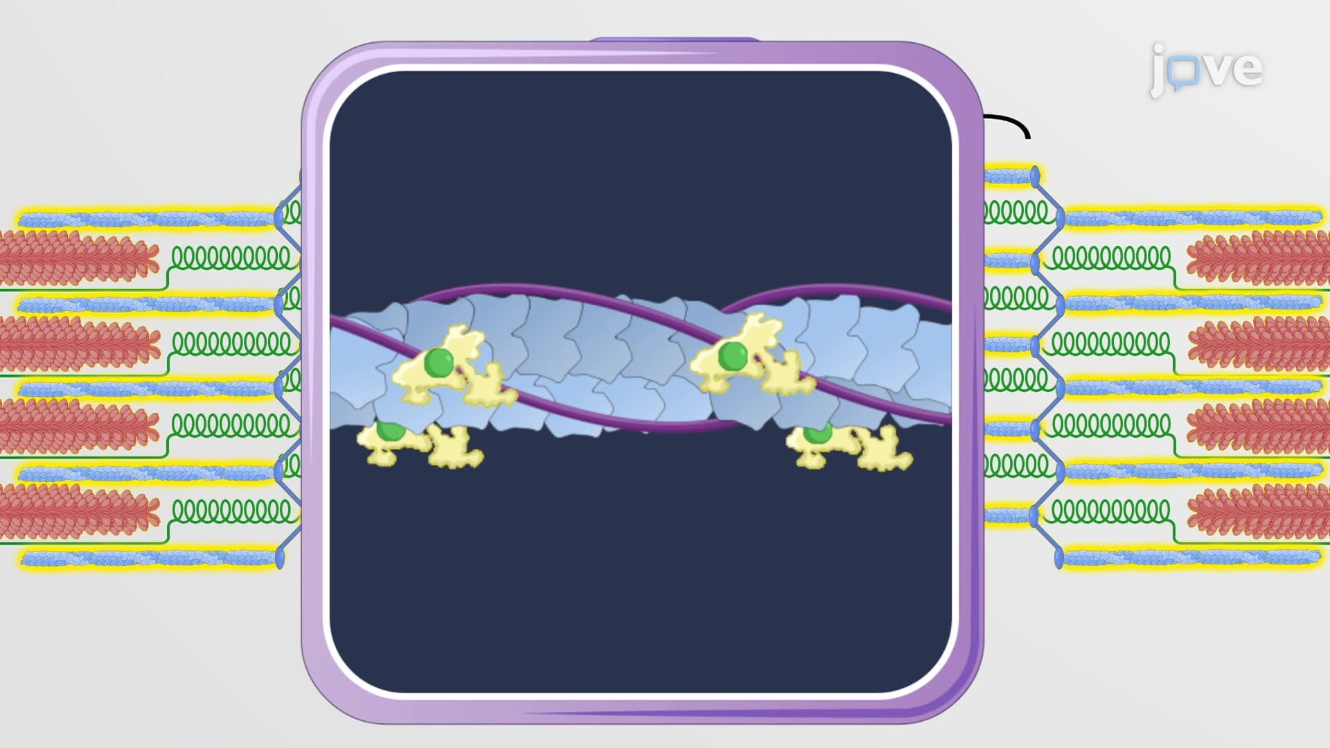
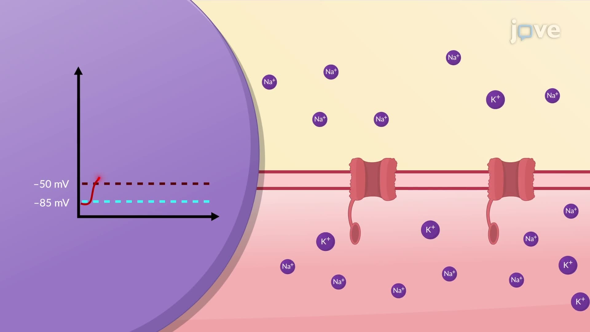
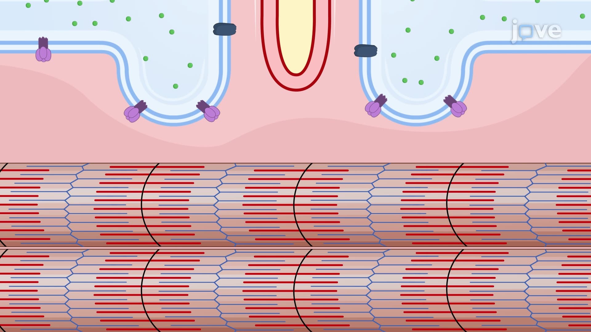
The synaptic cleft measures approximately 50-100 nanometers wide, creating an optimal distance for neurotransmitter diffusion. The motor end plate contains specialized nicotinic acetylcholine receptors that are structurally different from those found in the brain. These receptors are ligand-gated sodium channels that open when acetylcholine binds, allowing rapid depolarization of the muscle fiber membrane. Students preparing for the MCAT at institutions like Johns Hopkins University focus extensively on understanding how these receptors differ from muscarinic receptors found in autonomic synapses.
When studying NMJ physiology explained for AP Biology or college physiology courses, students must grasp the calcium-dependent release mechanism. Action potentials traveling down the motor axon trigger voltage-gated calcium channels to open at the synaptic end bulb. The resulting calcium influx causes synaptic vesicles containing acetylcholine to fuse with the presynaptic membrane through SNARE protein interactions. This process, called exocytosis, releases approximately 200-300 acetylcholine molecules per vesicle.
Understanding what is the neuromuscular junction in muscle physiology becomes crucial when examining diseases like myasthenia gravis, which affects over 200,000 Americans. In this autoimmune condition, antibodies block acetylcholine receptors on the motor end plate, leading to muscle weakness and fatigue. Medical students at institutions like Mayo Clinic's medical school study how drugs like pyridostigmine work by inhibiting acetylcholinesterase NMJ function, thereby prolonging acetylcholine action. This knowledge directly applies to USMLE Step 1 questions about neuromuscular transmission disorders.
Related Micro-courses