Loading video…


 22 Concepts
 178.8K views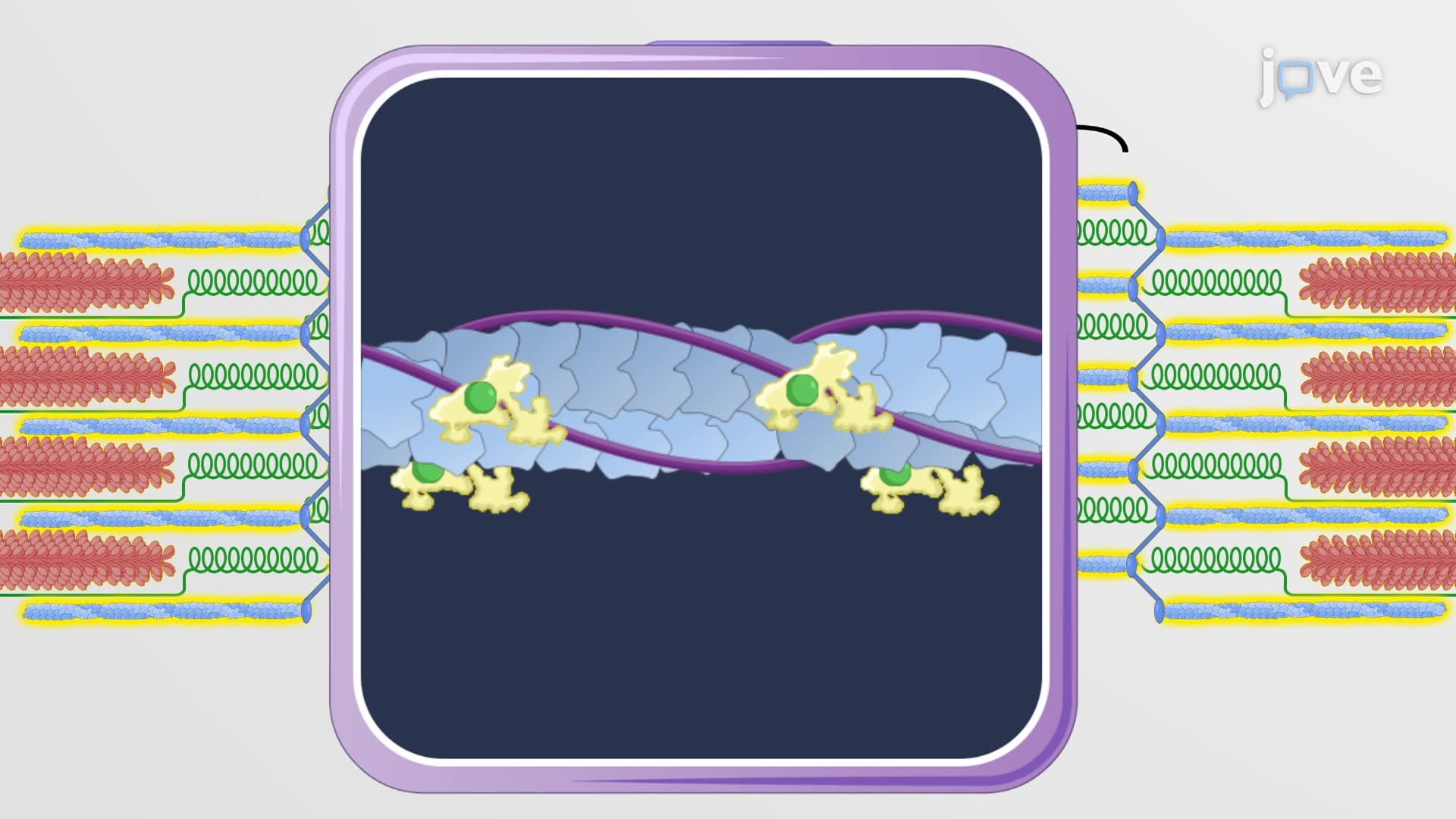

Video Summary: What Is the Sarcomere
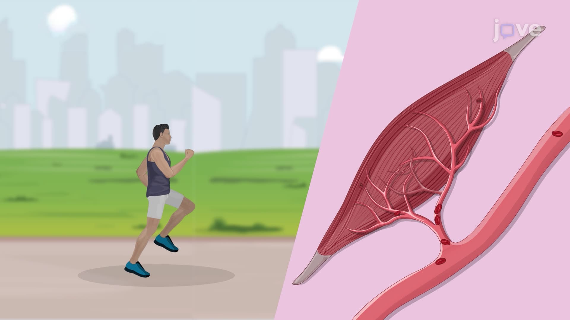
Ever wonder why your bicep can lift weights but your skin can't? The answer lies in sarcomere muscle structure-the microscopic machinery that powers every muscle contraction in your body. From a basketball player's explosive jump at Duke University to a surgeon's precise hand movements at Johns Hopkins, sarcomeres are the fundamental contractile units that make all voluntary movement possible. What is the sarcomere? It's a highly organized arrangement of protein filaments that transforms chemical energy into mechanical force through a sophisticated sliding mechanism. Watch the full video on JoVE Coach to master this concept with expert-led visuals and step-by-step explanations.
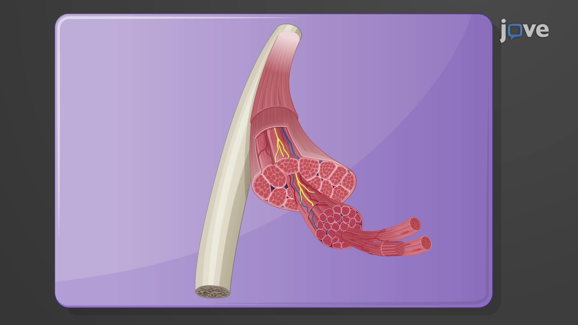
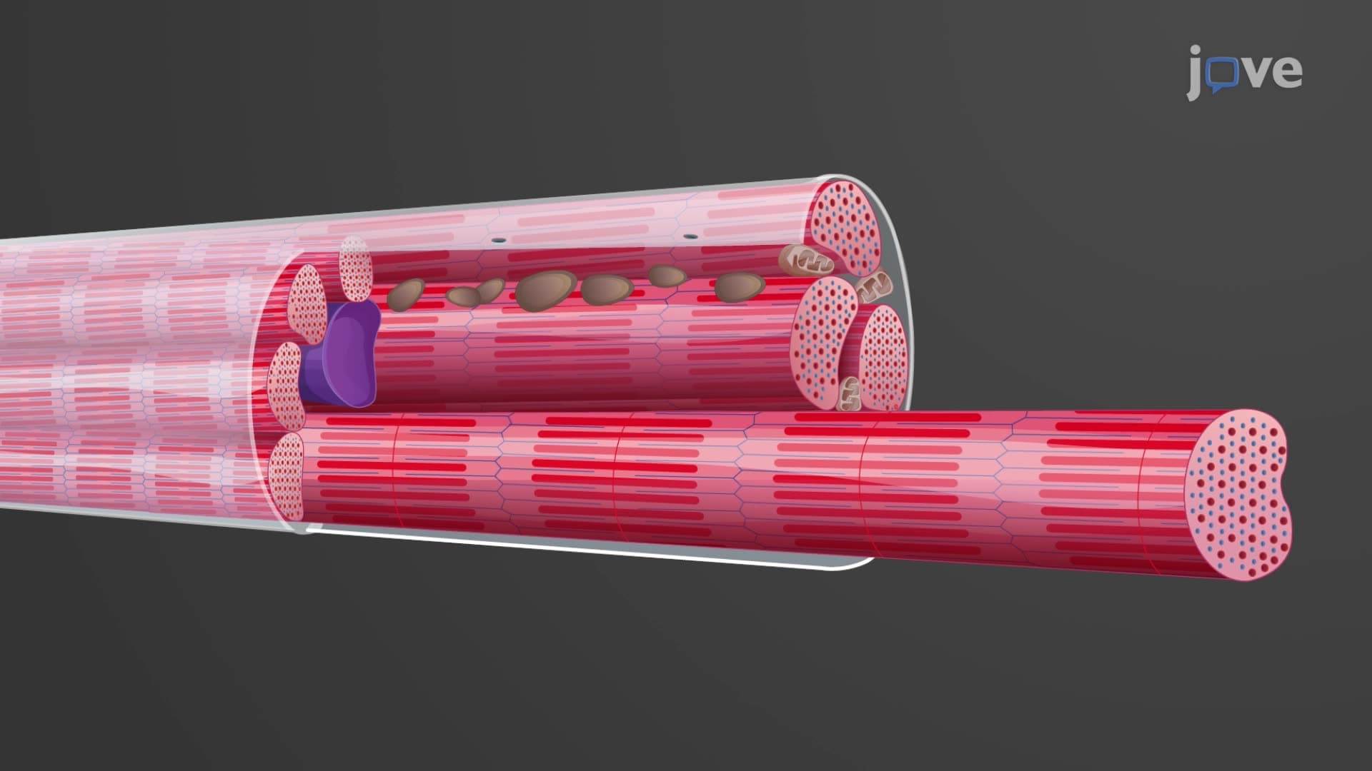
The sarcomere represents one of biology's most elegant examples of structure-function relationships. This microscopic unit, typically measuring 2-3 micrometers in length, serves as the fundamental contractile apparatus in skeletal and cardiac muscle. When students encounter sarcomere muscle structure in AP Biology or college physiology courses, they're studying the cellular foundation that enables everything from a marathon runner's endurance to a pianist's finger dexterity.
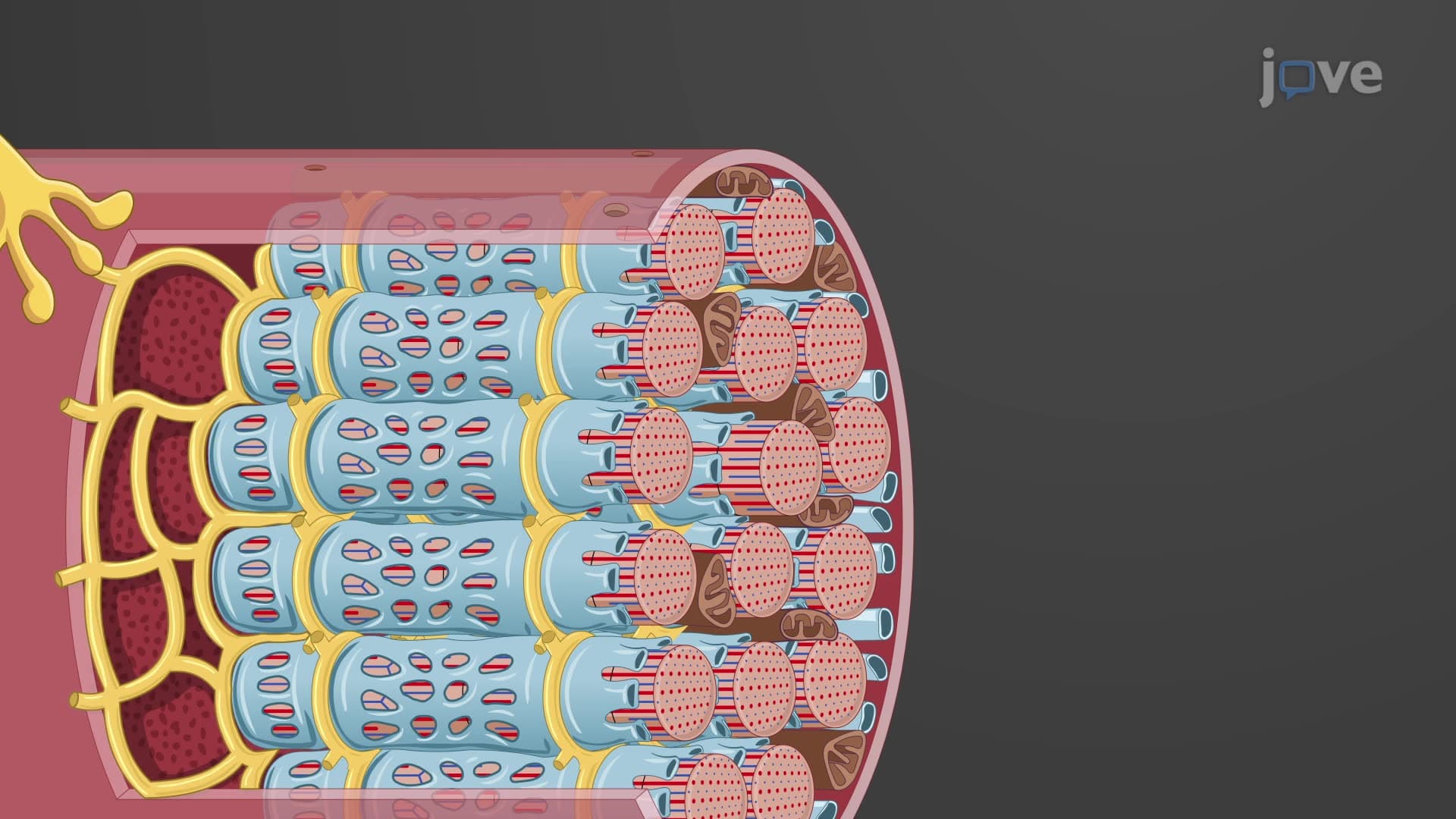
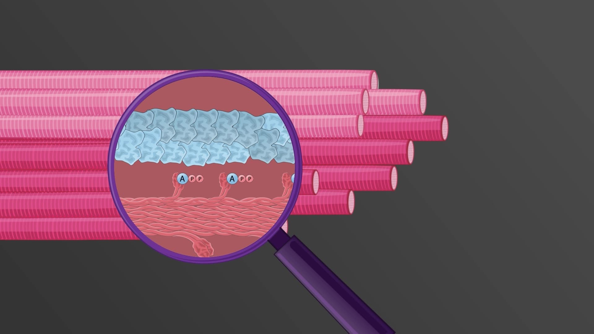
The sarcomere's sophisticated design centers on two primary filament types working in concert. Thick filaments, composed primarily of myosin protein, form the backbone of the contractile mechanism. These rod-shaped molecules feature distinctive head regions that act as molecular motors. Thin filaments contain actin, the track along which myosin heads "walk" during contraction. The regulatory proteins troponin and tropomyosin coat the actin filaments, controlling access to myosin binding sites based on calcium ion availability.
Titin, often called the body's largest protein, provides crucial structural support by spanning from Z disc to Z disc. This elastic protein maintains sarcomere integrity during stretching and contributes to muscle's passive tension properties. Medical students studying for the MCAT frequently encounter titin's role in conditions like dilated cardiomyopathy, where mutations affect heart muscle function.
The characteristic striations visible under microscopy result from the precise organization of these protein filaments. The A band represents the region containing thick myosin filaments, appearing dark due to protein density. Within the A band lies the H zone, where only thick filaments exist with no thin filament overlap. The lighter I band contains exclusively thin filaments, interrupted at its center by the dense Z disc-the sarcomere's boundary marker.
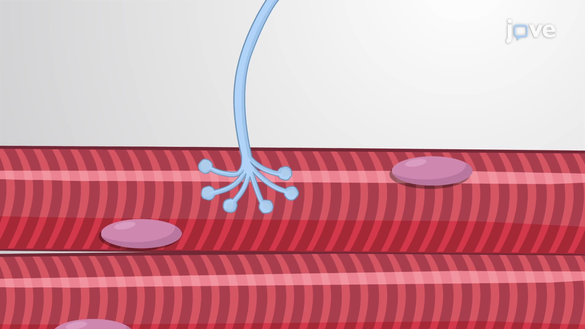
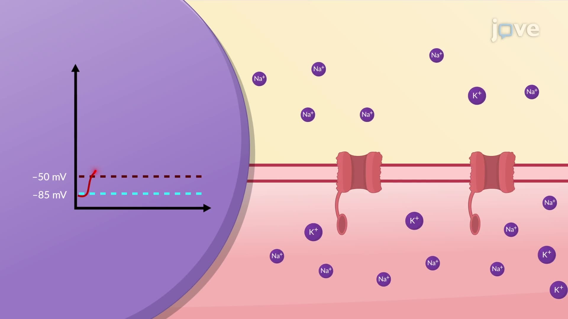
This banding pattern isn't merely structural; it reflects the functional organization essential for contraction. During muscle activation, thin filaments slide past thick filaments, causing the H zone to narrow while the A band length remains constant. Students preparing for college physiology exams should understand that this sliding filament mechanism explains how sarcomeres generate force without the proteins themselves changing length.
Understanding sarcomere anatomy function explained proves crucial for medical professionals diagnosing muscle diseases. Conditions like hypertrophic cardiomyopathy often involve mutations in sarcomere proteins, affecting cardiac muscle's ability to contract and relax properly. Research institutions like the National Institutes of Health continue investigating how sarcomere dysfunction contributes to muscle weakness, heart failure, and inherited myopathies affecting thousands of Americans annually.
Related Micro-courses