Loading subject page…

- Cell Biology
- Visualizing Cells, Tissues, and Molecules
- Cryo-electron Microscopy
Micro-course: Visualizing Cells, Tissues, and Molecules
138,515 views
Learn key concepts and earn your certificate.



Cryo-electron Microscopy


00:00
01:28
Video Summary: What Is Cryo Electron Microscopy
Ever wonder how scientists can see viruses in action without destroying them? Cryo electron microscopy revolutionizes biological imaging by flash-freezing samples to preserve their natural structures at near-atomic resolution. Unlike traditional electron microscopy that can damage delicate biological materials, this technique vitrifies specimens in liquid ethane at ultra-low temperatures. For example, researchers at Harvard Medical School used cryo-EM to reveal the detailed structure of the COVID-19 spike protein. Watch the full video on JoVE Coach to master this concept with expert-led visuals and step-by-step explanations.
- Understand what is cryo electron microscopy and how it differs from conventional electron microscopy techniques
- Identify the key advantages of vitrification in preserving native biological structures without chemical fixation
- Analyze why cryogenic temperatures below -150°C prevent radiation damage from high-energy electrons
- Compare vitrification methods for small samples versus thicker specimens like cells and tissues
- Recognize the glass-like state formation in vitrified water that prevents ice crystal artifacts
- Evaluate when to use cryo-EM of vitreous sections for studying larger biological samples
- Apply knowledge of sample preparation techniques including high-pressure freezing and ultrathin sectioning
- Explore real-world applications of cryo-EM in structural biology and drug discovery research
Understanding Cryo Electron Microscopy Fundamentals
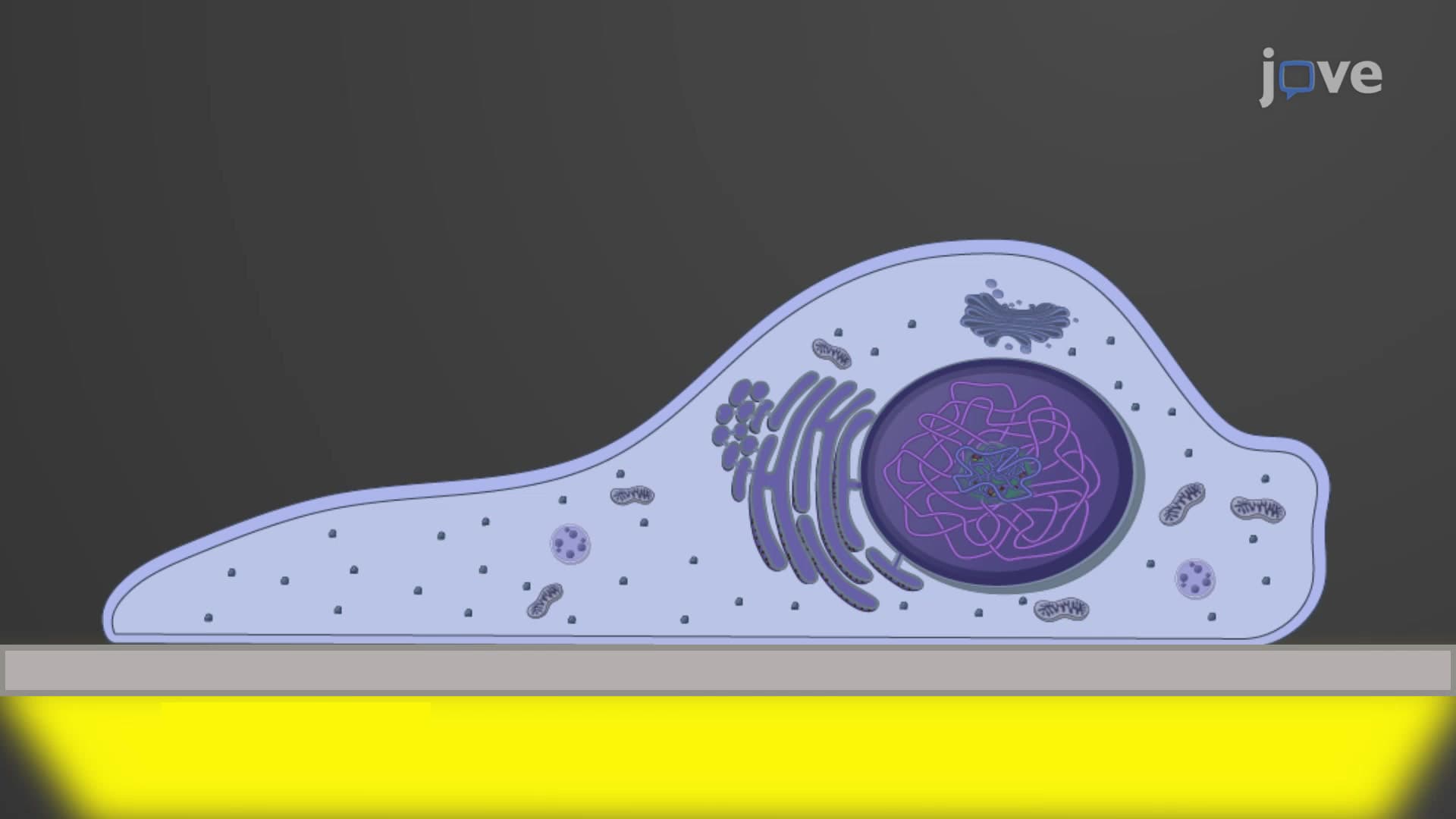
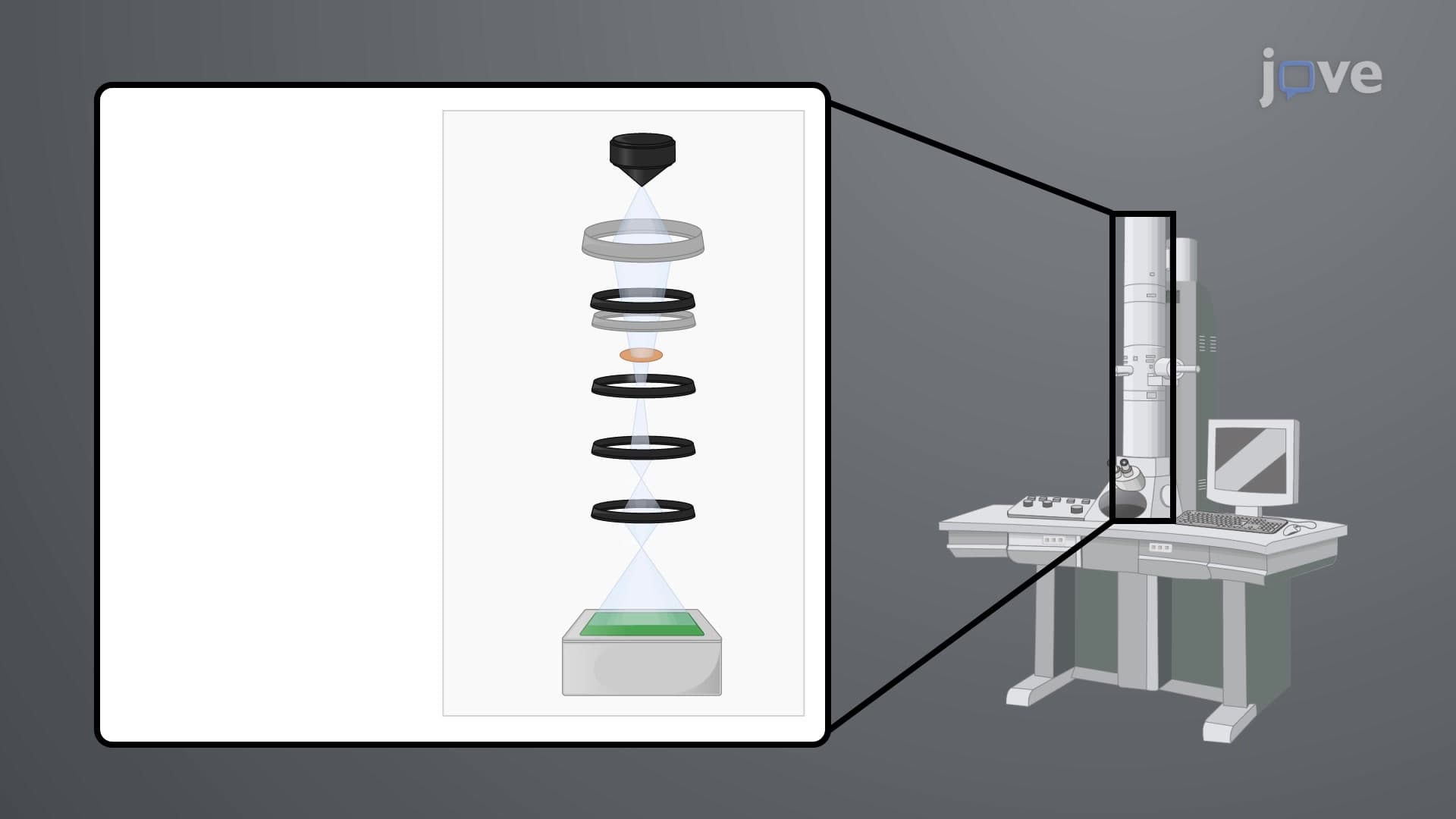
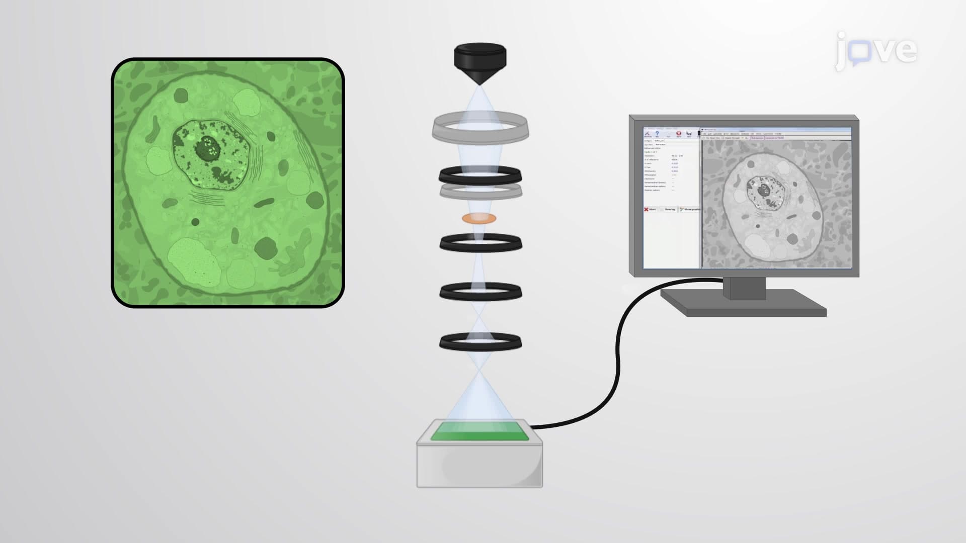
Cryo electron microscopy represents a groundbreaking advancement in biological imaging that has transformed our understanding of molecular structures. This technique addresses a critical limitation of traditional electron microscopy: the need to chemically fix and dehydrate samples, which often distorts their natural architecture. By operating at cryogenic temperatures below -150°C (-238°F), cryo-EM preserves biological specimens in their native, hydrated state while achieving remarkable resolution.
The Science Behind Vitrification
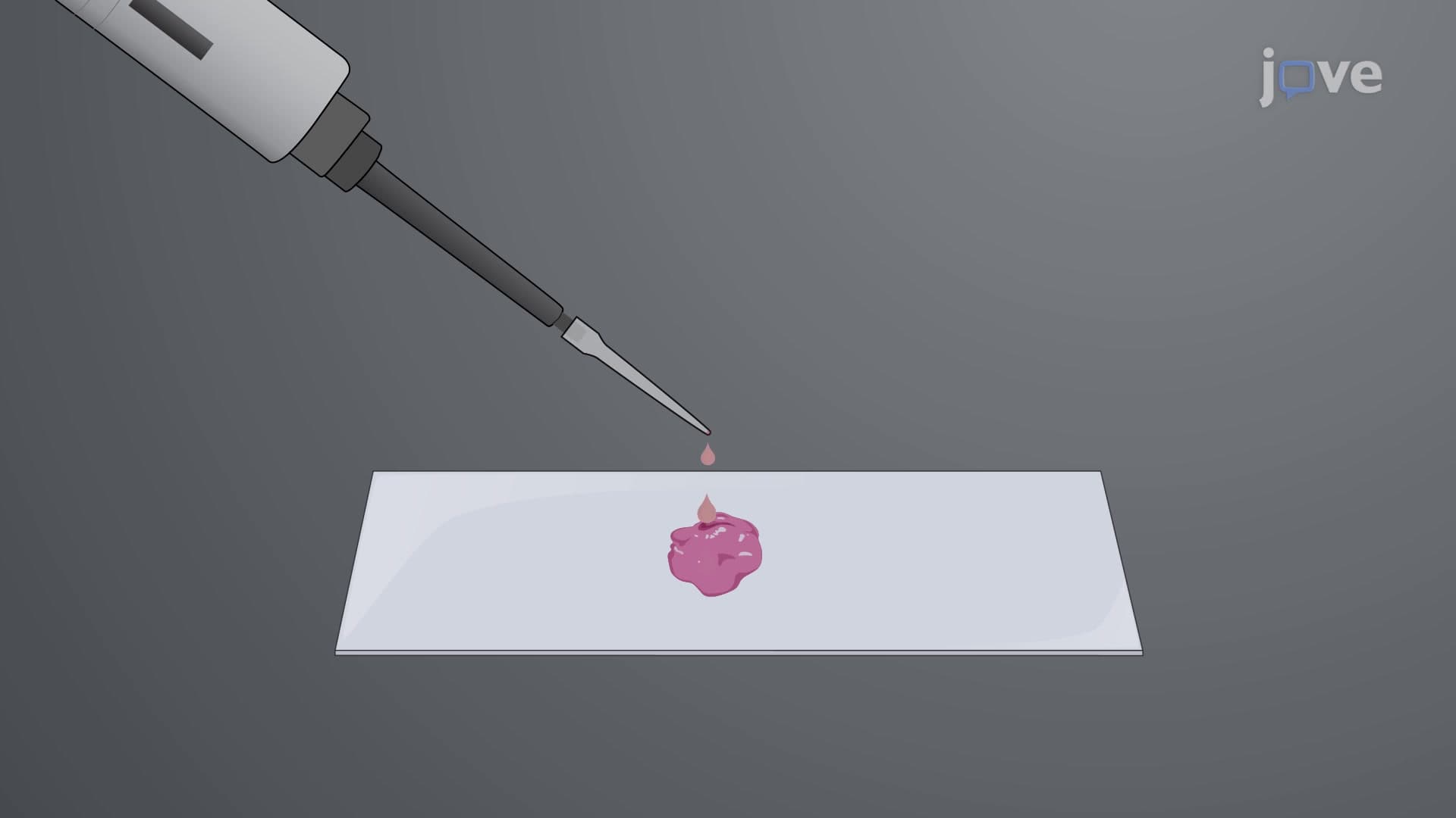
The cornerstone of cryo-EM success lies in vitrification, the rapid freezing process that transforms liquid water into a glass-like solid without forming destructive ice crystals. When biological samples are plunged into liquid ethane cooled by liquid nitrogen, they freeze so quickly (within milliseconds) that water molecules cannot organize into crystalline structures. This process is crucial for maintaining cellular integrity and protein conformations that would otherwise be lost in conventional preparation methods.
Advanced Techniques for Different Sample Types
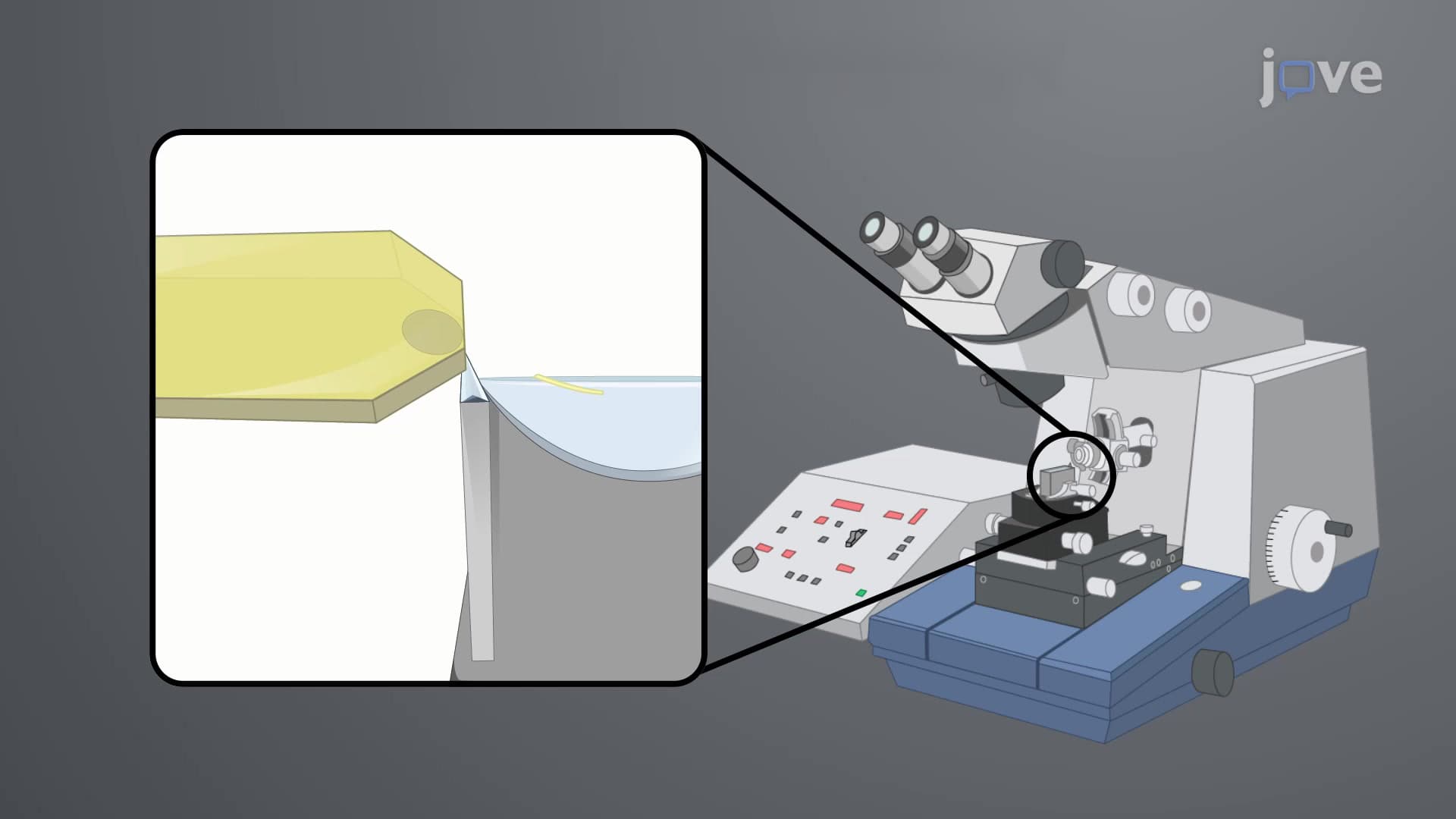
While small proteins and molecular complexes can be vitrified directly in thin layers, larger specimens require specialized approaches. The cryo-EM of vitreous sections method accommodates thicker samples like whole cells and tissue sections. This technique employs high-pressure freezing followed by precise sectioning at -140°C using specialized equipment called cryo-ultramicrotomes. Universities like Stanford and MIT utilize these advanced systems in their structural biology programs.
Real-World Applications and Academic Relevance
Cryo-EM has revolutionized fields from drug discovery to vaccine development. For instance, researchers at the University of Texas at Austin used cryo-EM to determine the structure of the Zika virus, leading to potential therapeutic targets. This technique frequently appears in AP Biology curricula when discussing protein structure and cellular organization. College-level biochemistry courses, particularly those preparing students for the MCAT, emphasize cryo-EM's role in structural determination. Understanding this technology is essential for students pursuing careers in biotechnology, pharmaceutical research, or academic medicine.
Frequently Asked Questions
Cryo electron microscopy is an imaging technique that examines flash-frozen biological samples at ultra-low temperatures to preserve their natural structures. The process involves rapidly freezing specimens in liquid ethane, creating a glass-like state that prevents ice crystal damage. High-energy electrons then pass through the sample to create detailed images at near-atomic resolution.
Yes, cryo-EM concepts frequently appear in MCAT biochemistry sections and AP Biology questions about protein structure and cellular organization. Students should understand the principles of sample preservation, vitrification, and how this technique advances our knowledge of molecular structures. Focus on comparing cryo-EM advantages over traditional electron microscopy methods.
College biochemistry programs use cryo-EM as a prime example of structural biology techniques when studying protein folding, enzyme mechanisms, and membrane complexes. Students analyze cryo-EM data to understand how molecular structures relate to biological function. Many universities incorporate cryo-EM case studies in their structural biology and biophysics coursework.
US pharmaceutical companies like Pfizer and Moderna used cryo-EM to develop COVID-19 vaccines by determining viral protein structures. Research institutions such as the NIH National Cancer Institute employ cryo-EM to study cancer-related proteins for drug development. The FDA also utilizes cryo-EM data for evaluating new therapeutic compounds and biologics.
While cryo-EM involves sophisticated technology, the basic concepts are accessible to high school students with a solid foundation in chemistry and biology. Focus on understanding the problems it solves rather than technical details. The key is grasping why preserving native structures matters for biological research and drug discovery.
Create comparison charts between cryo-EM and traditional electron microscopy, highlighting advantages and limitations. Practice drawing the vitrification process and explaining why ice crystals damage biological samples. Review specific examples of protein structures determined by cryo-EM, especially those relevant to human health and disease.
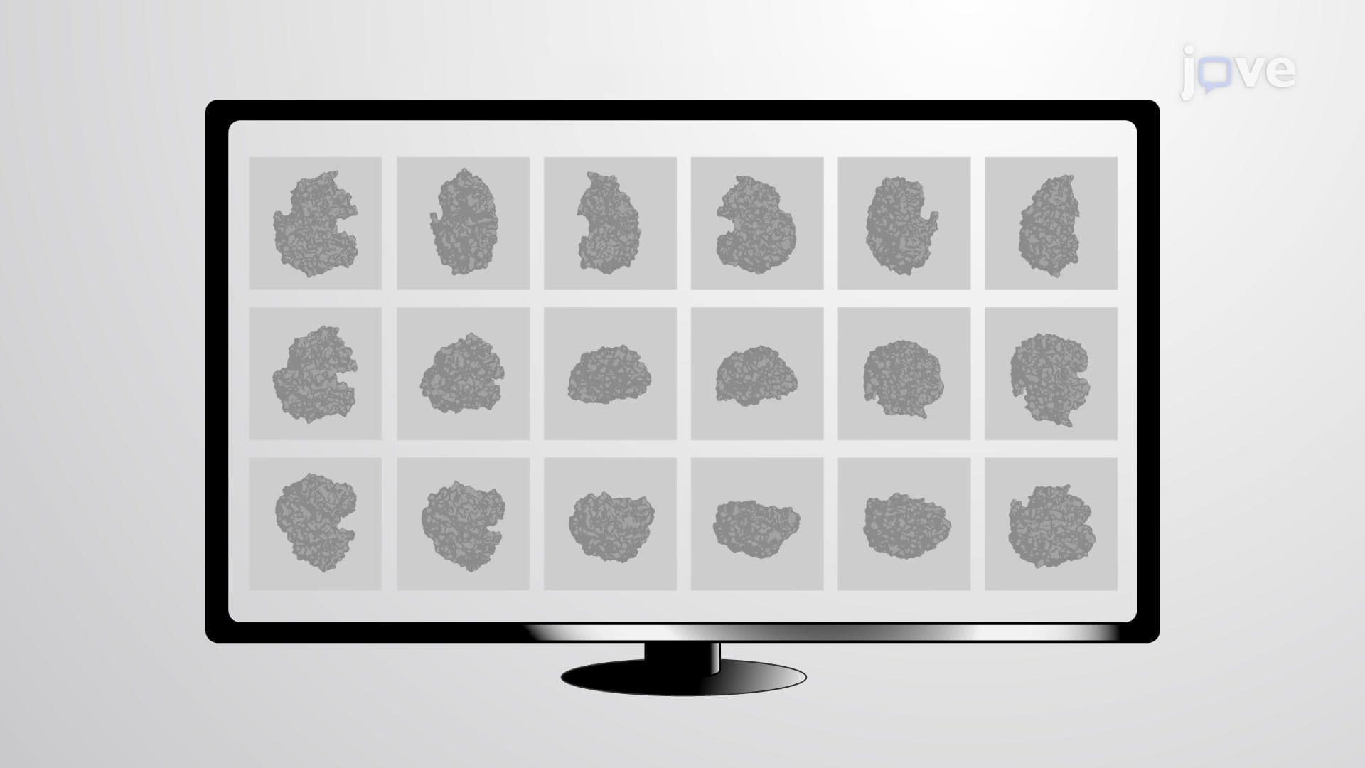
After mastering basic cryo-EM principles, explore single-particle analysis techniques used for determining protein structures and cryo-electron tomography for studying cellular architecture. Investigate how artificial intelligence and machine learning are revolutionizing cryo-EM image processing and structure determination in modern research laboratories.
Related Micro-courses