Loading subject page…

- Cell Biology
- Visualizing Cells, Tissues, and Molecules
- Fixation and Sectioning
Micro-course: Visualizing Cells, Tissues, and Molecules
138,515 views
Learn key concepts and earn your certificate.




Fixation and Sectioning

00:00
01:03
Video Summary: What Is Fixation and Sectioning
Ever wondered how pathologists at hospitals like Mayo Clinic examine cancer cells that are invisible to the naked eye? Fixation and sectioning transforms thick, opaque biological tissues into crystal-clear microscopic slides through a precise multi-step process. This fundamental laboratory technique preserves cellular structure while creating ultra-thin tissue slices that reveal disease patterns and cellular abnormalities. From high school biology labs to medical diagnostics, understanding what is fixation and sectioning unlocks the microscopic world that drives modern medicine. Watch the full video on JoVE Coach to master this concept with expert-led visuals and step-by-step explanations.
- Understand what is fixation and sectioning and why these processes are essential for microscopic tissue examination
- Identify the sequential steps involved in tissue preparation, from fixation through final slide mounting
- Analyze how chemical fixatives preserve cellular components and prevent tissue degradation
- Compare different embedding materials and their role in supporting fragile tissues during sectioning
- Evaluate the importance of proper dehydration gradients in tissue processing protocols
- Apply knowledge of refractive index matching to optimize microscopic image quality
- Recognize the critical role of microtomes in creating uniform tissue sections
- Explore how staining techniques enhance cellular visualization after sectioning
Understanding Fixation and Sectioning Fundamentals
Fixation and sectioning represents the cornerstone of histological preparation, transforming living tissues into permanent microscopic specimens. This process begins when surgeons at institutions like Johns Hopkins remove tissue samples during biopsies or surgeries. The race against time starts immediately-without proper fixation, cellular components begin degrading within minutes, potentially compromising diagnostic accuracy.
Chemical fixation serves as the first critical step, typically using formaldehyde-based solutions that cross-link proteins and nucleic acids. This process effectively "freezes" cellular architecture in its current state, preventing bacterial decomposition and enzyme activity that would otherwise distort tissue structure. The fixation process must penetrate completely through tissue thickness, requiring carefully timed exposure based on specimen size.
Dehydration and Embedding Process
Following fixation, tissues undergo systematic dehydration through increasing alcohol concentrations-typically progressing from 70% to 100% ethanol. This stepwise approach prevents cellular damage that would occur with rapid water removal. The alcohol extraction prepares tissues for embedding in paraffin wax, a hydrophobic medium that provides structural support during the sectioning process.
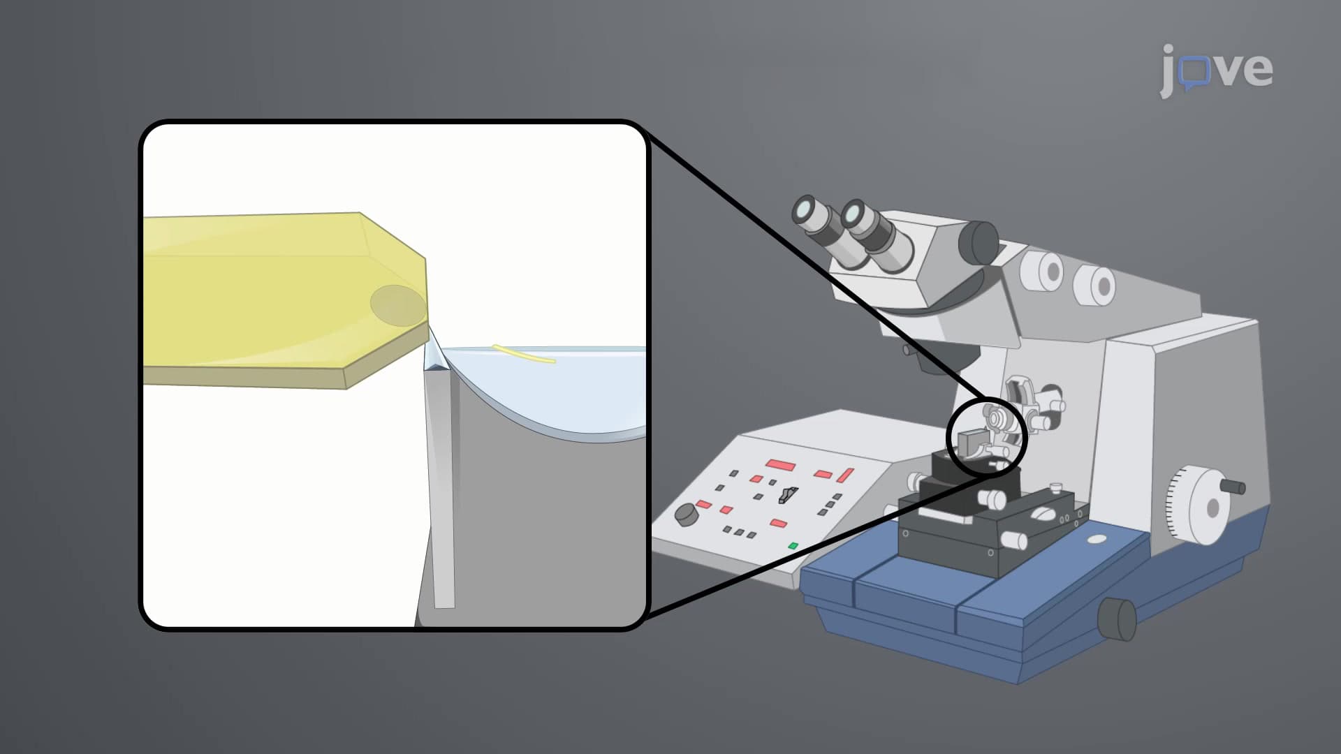
Paraffin embedding creates a solid matrix around the tissue, enabling precise cutting with specialized instruments called microtomes. These devices produce sections measuring just 3-5 micrometers thick-roughly 1/20th the diameter of a human hair. Such precision ensures light can pass through tissues while maintaining cellular detail visibility.
Sectioning and Slide Preparation
The sectioning process requires extraordinary skill, as histotechnologists manipulate microtome blades to create ribbon-like tissue strips. These delicate sections are carefully transferred to glass slides and subjected to dewaxing procedures using organic solvents like xylene. The removal of embedding material allows subsequent staining procedures to penetrate cellular components effectively.
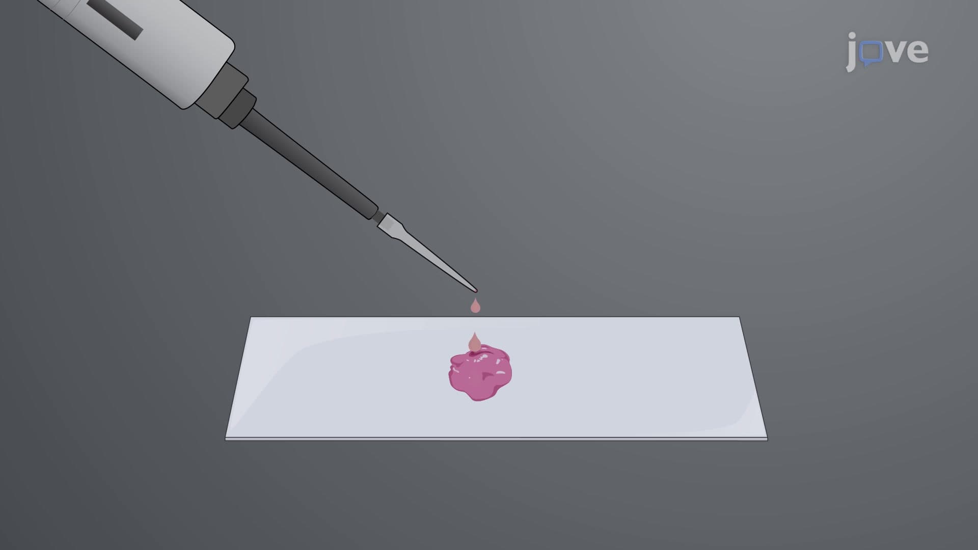
Final slide preparation involves mounting media application and coverslip placement. The refractive index matching between slide, mounting medium, and coverslip eliminates optical distortions that would compromise image clarity. This attention to optical physics ensures pathologists can accurately identify cellular abnormalities during diagnostic examinations.
Students preparing for AP Biology or college-level histology courses should understand that these techniques directly impact medical diagnoses. From cancer detection to infectious disease identification, the quality of fixation and sectioning procedures can literally mean the difference between accurate diagnosis and missed pathology. Mastering these concepts provides essential foundation knowledge for healthcare careers and advanced biological sciences.
Frequently Asked Questions
Fixation and sectioning is a laboratory process that prepares biological tissues for microscopic examination by preserving cellular structure and creating ultra-thin tissue slices. The process involves chemically "fixing" tissues to prevent decay, removing water content, embedding in supportive material, and cutting into sections thin enough for light to pass through. This technique is essential for medical diagnoses and biological research.
MCAT questions often focus on the chemical principles behind fixation, particularly protein cross-linking and osmotic effects during dehydration. AP Biology exams may test understanding of why tissues require processing before microscopic observation and the relationship between section thickness and light transmission. Students should understand both the procedural steps and underlying scientific rationales.
Focus on fixative chemistry (formaldehyde cross-linking mechanisms), dehydration gradients and their cellular effects, embedding media properties, microtome operation principles, and refractive index concepts in microscopy. Many college exams also test troubleshooting skills-understanding what causes common preparation artifacts and how to prevent them.
Hospital pathology departments use these techniques daily for cancer diagnosis, surgical margin assessment, and disease identification. When surgeons at institutions like Cleveland Clinic remove suspicious tissues, pathologists must process samples within hours to provide accurate diagnoses. The quality of fixation and sectioning directly impacts patient treatment decisions and surgical outcomes.
Basic high school chemistry concepts are sufficient to understand fixation and sectioning principles. Knowledge of protein structure, chemical bonding, and solution properties provides helpful background, but the core concepts are accessible to students with standard biology and chemistry coursework. Focus on understanding the purpose behind each step rather than memorizing complex chemical formulas.
Create a step-by-step flowchart showing the entire process from fresh tissue to finished slide, including the scientific rationale for each step. Practice identifying potential problems at each stage and their solutions. Many instructors test procedural knowledge through lab practicals where you must explain why specific steps are necessary or predict outcomes of protocol modifications.
Build upon this foundation by studying histochemical staining techniques, immunohistochemistry principles, and advanced microscopy methods like fluorescence and electron microscopy preparation. Understanding tissue preparation naturally leads to histopathology, where you learn to identify diseased tissues, and research applications in cell biology and molecular pathology.
Related Micro-courses