Loading subject page…

- Cell Biology
- Visualizing Cells, Tissues, and Molecules
- Imaging Biological Samples with Optical Microscopy
Micro-course: Visualizing Cells, Tissues, and Molecules
138,515 views
Learn key concepts and earn your certificate.


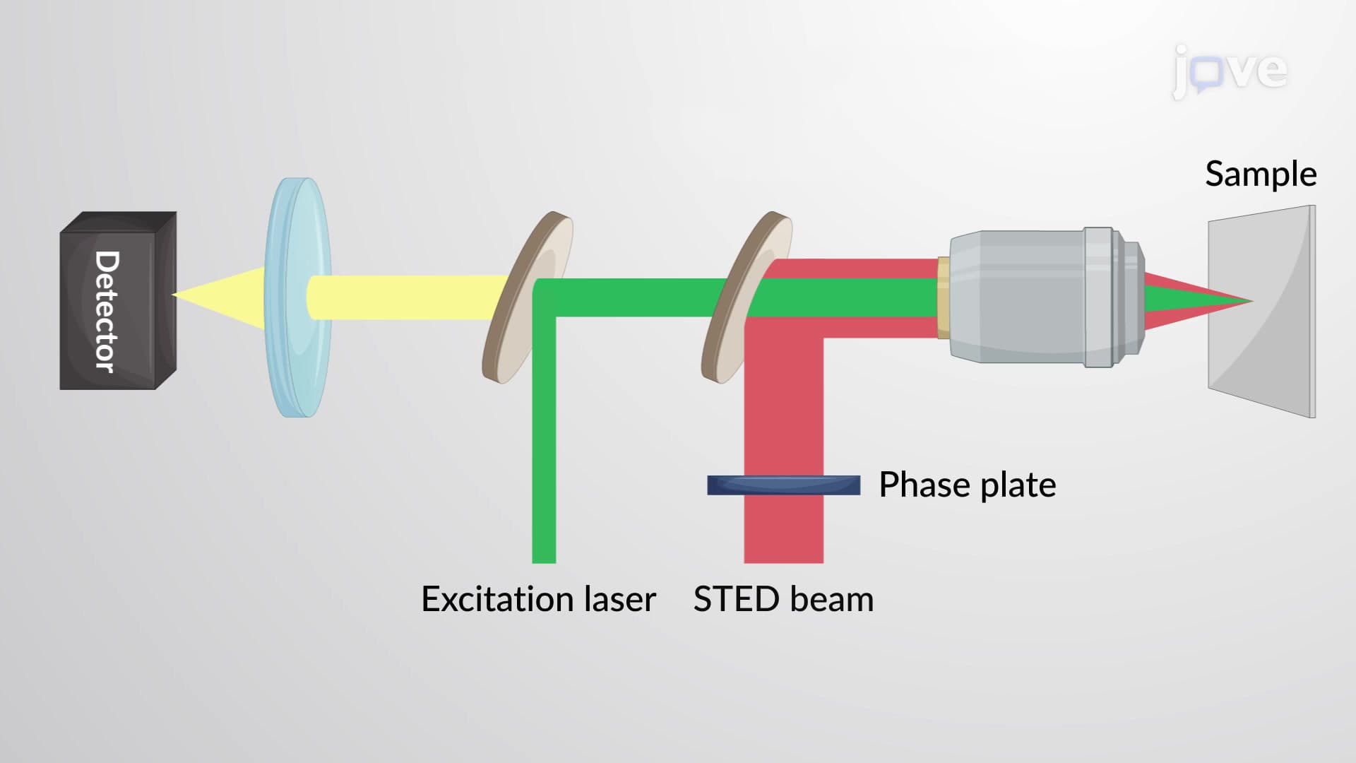
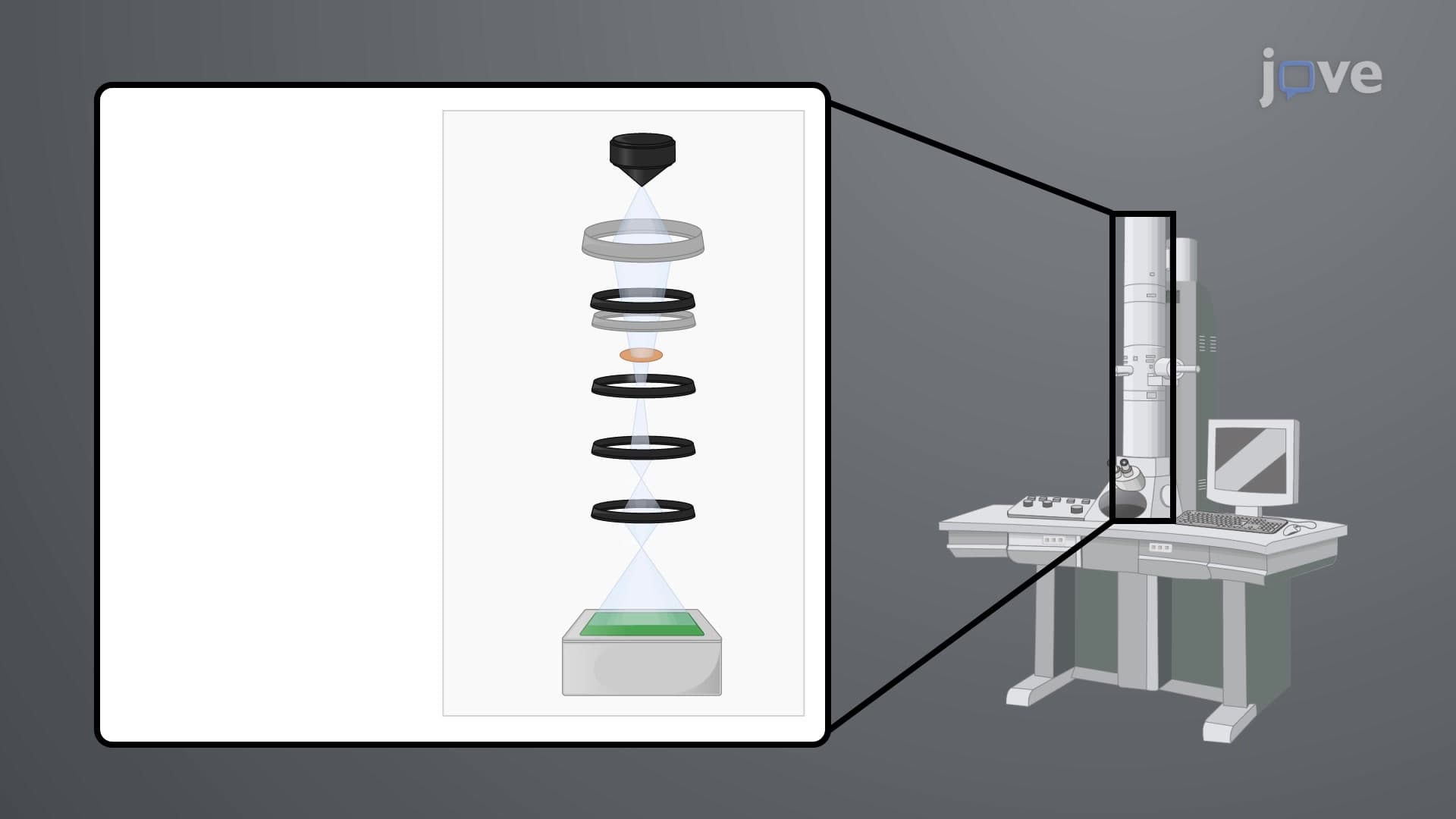
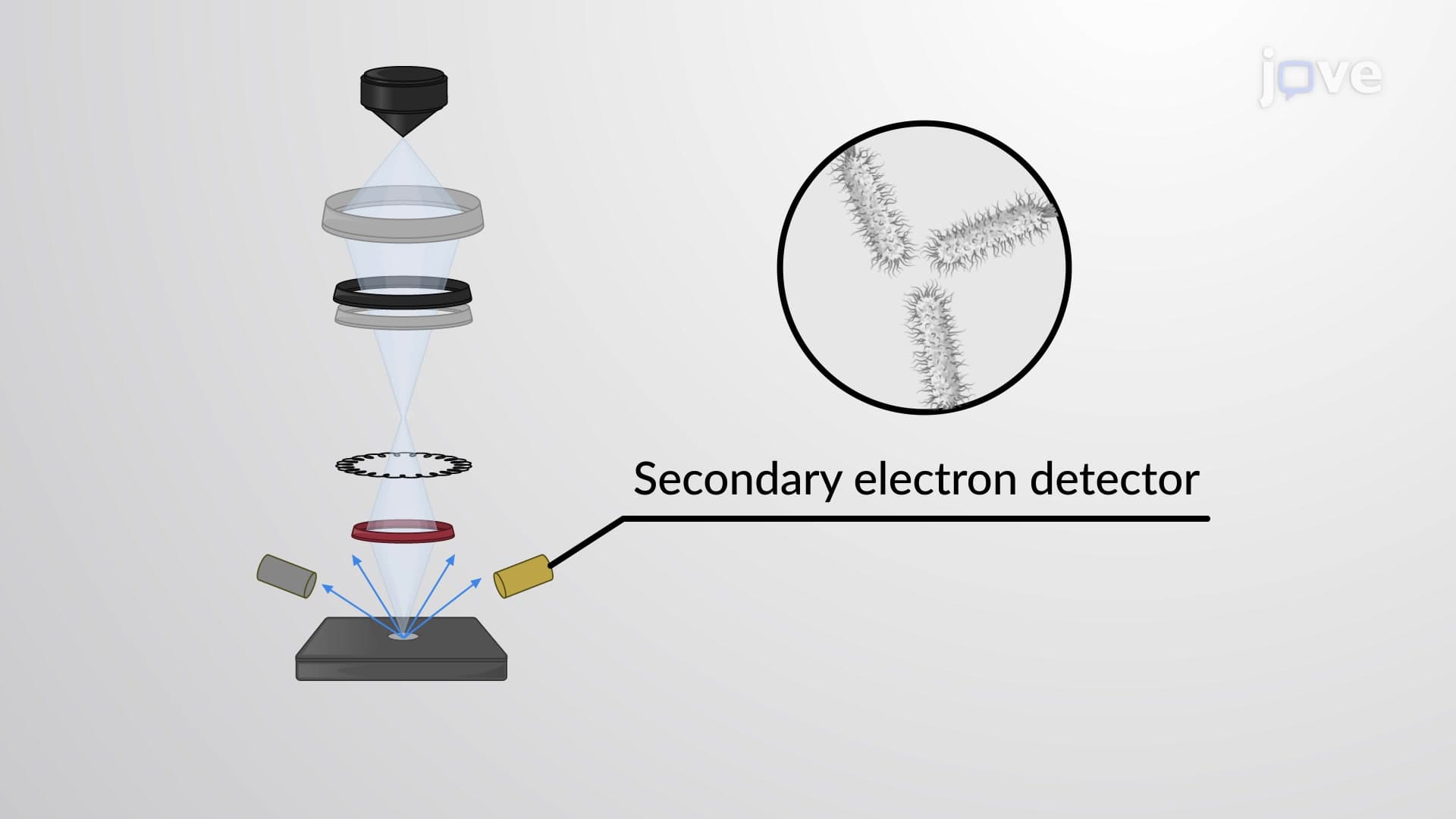
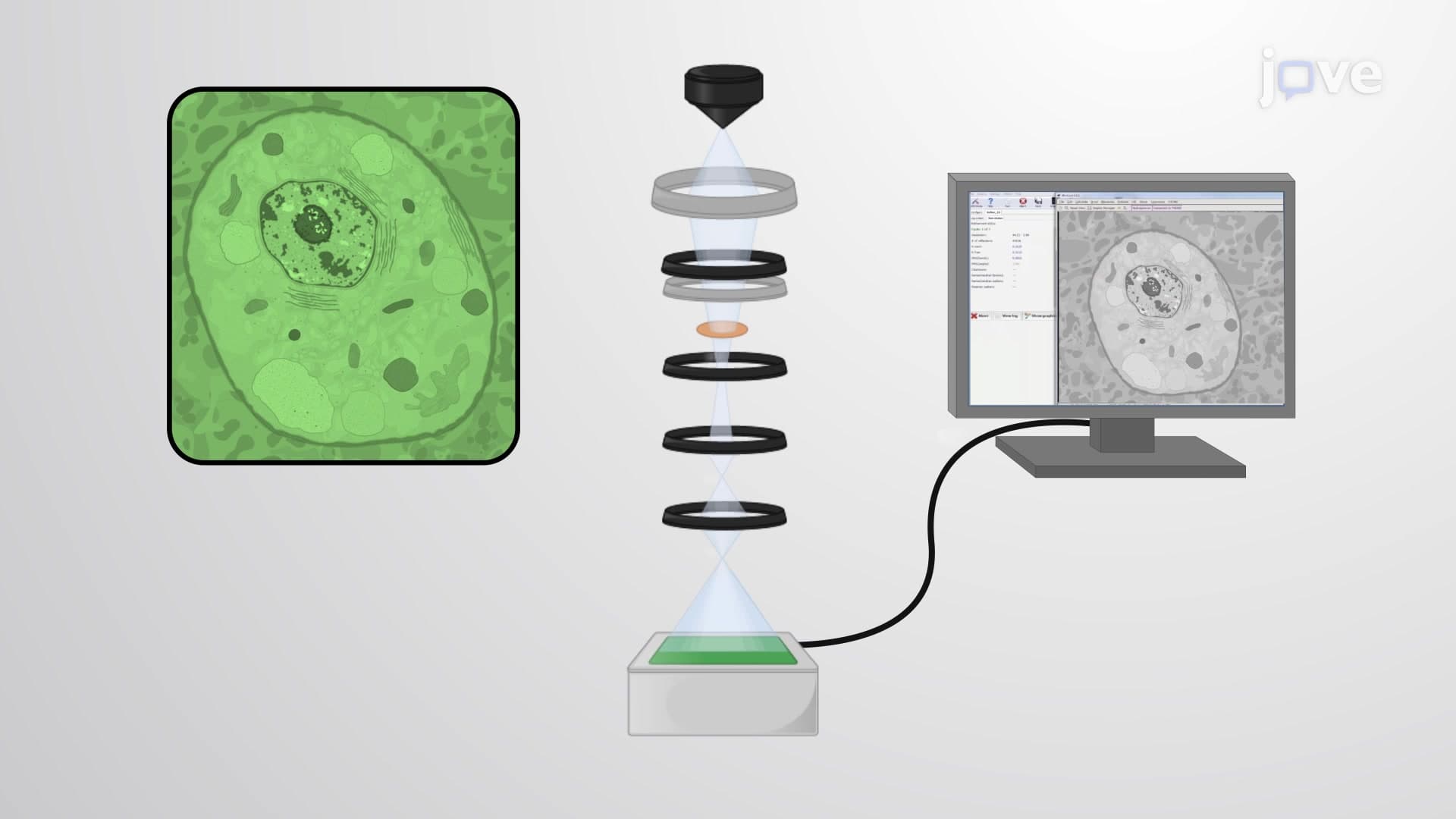


Imaging Biological Samples with Optical Microscopy


00:00
01:18
Video Summary: What Is Imaging Biological Samples
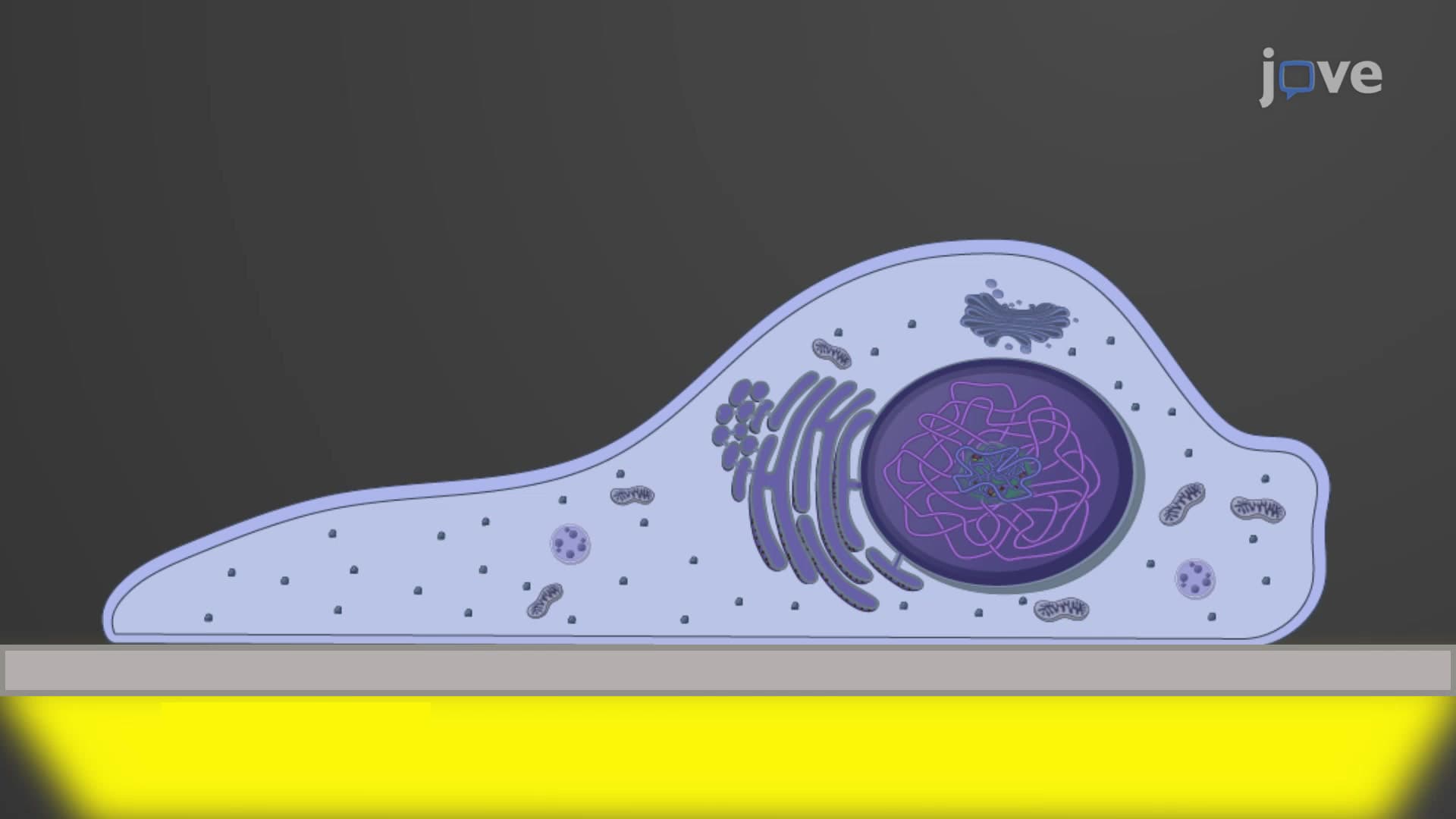
Did you know that the human eye can't see anything smaller than 100 micrometers, yet a single bacterial cell measures only 1-5 micrometers? Imaging biological samples allows scientists to visualize microscopic life forms and cellular structures invisible to the naked eye. From diagnosing infections at the CDC to studying cancer cells at Johns Hopkins University, optical microscopy remains fundamental to biological research and medical diagnosis. Watch the full video on JoVE Coach to master this concept with expert-led visuals and step-by-step explanations.
- Understand what is imaging biological samples and how optical microscopy enables visualization of microscopic structures
- Identify the key components of optical microscopes including condensers, objective lenses, and ocular lenses
- Analyze how numerical aperture determines light-gathering ability and image resolution in biological imaging
- Compare different magnification calculations by multiplying objective and ocular lens powers
- Evaluate resolution limits of 0.2 micrometers and their impact on visualizing cellular components
- Apply knowledge of refractive indices when selecting immersion media (air, water, oil) for optimal imaging
- Recognize the relationship between wavelength, numerical aperture, and image resolution quality
- Explore practical applications of 1000x maximum magnification in biological sample analysis
Understanding What is Imaging Biological Samples
Imaging biological samples represents the cornerstone of modern biological research and medical diagnostics. This fundamental technique allows scientists, researchers, and medical professionals to observe and analyze living organisms and their components at magnifications far beyond human visual capability. From high school AP Biology labs to cutting-edge research at Harvard Medical School, optical microscopy serves as the primary gateway to understanding cellular structure and function.
The Science Behind Optical Microscopy Systems
Optical microscopes operate through a sophisticated light manipulation system that transforms invisible microscopic details into observable images. The process begins with illumination from various light sources, which passes through a condenser lens system that focuses light precisely onto the biological specimen. This focused illumination then travels through the sample, where different cellular components absorb, reflect, or transmit light differently based on their density and composition.
The objective lens, positioned closest to the specimen, collects this transmitted light and creates a primary magnified image. Subsequently, the ocular lens (eyepiece) further magnifies this image, typically by 10x, creating the final composite magnification visible to the observer. This dual-lens system enables magnifications ranging from 40x to 1000x, calculated by multiplying the individual lens magnifications.
Numerical Aperture and Resolution Fundamentals
The numerical aperture (NA) concept proves critical for understanding image quality in biological sample imaging. This value determines both the light-gathering capacity and resolving power of microscope lenses. Higher numerical aperture values indicate superior light collection efficiency and finer resolution capabilities. The mathematical relationship involves the refractive index of the medium between the lens and specimen, along with the half-angle of the maximum light cone captured by the lens.
Students preparing for the MCAT or AP Biology exams should understand that resolution limits of approximately 0.2 micrometers define what structures remain visible through optical microscopy. This limitation means prokaryotic cells (bacteria) measuring 1-5 micrometers appear clearly, while smaller structures like viruses (20-300 nanometers) require electron microscopy for visualization.
Practical Applications in US Healthcare and Research
Major US institutions utilize biological sample imaging extensively. The Mayo Clinic employs optical microscopy for cancer diagnosis through tissue biopsy analysis. The NIH conducts infectious disease research using advanced microscopy techniques to study pathogen behavior. University research programs, from UC Berkeley to MIT, rely on these imaging principles for breakthrough discoveries in cell biology, genetics, and biomedical engineering.
For college students, mastering these concepts proves essential for success in biology, pre-medical courses, and laboratory sciences. Understanding magnification calculations, resolution limitations, and proper specimen preparation techniques directly applies to practical laboratory work and standardized testing scenarios.
Frequently Asked Questions
Imaging biological samples is the process of using microscopes to visualize and study living organisms, cells, and biological structures too small for the naked eye to see. It's fundamental to biology because it allows scientists to observe cellular processes, diagnose diseases, conduct research, and understand life at the microscopic level. This technique forms the foundation for fields like microbiology, pathology, and cellular biology.
For AP Biology, imaging biological samples involves understanding optical microscopy principles including light pathways, lens systems, and magnification calculations. Students must know resolution limits (0.2 micrometers), numerical aperture concepts, and how to calculate total magnification by multiplying objective and eyepiece powers. This knowledge directly applies to laboratory practicals and multiple-choice questions on the AP exam.
MCAT questions often test microscopy principles within biological sciences passages, focusing on resolution calculations, magnification formulas, and practical applications in medical research. Expect questions about numerical aperture, refractive indices, and how microscopy limitations affect cellular structure visualization. Understanding these concepts helps with both standalone questions and passage-based analytical reasoning.
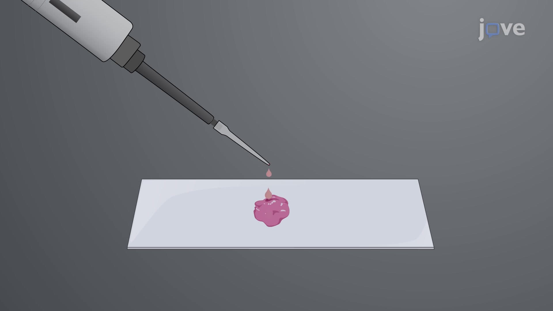
US hospitals and clinics use biological sample imaging for cancer diagnosis through tissue biopsies, bacterial infection identification in blood samples, and fertility treatments through sperm analysis. The CDC employs advanced microscopy for disease outbreak investigations, while pharmaceutical companies use these techniques for drug development and quality control testing.
Not at all! While the physics concepts may seem complex initially, imaging biological samples builds logically from basic light and lens principles you already understand. Start with simple magnification calculations, then gradually explore resolution and numerical aperture concepts. Most students find hands-on laboratory experience makes these theoretical concepts much clearer and more intuitive.
Focus on three key areas: magnification calculations (multiply objective and eyepiece powers), resolution limits (0.2 micrometers for optical microscopes), and numerical aperture relationships. Practice solving problems involving different lens combinations and understand when optical microscopy limitations require alternative techniques. Create diagrams showing light pathways through microscope components to reinforce your understanding.
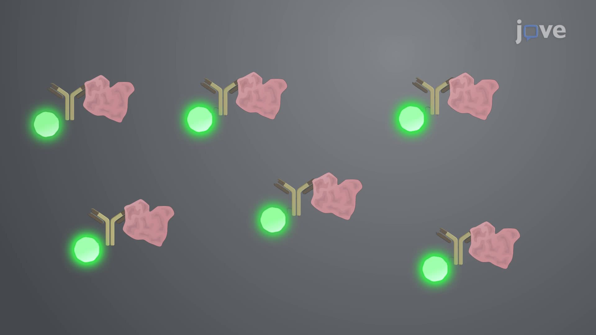
Consider studying electron microscopy (transmission and scanning), fluorescence microscopy techniques, and confocal imaging systems. These advanced methods overcome optical microscopy limitations and enable visualization of subcellular structures, protein interactions, and dynamic cellular processes. Many pre-med students find these topics particularly valuable for research opportunities and graduate school preparation.
Related Micro-courses