Loading subject page…


138,516 views

Video Summary: What Is Immunocytochemistry and Immunohistochemistry
Ever wonder how researchers at Johns Hopkins can pinpoint cancer cells hiding among millions of healthy tissue cells? Immunocytochemistry and immunohistochemistry are powerful laboratory techniques that use antibody-enzyme systems to make specific proteins glow like biological spotlights in cells and tissues. These methods help pathologists diagnose diseases and guide treatment decisions across major US medical centers. Watch the full video on JoVE Coach to master this concept with expert-led visuals and step-by-step explanations.
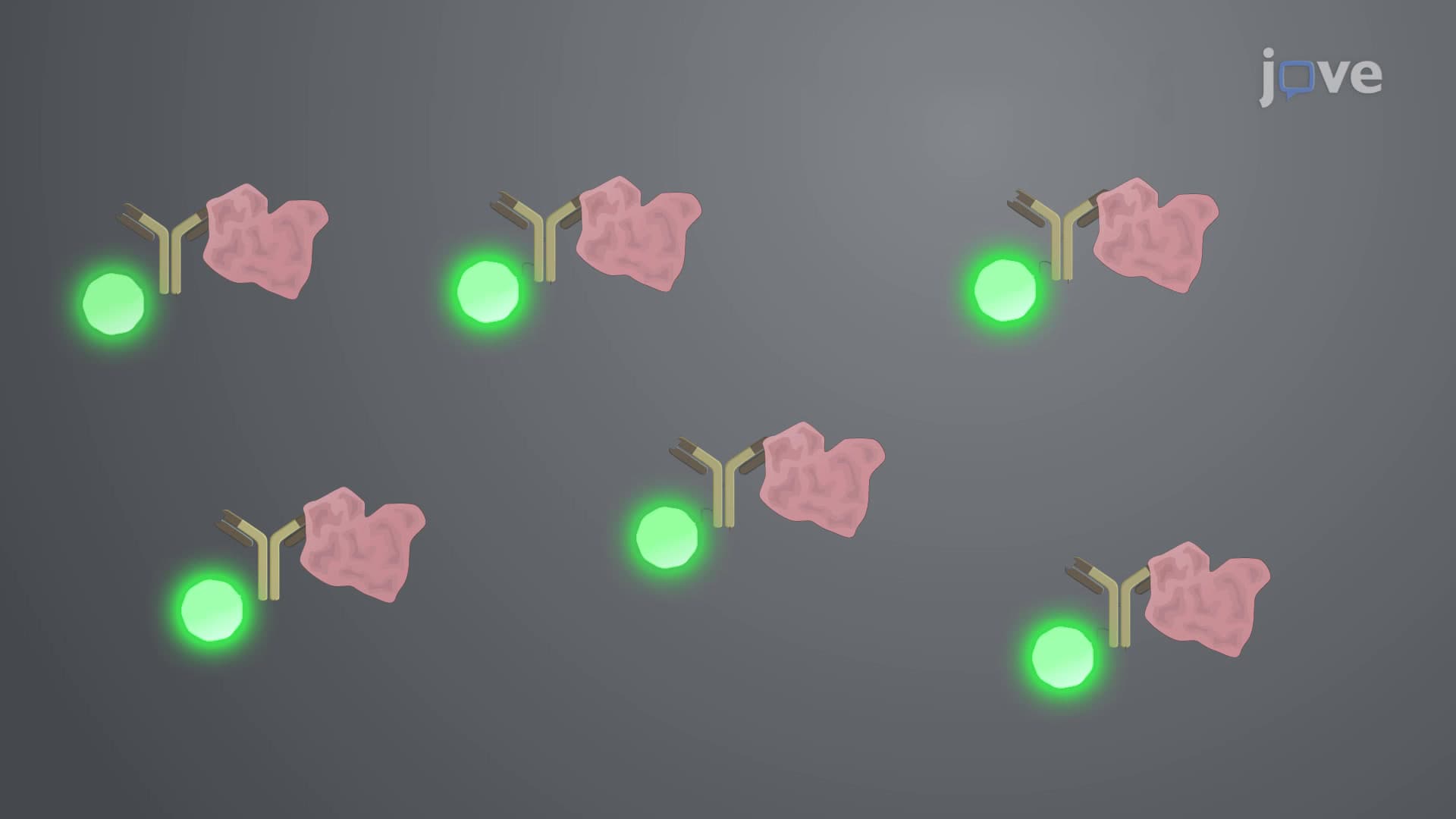
Immunocytochemistry and immunohistochemistry represent cornerstone techniques in modern biomedical research and clinical diagnostics. Both methods harness the exquisite specificity of antibodies to detect and visualize target proteins within biological samples. The fundamental principle relies on antibody-antigen binding, where highly specific antibodies recognize and attach to particular protein sequences, creating a molecular "lock-and-key" system that enables precise protein identification.
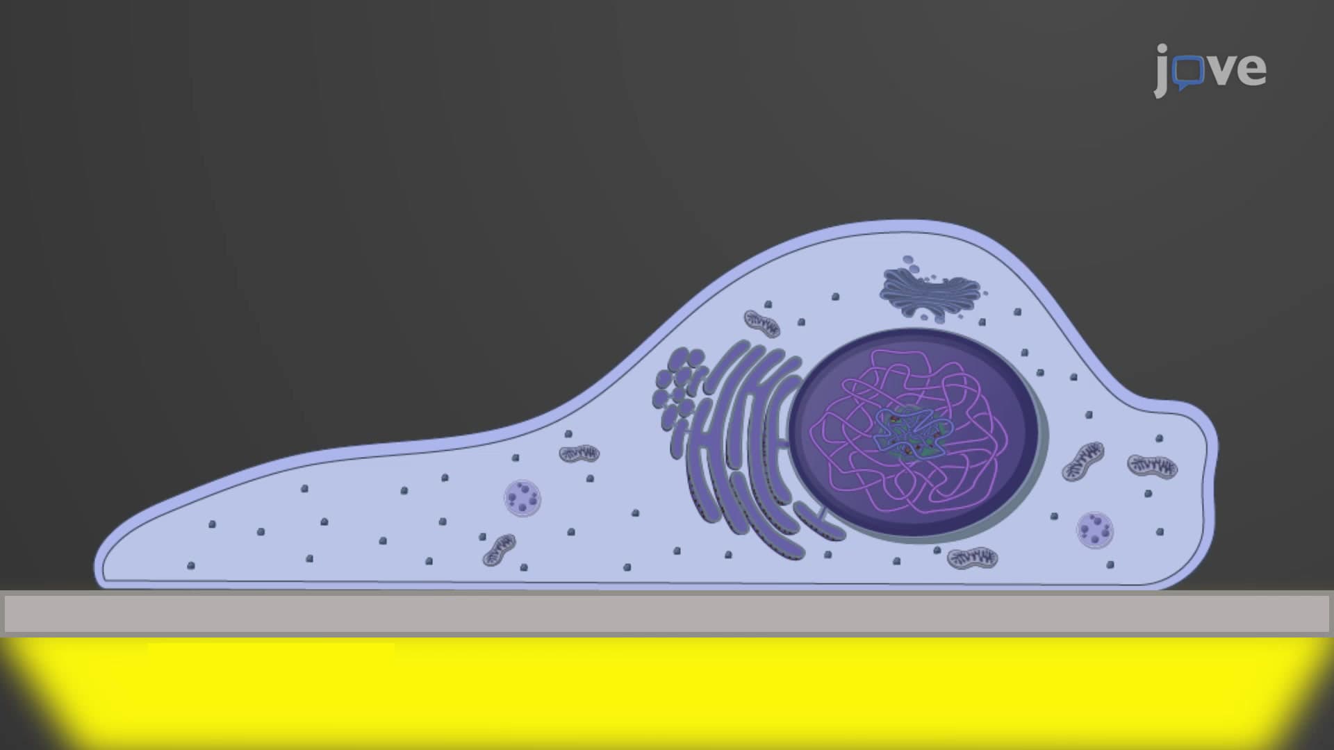
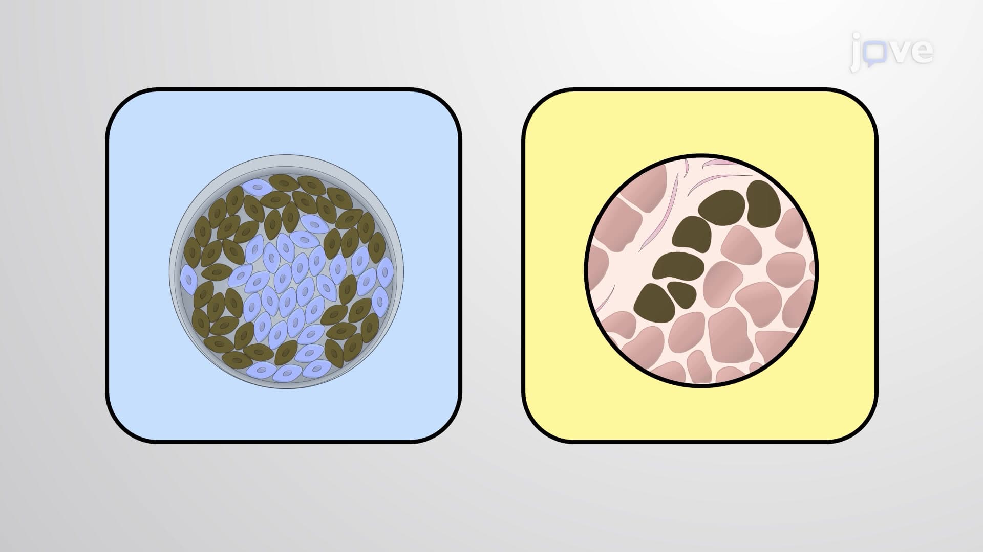
The primary difference lies in sample preparation and application scope. Immunocytochemistry (ICC) works with cultured cells grown as monolayers on glass coverslips-imagine studying individual actors on a stage. Researchers at Stanford University commonly use ICC to examine how cancer cells respond to experimental drugs in controlled laboratory conditions. The cells undergo fixation with cross-linking agents like formaldehyde, preventing protein degradation while maintaining cellular architecture.
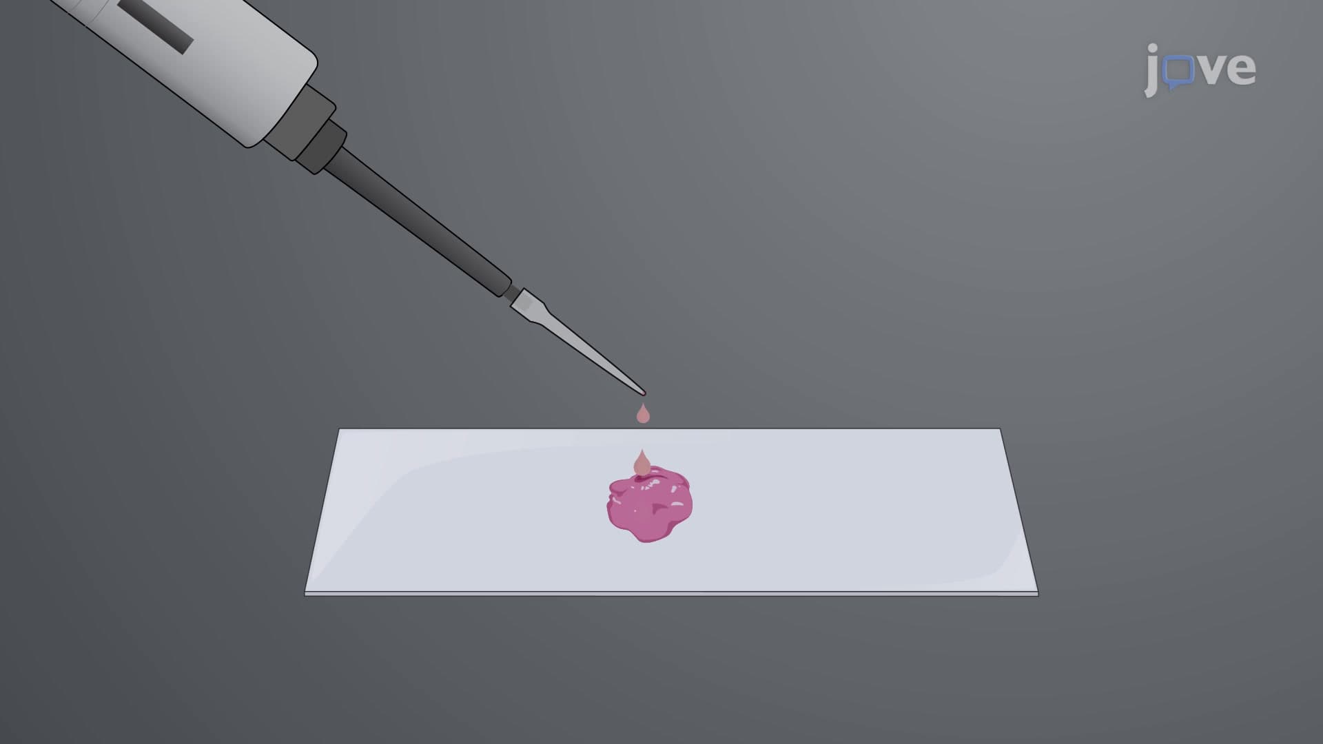
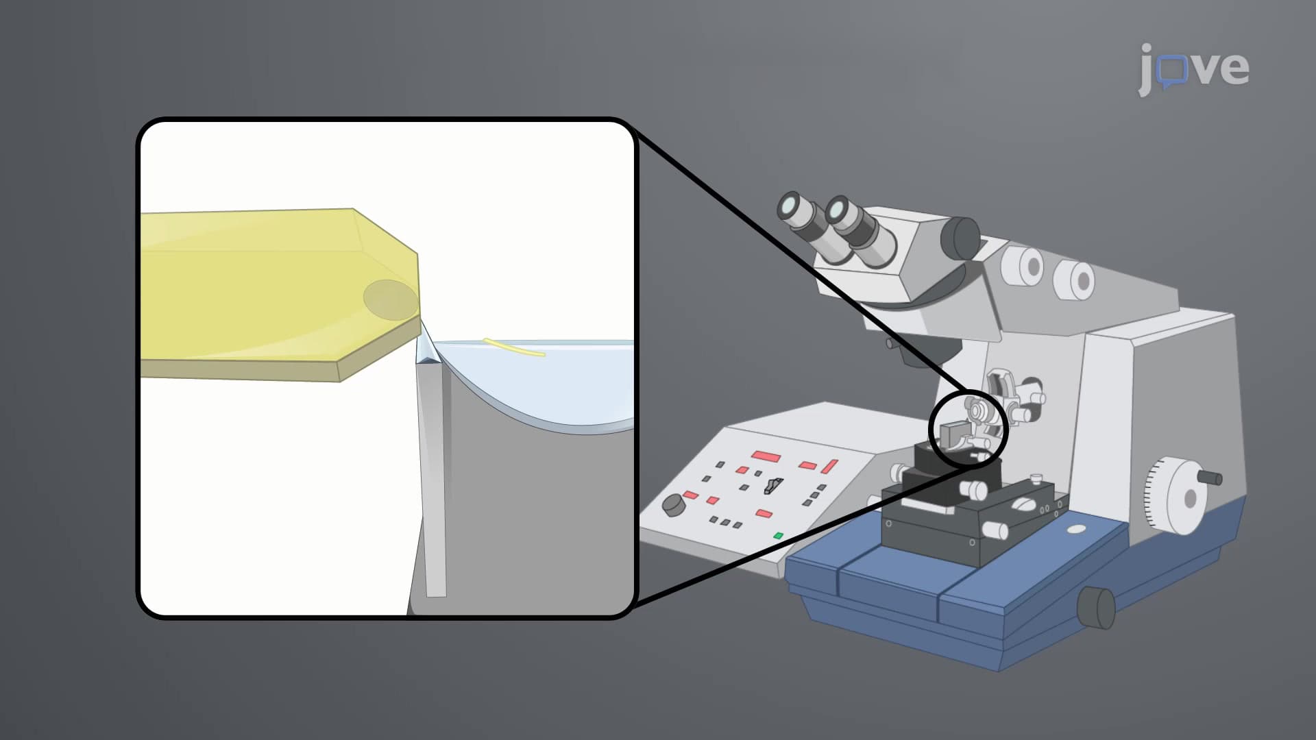
Immunohistochemistry (IHC), conversely, examines whole tissue sections-like studying entire theater audiences. At Mayo Clinic, pathologists routinely use IHC on patient biopsy samples embedded in paraffin blocks. These samples are sectioned into ultra-thin slices (typically 4-6 micrometers) using specialized microtomes, then mounted on microscope slides for analysis.
Both techniques employ enzyme-linked detection systems for visualization. Horseradish peroxidase (HRP) serves as the workhorse enzyme, catalyzing the oxidation of 3,3'-diaminobenzidine (DAB) substrate into an insoluble brown precipitate. This reaction creates visible deposits exactly where target proteins exist, functioning like a biological GPS system.
When initial signals prove insufficient, secondary antibody amplification becomes crucial. Primary antibodies bind directly to target antigens, while enzyme-conjugated secondary antibodies recognize and bind to primary antibodies, multiplying the signal intensity. This two-step process resembles using an amplifier to boost weak radio signals-essential for detecting low-abundance proteins.
These techniques prove invaluable for AP Biology students studying cellular processes and MCAT preparation covering molecular biology concepts. In clinical practice, pathologists at institutions like MD Anderson Cancer Center use IHC to identify specific cancer markers, guiding personalized treatment decisions. Research laboratories across US universities employ both methods to investigate disease mechanisms, drug effectiveness, and basic cellular functions.
Related Micro-courses