Loading subject page…

138,515 views

Video Summary: What Is Super Resolution Fluorescence Microscopy
Did you know that scientists can now see cellular structures smaller than 200 nanometers-beyond what traditional microscopes can resolve? Super resolution fluorescence microscopy breaks through the fundamental limits of light diffraction, enabling researchers at institutions like MIT and Stanford to visualize individual proteins and DNA molecules within living cells. This revolutionary technique uses clever light patterns and specialized fluorescent probes to achieve unprecedented detail in biological imaging. Watch the full video on JoVE Coach to master this concept with expert-led visuals and step-by-step explanations.
Super resolution fluorescence microscopy represents a paradigm shift in biological imaging, allowing scientists to visualize cellular structures with unprecedented clarity. Traditional light microscopy hits a fundamental barrier called the diffraction limit-approximately 200 nanometers-which prevents clear imaging of closely spaced objects. When fluorescent molecules are closer than this limit, their light signals blur together, creating overlapping spots that obscure fine details.
This limitation posed significant challenges for researchers studying cellular processes. Consider trying to understand how proteins interact within a synapse, or how DNA organizes within the nucleus-structures often smaller than 200nm. Super resolution techniques ingeniously circumvent this barrier through sophisticated light manipulation and computational analysis.
Structured illumination microscopy (SIM) employs a clever optical trick using patterned illumination. Instead of uniformly lighting the sample, SIM projects striped patterns of light at different angles and phases. When this patterned light interacts with fluorescent structures, it creates moiré interference patterns containing high-resolution information normally lost to diffraction.
Researchers at the University of California San Francisco have used SIM to study mitochondrial dynamics in living cells, revealing previously invisible organizational patterns. The technique captures multiple images with different stripe orientations, then computationally processes these to reconstruct a final image with doubled resolution-achieving approximately 100nm precision.
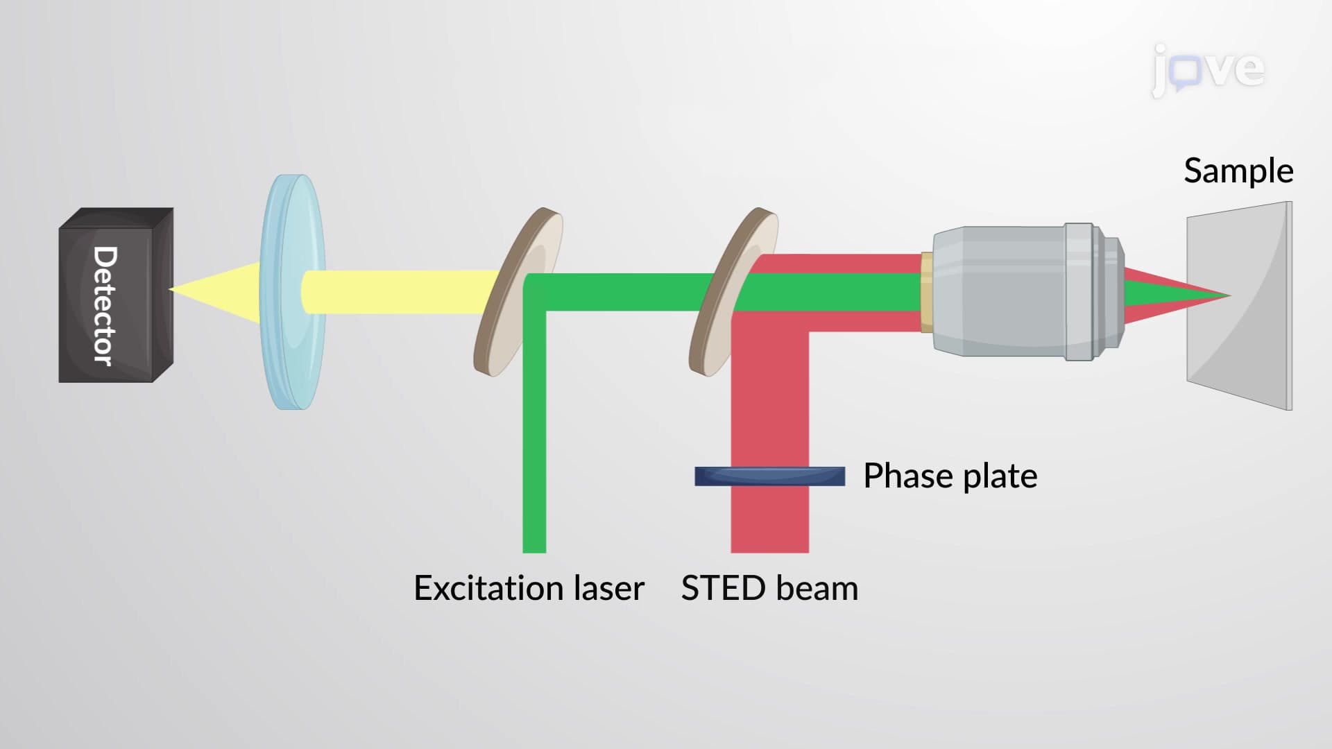
Stimulated emission depletion (STED) microscopy takes a radically different approach. This technique uses two synchronized laser beams: an excitation laser that activates fluorophores normally, and a specialized "depletion" laser shaped like a donut. The depletion beam surrounds the excitation point, forcing fluorescent molecules in the outer regions to return to their ground state without emitting light.
This process effectively shrinks the fluorescent spot size well below the diffraction limit. Researchers at Harvard Medical School have achieved resolutions as fine as 20-30 nanometers using STED, enabling detailed studies of synaptic protein arrangements in brain tissue samples.
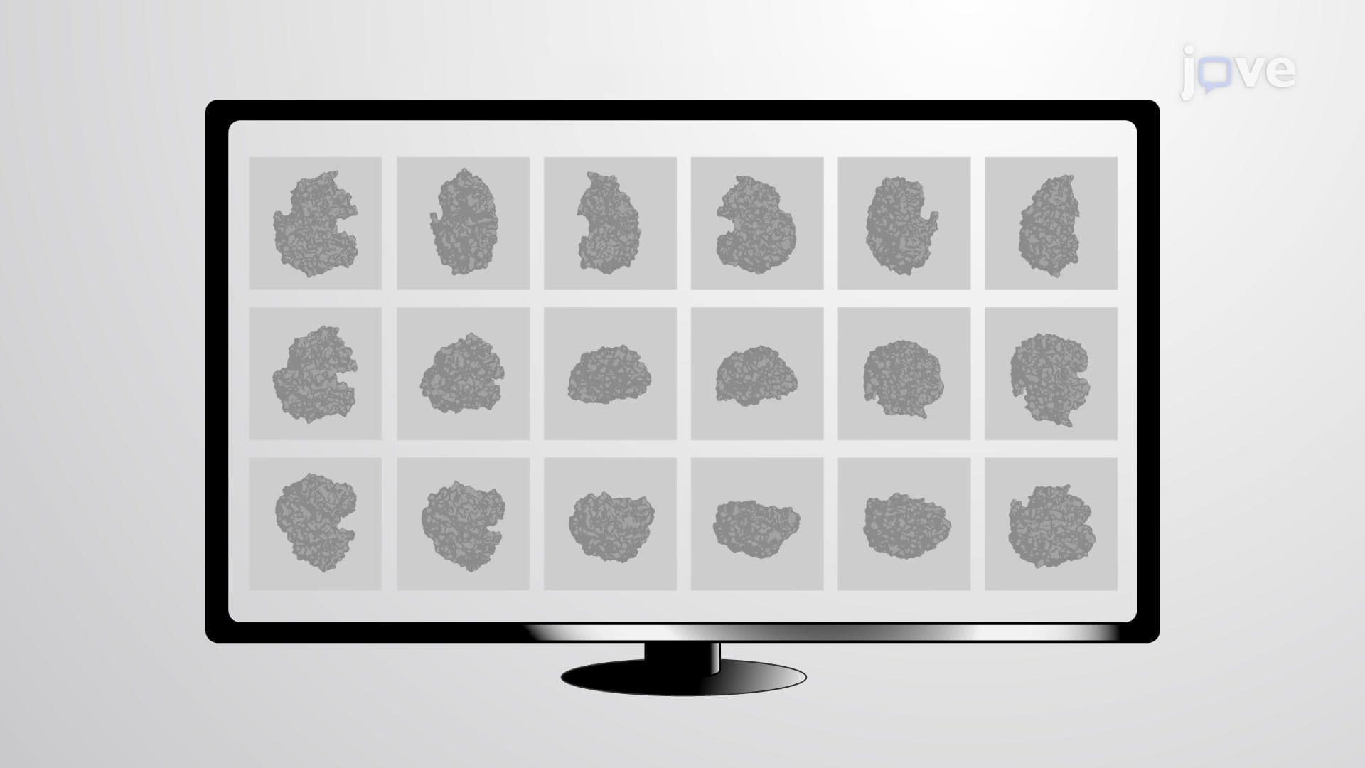
The third major approach-including techniques like PALM (photoactivated localization microscopy) and STORM (stochastic optical reconstruction microscopy)-uses temporally separated activation of fluorescent probes. Instead of illuminating all fluorophores simultaneously, these methods activate only small subsets at any given time, ensuring individual molecules don't overlap spatially.
Advanced algorithms then precisely determine each molecule's position, building up a comprehensive high-resolution image over time. This approach proves particularly valuable for studying dynamic cellular processes and has applications in AP Biology coursework covering cellular transport and membrane organization.
Related Micro-courses