Loading subject page…

- Molecular Biology
- Cell Division
- Determining the Plane of Cell Division
Micro-course: Cell Division
101,001 views
Learn key concepts and earn your certificate.




Determining the Plane of Cell Division

00:00
02:13
Video Summary: Determining the Plane of Cell Division Explained
Every time your body heals a cut or grows during adolescence, millions of cells must divide with pinpoint accuracy to avoid developmental disorders. Determining the plane of cell division is the critical process that ensures chromosomes separate correctly and cellular contents distribute evenly between daughter cells. In medical research at institutions like Johns Hopkins, scientists study how errors in this process contribute to cancer cell formation and genetic abnormalities. Watch the full video on JoVE Coach to master this concept with expert-led visuals and step-by-step explanations.
- Understand how determining the plane of cell division ensures accurate chromosome separation and proper distribution of cytoplasmic components during mitosis
- Identify the three primary models that explain division plane determination: astral stimulation, polar relaxation, and central spindle stimulation
- Analyze the role of microtubules in interacting with the cell cortex to specify cleavage furrow formation at the cellular equator
- Compare how astral microtubules function differently in stimulation versus inhibition mechanisms during cell division
- Evaluate why the multi-pronged approach of combining all three models increases cell division accuracy across different cell types
- Apply knowledge of central spindle formation from interpolar microtubules during anaphase to predict division outcomes
- Recognize how cell size and protein concentration variations affect the relative contribution of each division plane model
- Explore species-specific examples like C. elegans embryos to understand how astral microtubule arrays influence division plane determination
Understanding Determining the Plane of Cell Division Fundamentals
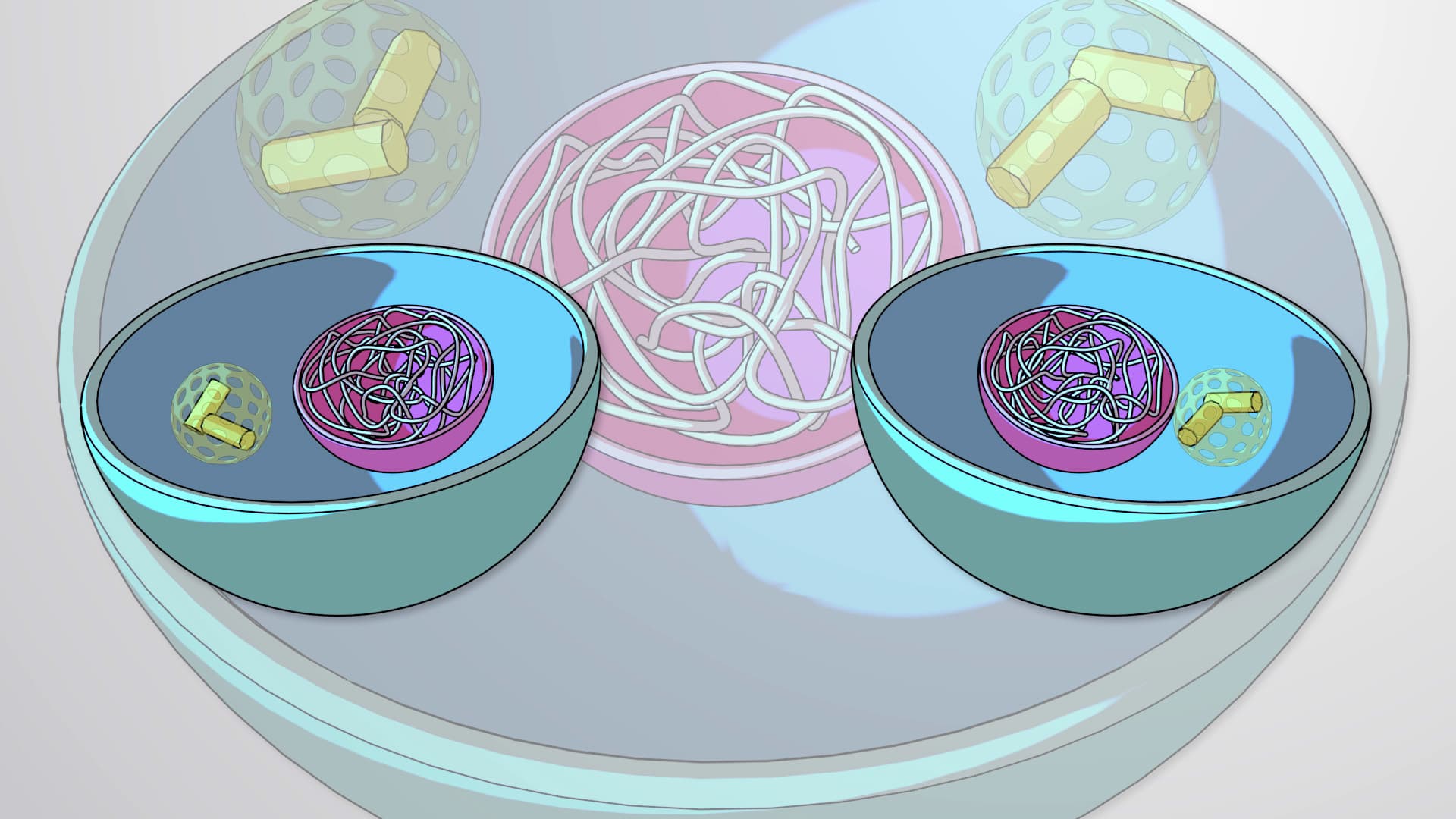
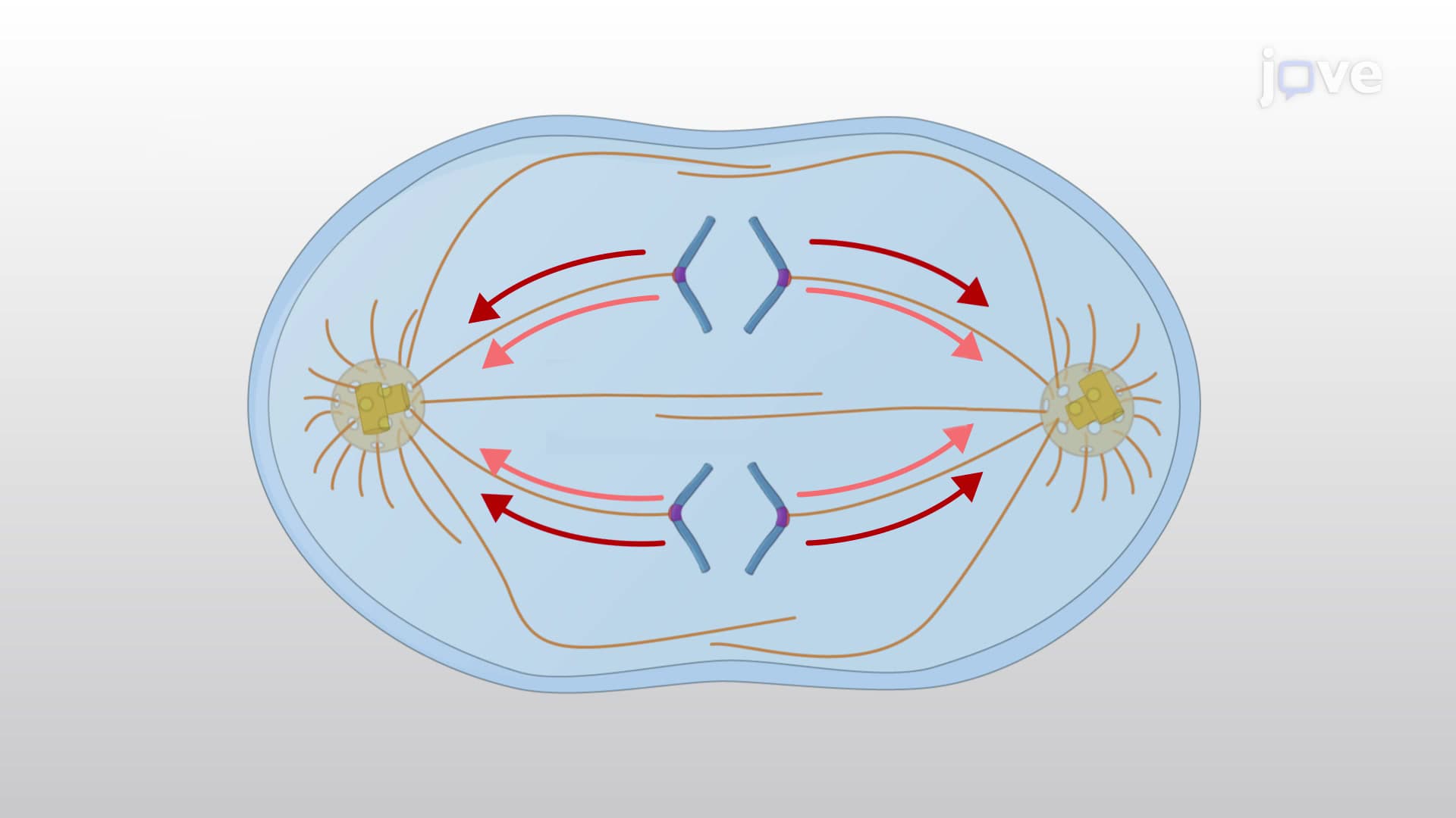
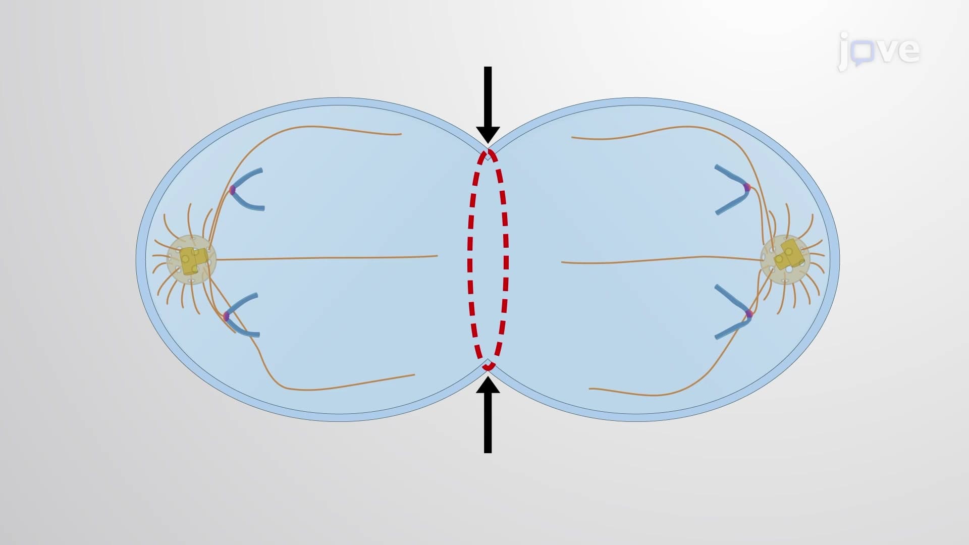
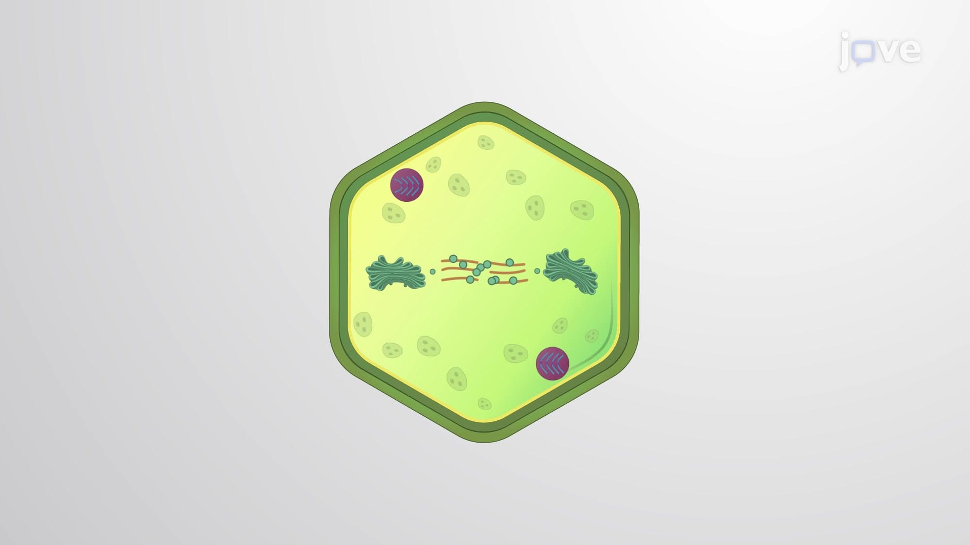
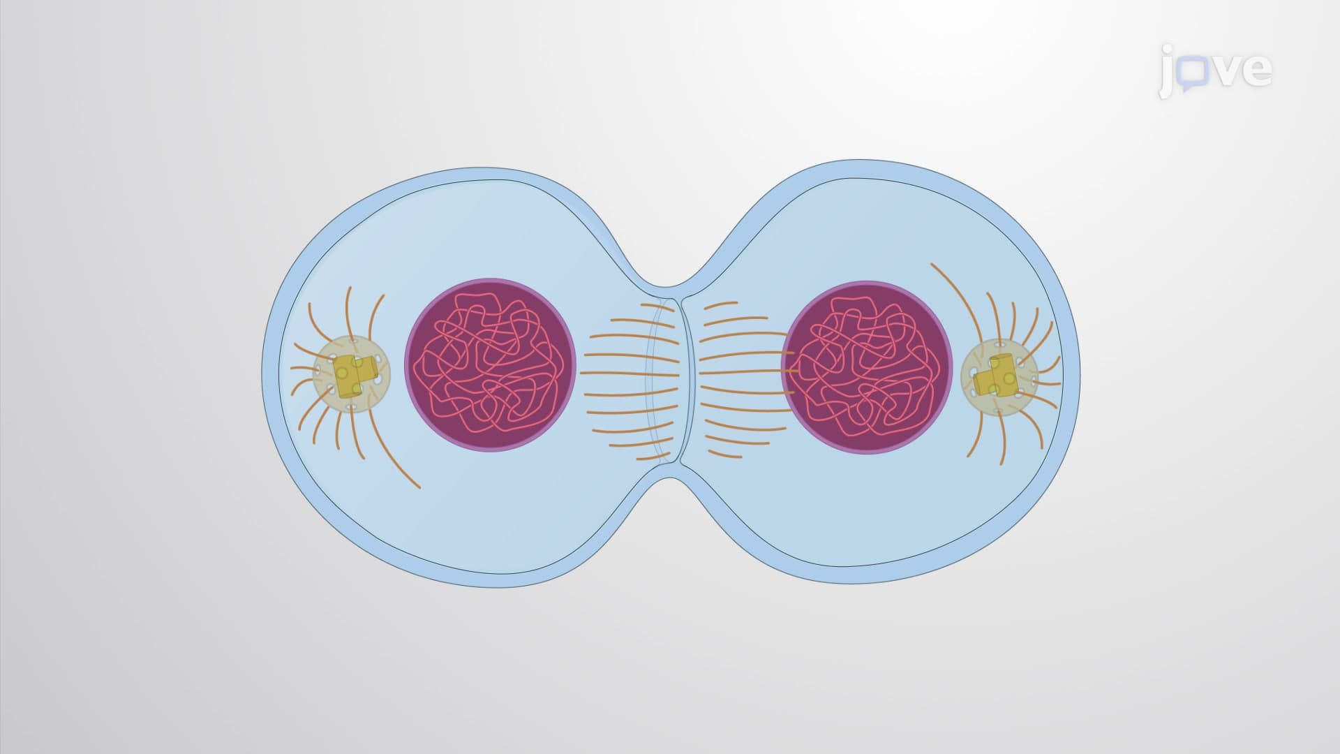
Determining the plane of cell division represents one of cell biology's most precisely orchestrated events, where a single miscalculation can lead to chromosome loss, unequal cell division, or developmental abnormalities. This process occurs during anaphase when the cell must establish exactly where to form the cleavage furrow-the contractile ring that will physically separate the cell into two daughter cells. The division plane always forms at the cell's equator, positioned precisely between the segregated chromosome sets.
The stakes couldn't be higher: errors in division plane determination contribute to aneuploidy (abnormal chromosome numbers) found in many cancers studied at research institutions like Memorial Sloan Kettering Cancer Center. Students preparing for the AP Biology exam frequently encounter questions about this topic, particularly regarding how microtubule dynamics influence cell division outcomes.
The Three-Model Framework for Division Plane Control
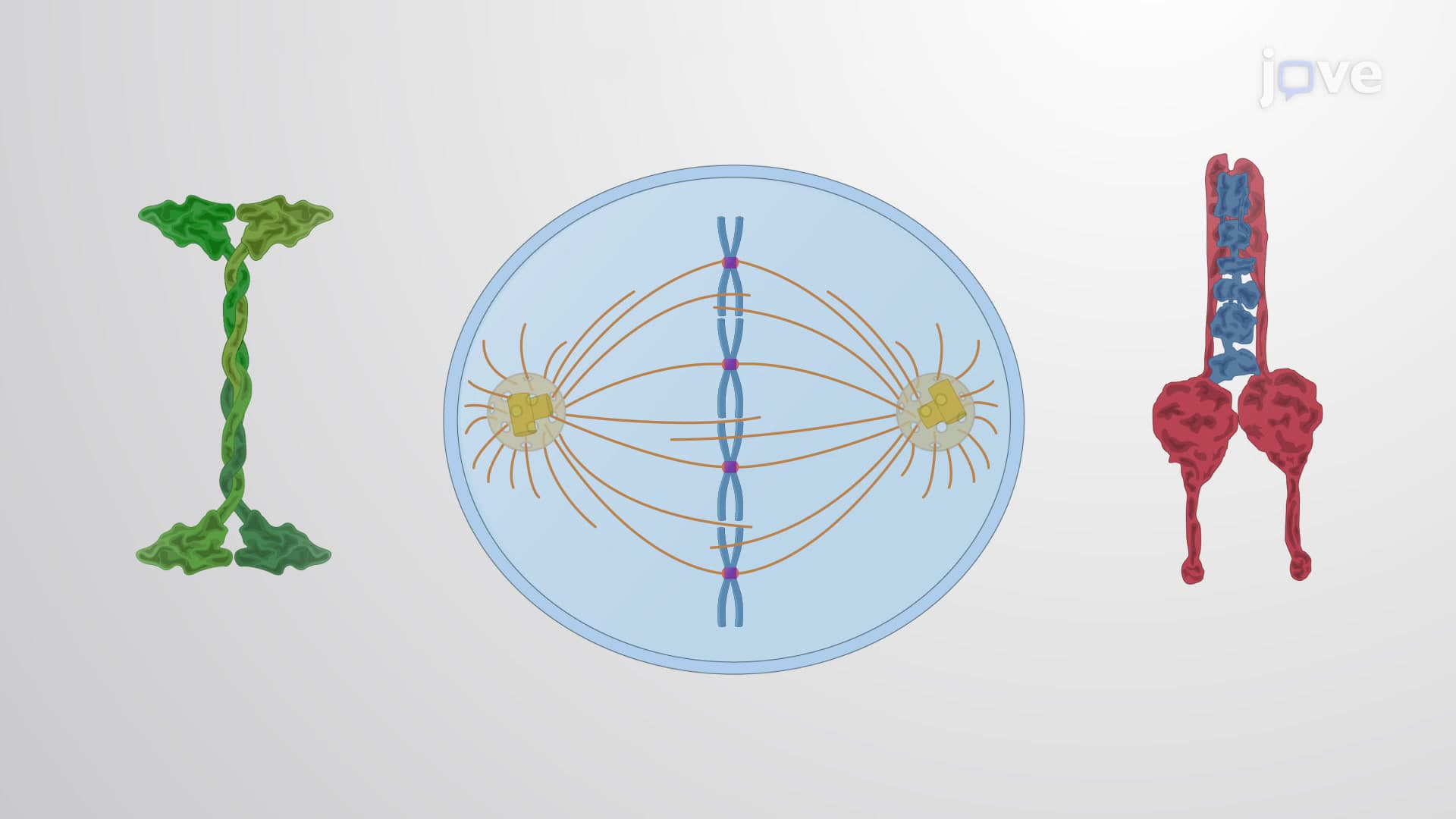
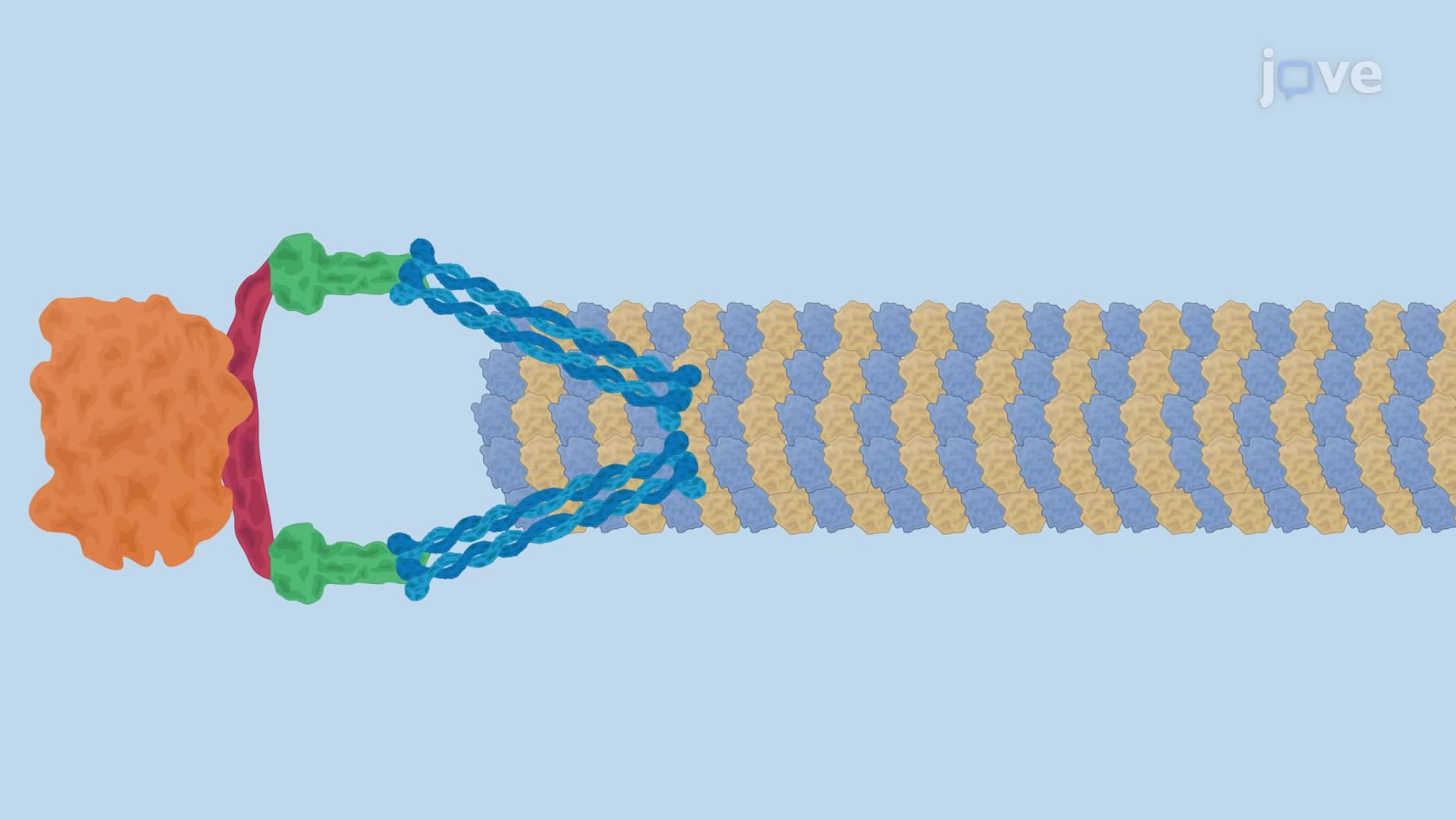
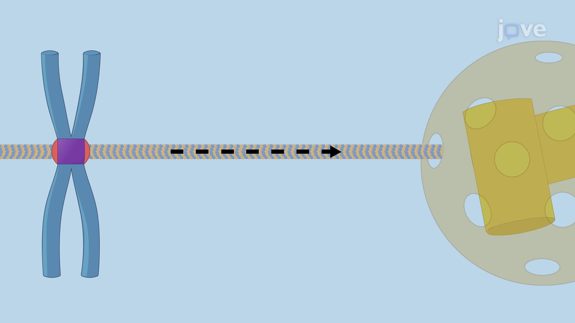
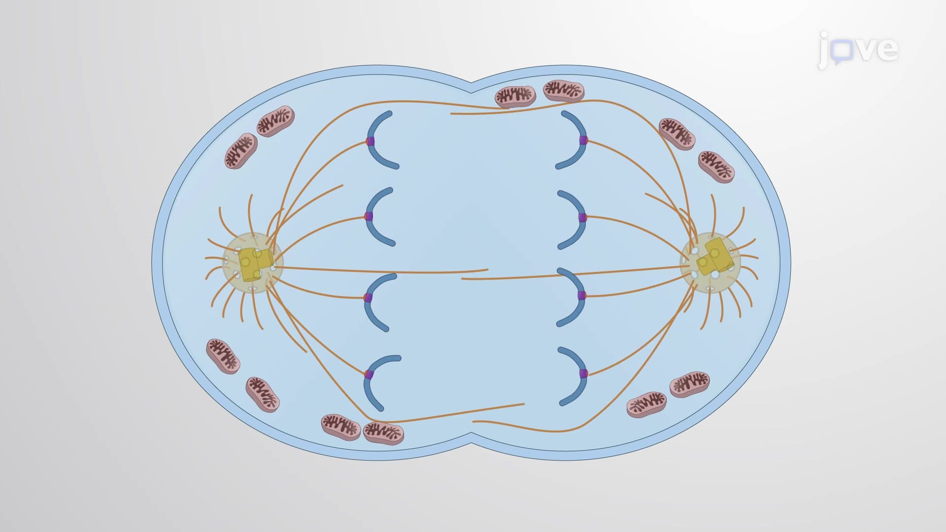
Scientists have identified three complementary models explaining how cells achieve such remarkable precision. The astral stimulation model proposes that radial arrays of astral microtubules-those extending outward from centrosomes-directly contact and activate the equatorial cortex, triggering cleavage furrow formation. Think of these microtubules as cellular GPS signals pinpointing the exact division location.
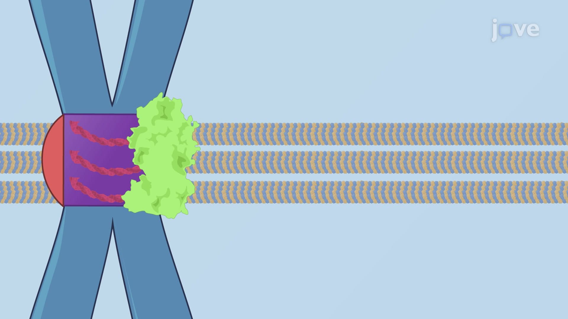
The polar relaxation model operates through inhibition rather than stimulation. Here, astral microtubules send suppressive signals to both cell poles, preventing cleavage furrow formation at these locations. This creates a "process of elimination" scenario where only the equatorial region remains available for division. The central spindle stimulation model involves interpolar microtubules that reorganize during anaphase into overlapping, antiparallel structures called the central spindle, which then stimulates equatorial cortex contraction.
Integrated Control Mechanisms and Clinical Relevance
Modern cell biology recognizes that all three models work simultaneously, creating a robust system resistant to molecular fluctuations. This redundancy explains why cell division succeeds even when individual proteins vary in concentration or when cell sizes differ dramatically. Medical schools teaching courses like those at Harvard Medical School emphasize how this multi-layered control prevents the chromosome segregation errors that characterize many human diseases.
The relative importance of each mechanism varies by cell type and size. In C. elegans embryos-a favorite research model at institutions like Rockefeller University-extensive astral microtubule arrays play dominant roles in division plane determination. Conversely, smaller mammalian cells may rely more heavily on central spindle mechanisms.
Students preparing for MCAT Cell Biology sections should understand that this topic connects directly to cancer biology, developmental disorders, and fertility issues, making it relevant for multiple medical specialties from oncology to reproductive medicine.
Frequently Asked Questions
Determining the plane of cell division is the cellular process that establishes exactly where a dividing cell will split into two daughter cells, ensuring chromosomes separate accurately and cytoplasmic contents distribute evenly. This process prevents genetic abnormalities and maintains proper cell function, making it essential for healthy growth, development, and tissue repair throughout your body.
AP Biology frequently tests this concept through questions about mitotic spindle function, microtubule organization, and cell division errors. Expect to analyze diagrams showing astral microtubules, identify cleavage furrow formation mechanisms, and explain how division plane errors contribute to aneuploidy-particularly in free-response questions about cell cycle regulation and cancer biology.
MCAT Cell Biology sections often link this concept to cancer development, chromosome segregation disorders, and developmental biology pathways. You'll encounter questions about microtubule dynamics, centrosome function, and how division plane errors create the chromosomal instability characteristic of tumor cells studied in medical research.
Division plane errors contribute to chromosomal abnormalities seen in many cancers treated at major medical centers like MD Anderson and Cleveland Clinic. These errors can cause unequal chromosome distribution, leading to tumor cell formation, developmental disorders in embryos, and fertility issues that reproductive specialists encounter regularly in clinical practice.
No advanced math is required-this topic focuses on biological mechanisms and spatial relationships rather than complex calculations. Students with basic high school biology and chemistry backgrounds can master these concepts by understanding protein interactions, cellular structures, and cause-and-effect relationships in cell division processes.
Create acronyms like "APC" (Astral stimulation, Polar relaxation, Central spindle) and associate each with its function: Astral "activates," Polar "prevents," Central "concentrates." Practice drawing diagrams showing microtubule arrangements for each model, since visual questions frequently appear on biology exams testing this material.
Consider Cell Biology, Developmental Biology, or Cancer Biology courses that build on these foundations. Many pre-med students find Molecular Biology particularly valuable since it explores the genetic regulation controlling division plane determination, preparing you for advanced topics in medical school cell biology coursework.
College exams often present experimental scenarios where you must predict division outcomes when specific proteins are inhibited or cell conditions change. Expect to analyze microscopy images, interpret data from cell division timing experiments, and explain how the three-model system provides redundancy against molecular perturbations.
Related Micro-courses