Loading subject page…


101,001 views

Video Summary: What Is the Contractile Ring
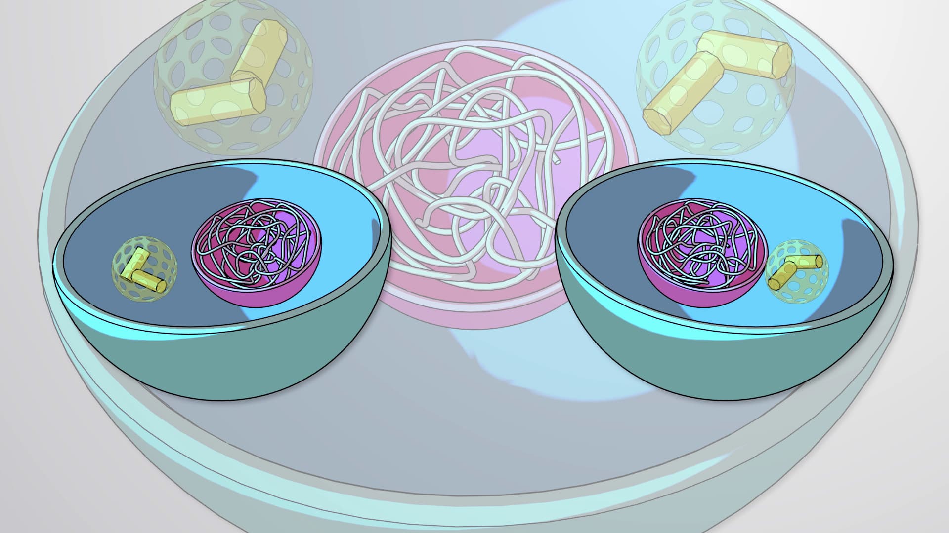
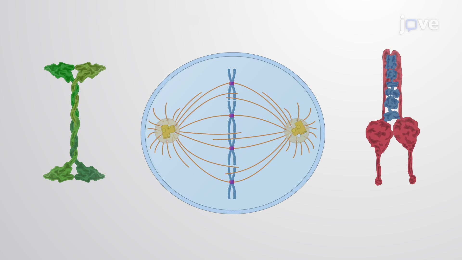
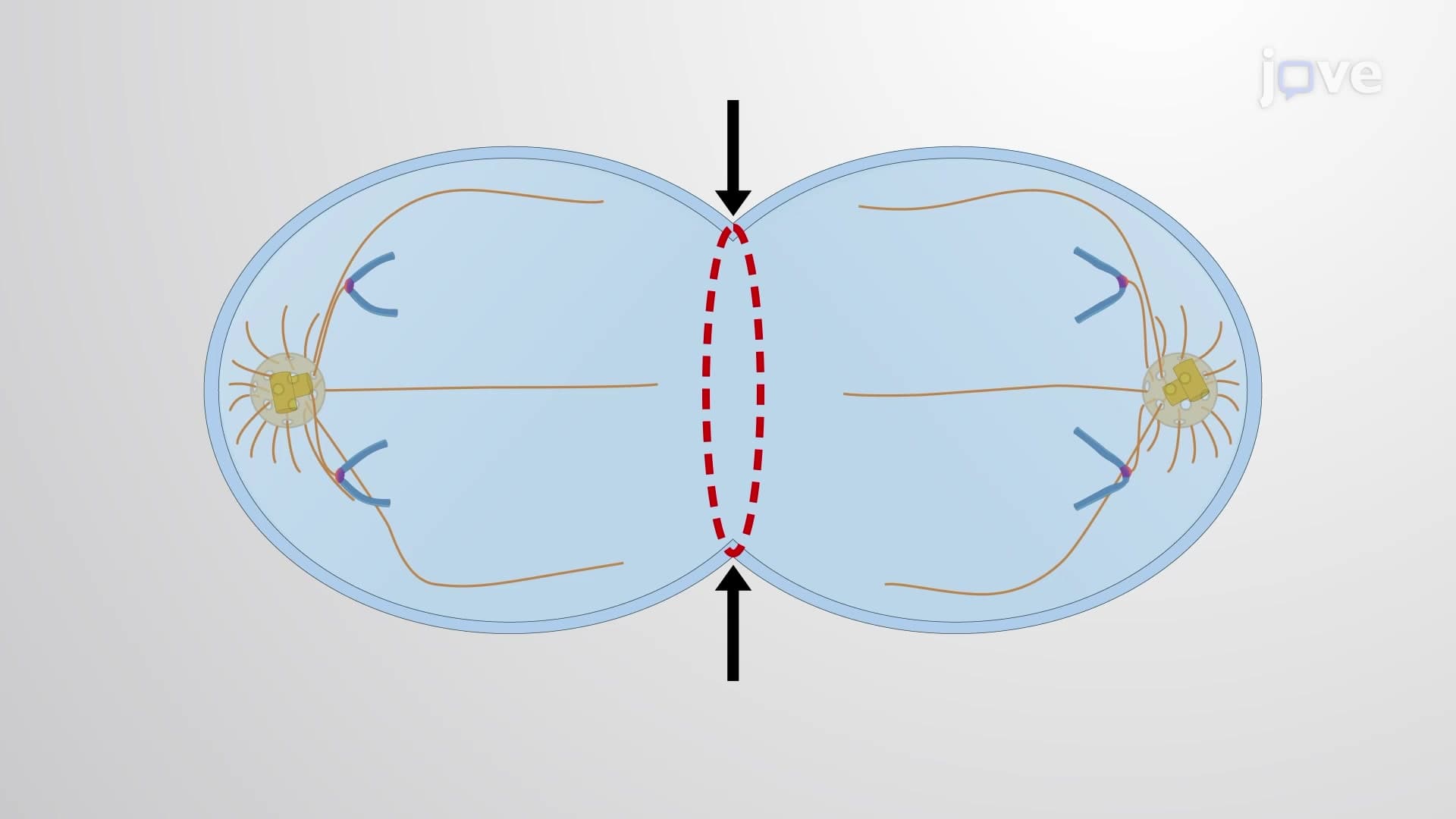
Did you know that every single cell division in your body-from healing a scraped knee to growing taller-depends on a microscopic "molecular zipper" called the contractile ring? The contractile ring acts like a biological rubber band that tightens around dividing cells, physically pinching them apart to create two identical daughter cells. This process is essential for everything from wound healing after a sports injury to the growth spurts experienced by American teenagers. The contractile ring formation involves precisely coordinated proteins that work together like a cellular construction crew, ensuring proper cell division timing and placement. Watch the full video on JoVE Coach to master this concept with expert-led visuals and step-by-step explanations.
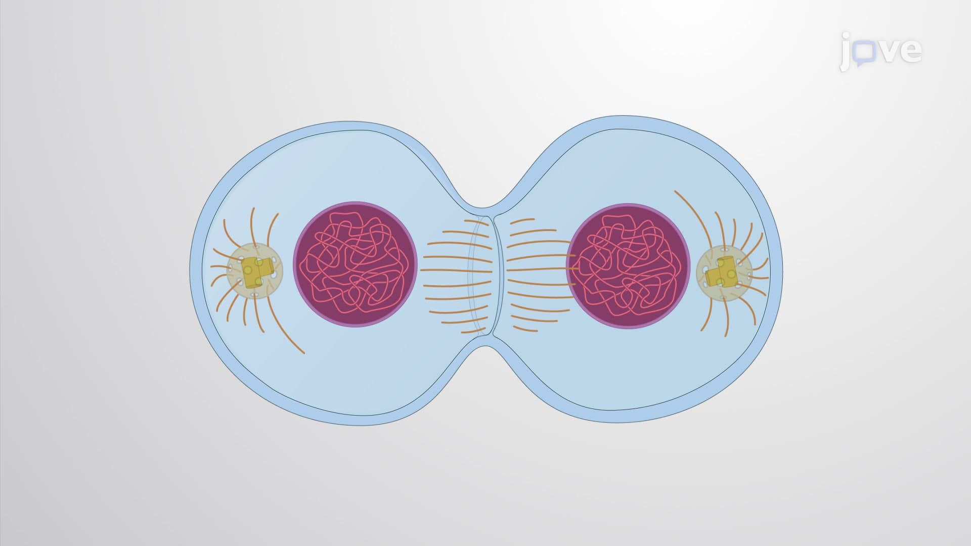
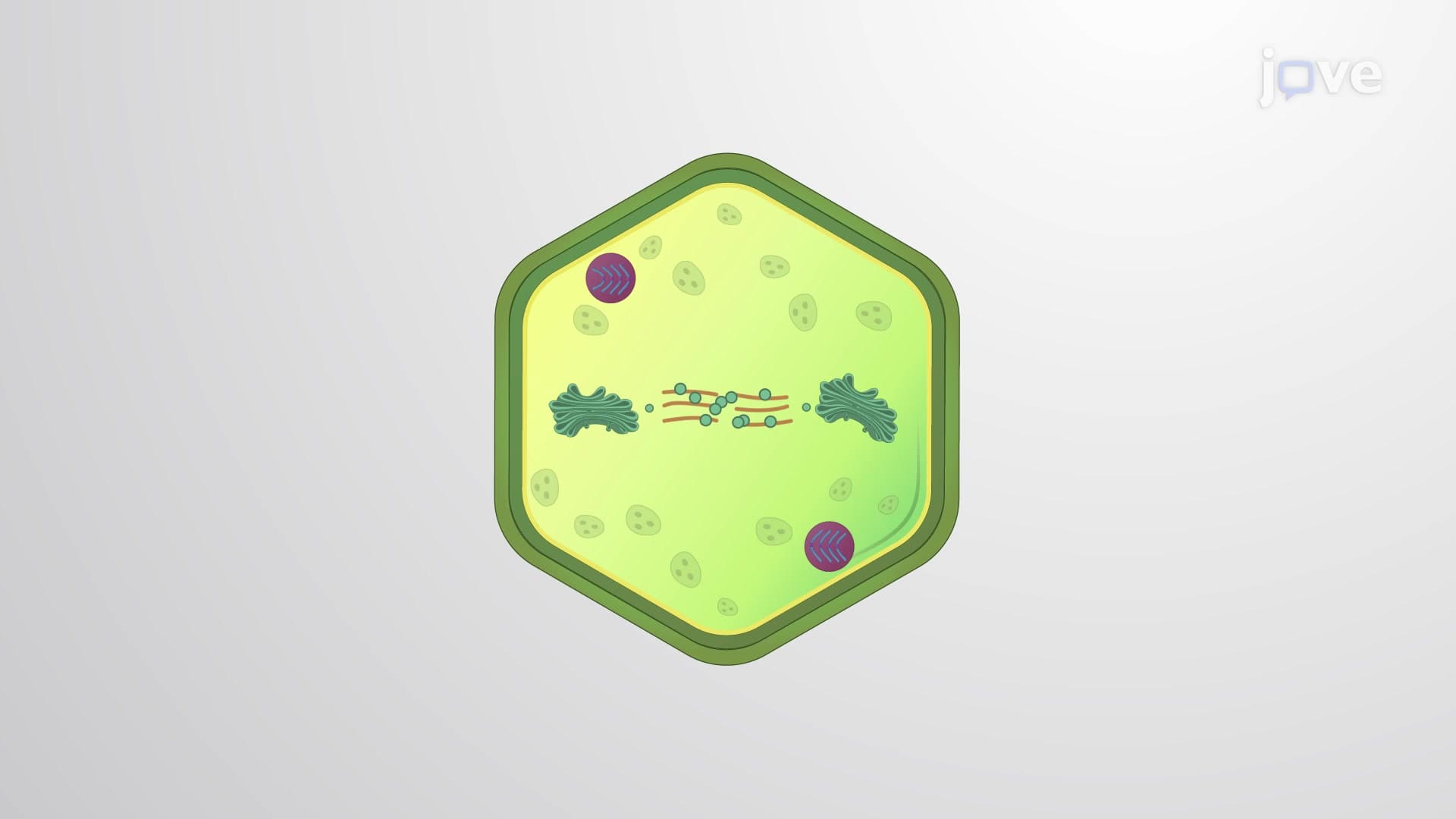
The contractile ring represents one of biology's most elegant molecular machines, serving as the primary mechanism that physically separates one cell into two during cytokinesis. This dynamic structure forms specifically at the cell's equator, creating a constricting band that gradually tightens until complete cellular division occurs. Understanding the contractile ring is fundamental for students preparing for AP Biology exams, where cell cycle questions frequently appear, and forms the basis for more advanced topics in college-level cell biology courses.
The contractile ring's effectiveness stems from its sophisticated protein composition, primarily featuring actin filaments arranged in antiparallel configurations and myosin II motor proteins. These structural elements work in concert under the regulation of RhoA, a small GTPase that acts as the master coordinator of ring assembly. The actin filaments provide the scaffolding structure, while myosin II generates the mechanical force necessary for contraction. This protein interaction exemplifies the cytoskeletal dynamics that medical students encounter on the MCAT, particularly in questions about cellular mechanics and protein function.
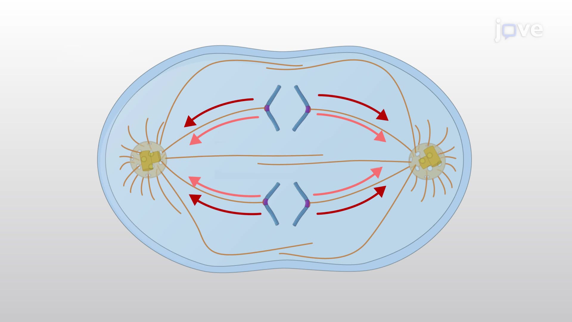
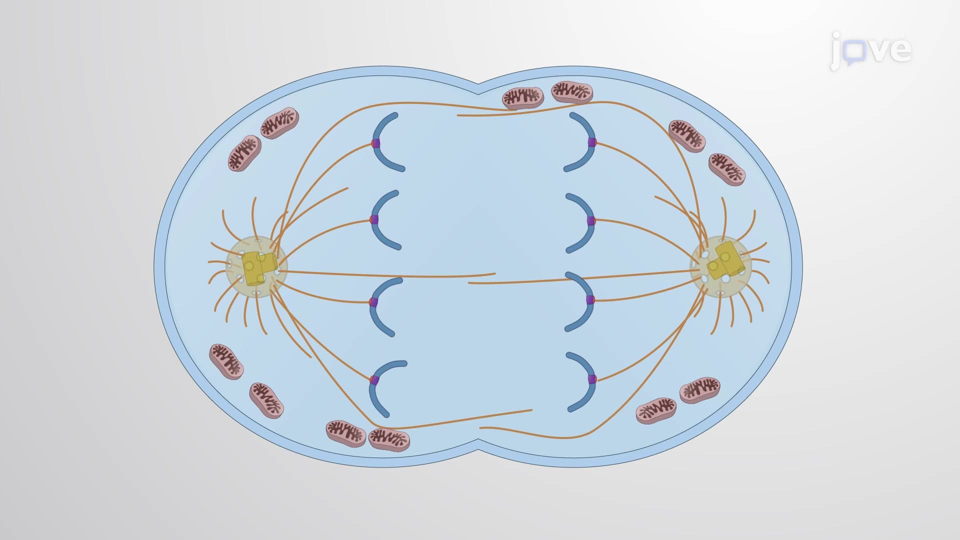
RhoA's regulatory role extends beyond simple protein activation-it ensures both spatial and temporal precision in contractile ring formation. The protein becomes activated specifically at the cell cortex near the cellular equator, preventing inappropriate ring formation at other locations. Additionally, RhoA coordinates with anaphase spindle fibers to guarantee that contractile ring assembly occurs only after proper chromosome segregation. This timing mechanism prevents premature cell division that could result in genetic abnormalities, a concept frequently tested in undergraduate genetics courses and relevant to understanding chromosomal disorders studied in American medical schools.
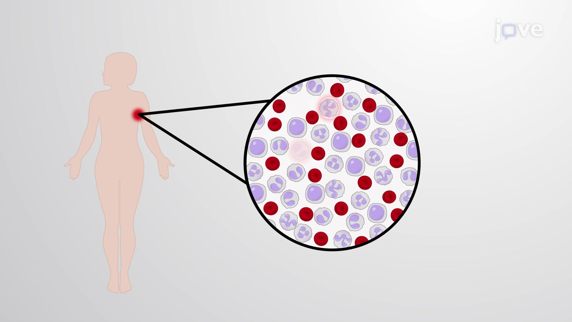
Contractile ring dysfunction has profound implications for human health, particularly in cancer development where abnormal cell division contributes to tumor formation. Research at institutions like the National Cancer Institute has revealed that defects in contractile ring proteins can lead to failed cytokinesis, resulting in polyploid cells with abnormal chromosome numbers. Understanding these mechanisms helps explain how certain chemotherapy drugs target dividing cells and provides insight into why rapidly dividing tissues, such as hair follicles and intestinal lining, are particularly affected by cancer treatments. For pre-med students, this knowledge connects basic cell biology to clinical applications they'll encounter in medical practice.
Related Micro-courses