Loading concepts…


10 Concepts
11 Concepts
11 Concepts
17 Concepts
20 Concepts
16 Concepts
16 Concepts
21 Concepts
11 Concepts
15 Concepts
14 Concepts
21 Concepts
8 Concepts
15 Concepts
17 Concepts
19 Concepts
11 Concepts
18 Concepts
6 Concepts
21 Concepts
 18 Concepts
 101K Views
 Popular topics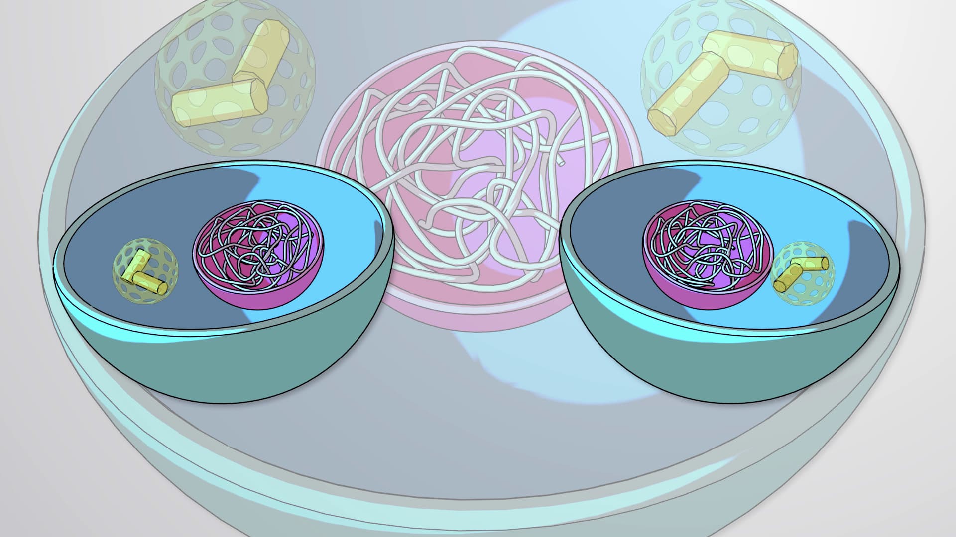
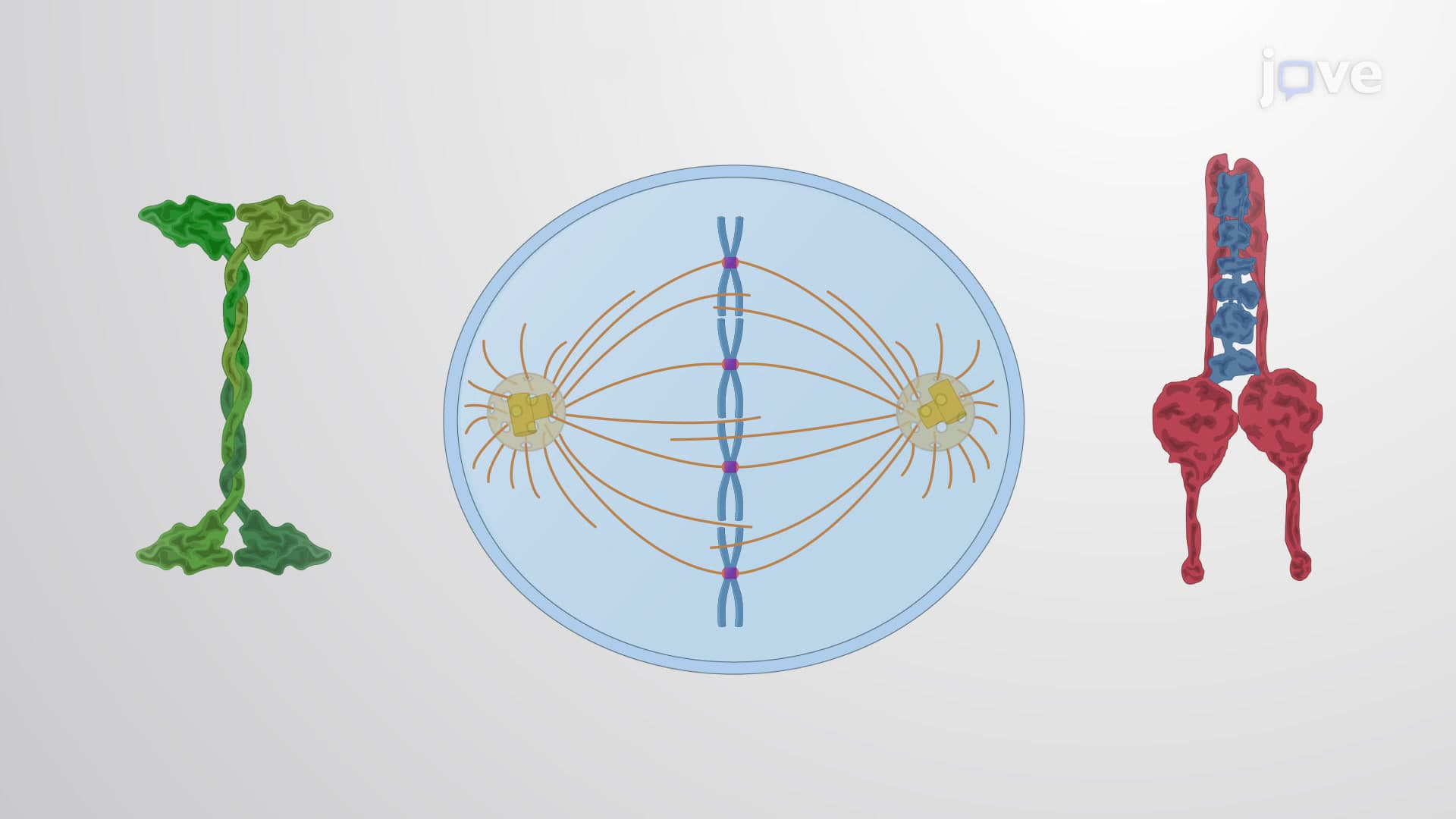



Cell division is the fundamental biological process by which a single cell creates two genetically identical daughter cells through mitosis and cytokinesis. This essential mechanism drives growth, tissue repair, and reproduction in all eukaryotic organisms, from human development to plant regeneration. Understanding cell division is crucial for comprehending cancer biology, genetic disorders, and regenerative medicine applications throughout the United States healthcare system. JoVE Coach provides comprehensive coverage of this vital cellular process.
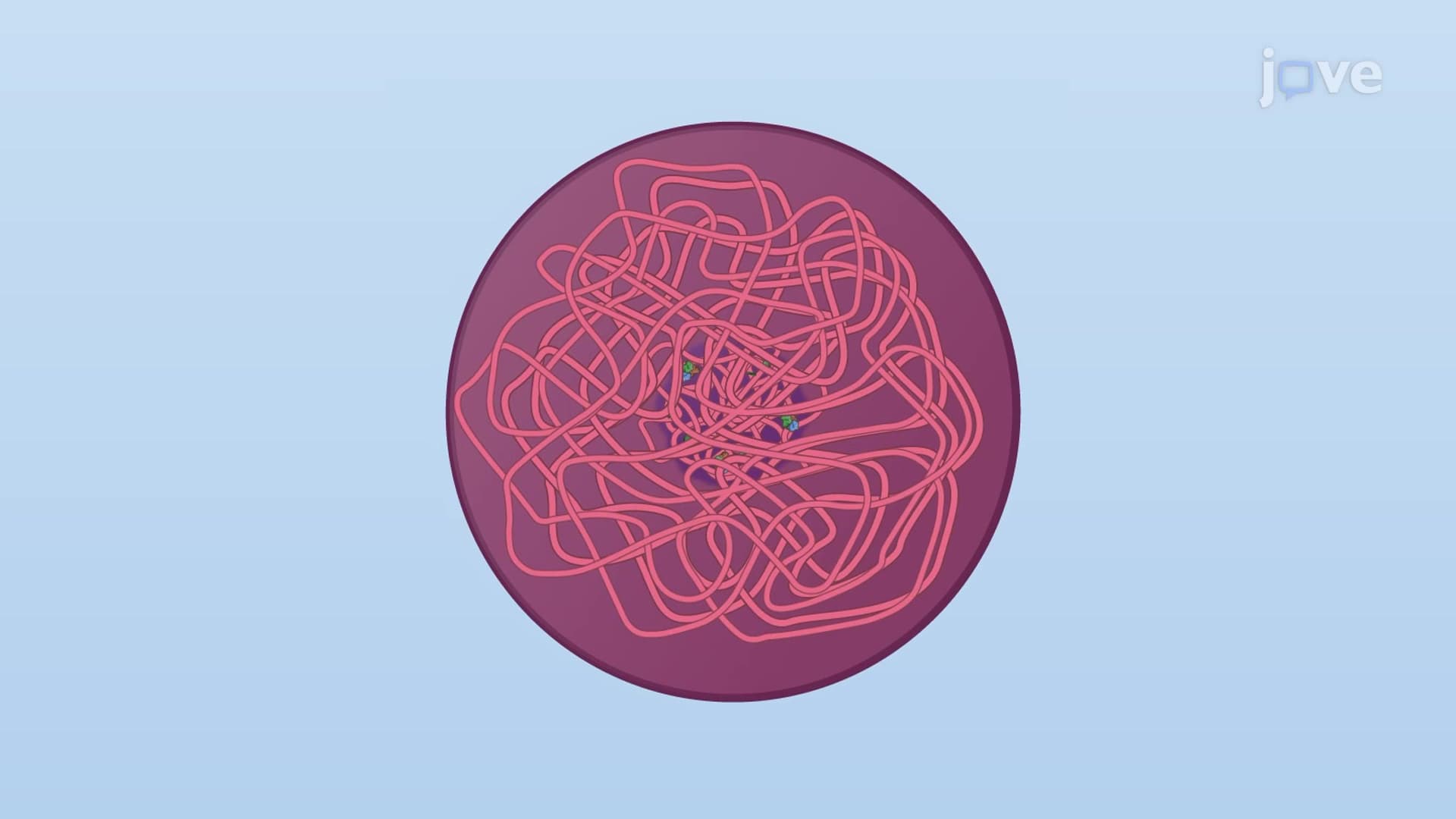
1. Chromosome Duplication and Sister Chromatid Formation During S-phase of the cell cycle, DNA replication creates identical copies of each chromosome called sister chromatids. This semi-conservative process involves DNA helicase unwinding the double helix, DNA polymerase synthesizing new strands, and ligase sealing DNA fragments. Sister chromatids remain attached at the centromere through cohesin protein complexes until separation during anaphase. Understanding this process is essential for comprehending genetic diseases, cancer development, and the mechanisms targeted by chemotherapy drugs used in American cancer treatment centers.
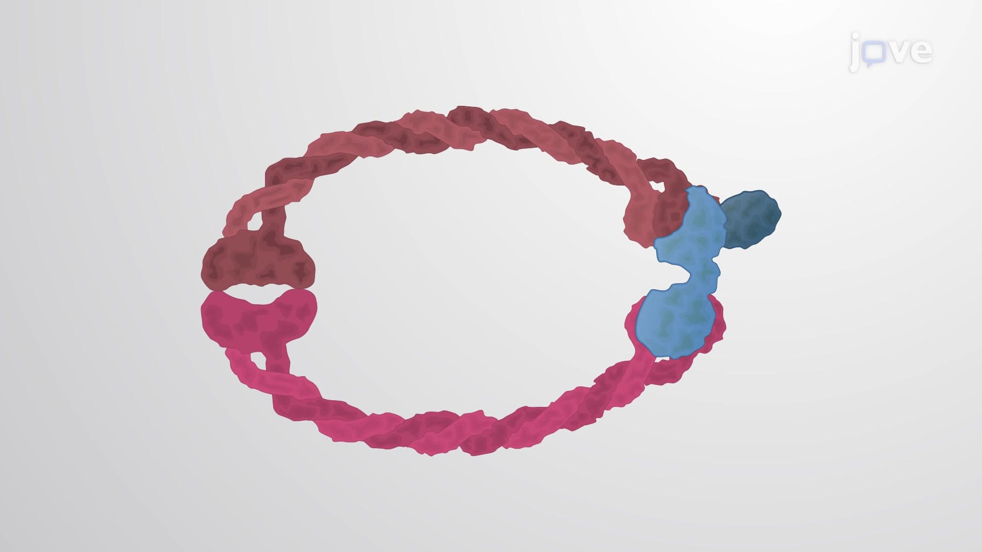
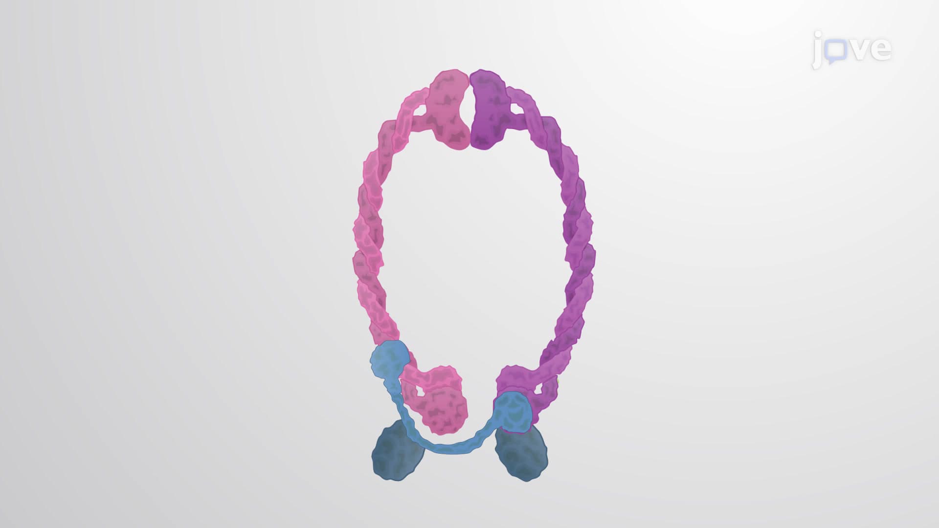
2. Cohesin and Condensin Protein Complexes Cohesin proteins form ring-like structures that hold sister chromatids together from S-phase through metaphase. These complexes contain SMC1, SMC3, SCC1, and SCC3 subunits that create a molecular clamp around DNA. Condensin proteins reorganize long, tangled chromatin into compact, separable chromosomes during mitosis. These processes prevent chromosome breaks and ensure proper segregation. Defects in these systems contribute to chromosomal instability observed in various cancers treated at major US medical institutions like MD Anderson and Memorial Sloan Kettering.
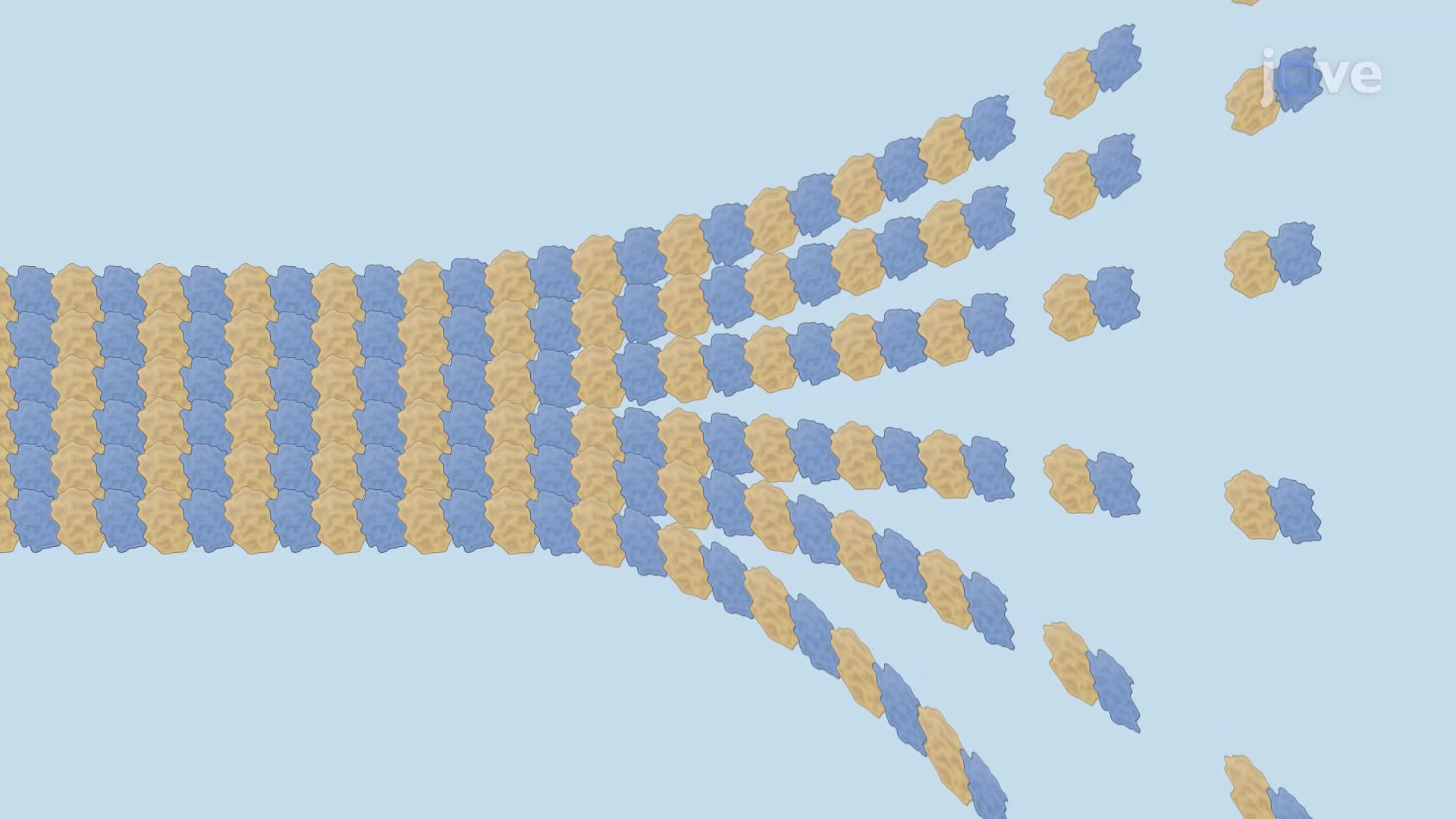
3. Mitotic Spindle Assembly and Organization The mitotic spindle consists of microtubule arrays organized around centrosomes that separate chromosomes during cell division. Three types of microtubules form the spindle: kinetochore microtubules that attach to chromosomes, interpolar microtubules that overlap at the spindle midzone, and astral microtubules that position the spindle within the cell. Motor proteins including kinesin and dynein generate forces that assemble and operate the spindle. This intricate machinery is targeted by cancer drugs like paclitaxel, commonly used in American oncology practices.
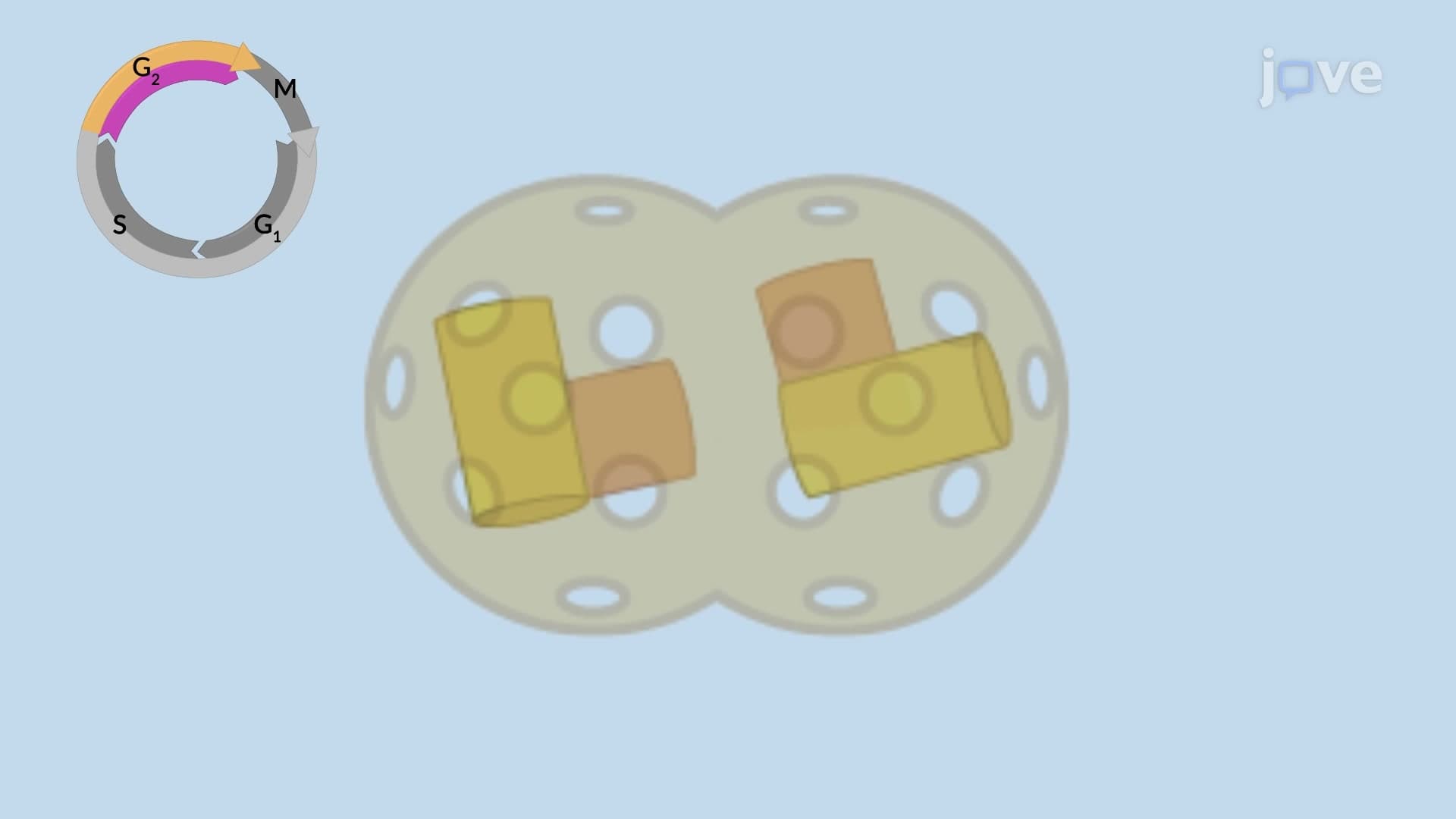
4. Centrosome Duplication Cycle Centrosomes serve as microtubule organizing centers and duplicate once per cell cycle to form the two spindle poles required for mitosis. Each centrosome contains two centrioles surrounded by pericentriolar matrix proteins. Duplication begins in G1-phase with centriole disengagement, followed by daughter centriole formation during S-phase. Errors in centrosome duplication can cause chromosomal instability and contribute to cancer development. Research at institutions like Harvard Medical School and Stanford University continues to investigate centrosome abnormalities in human diseases.
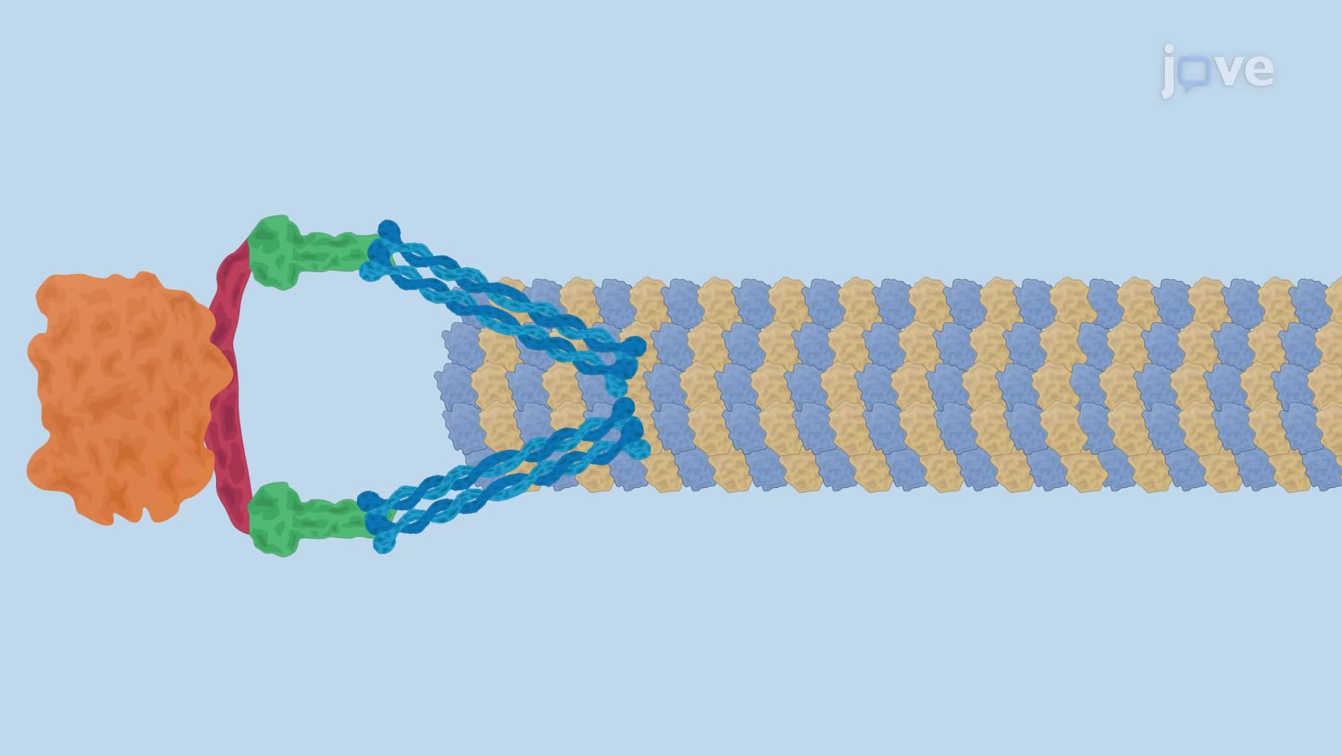
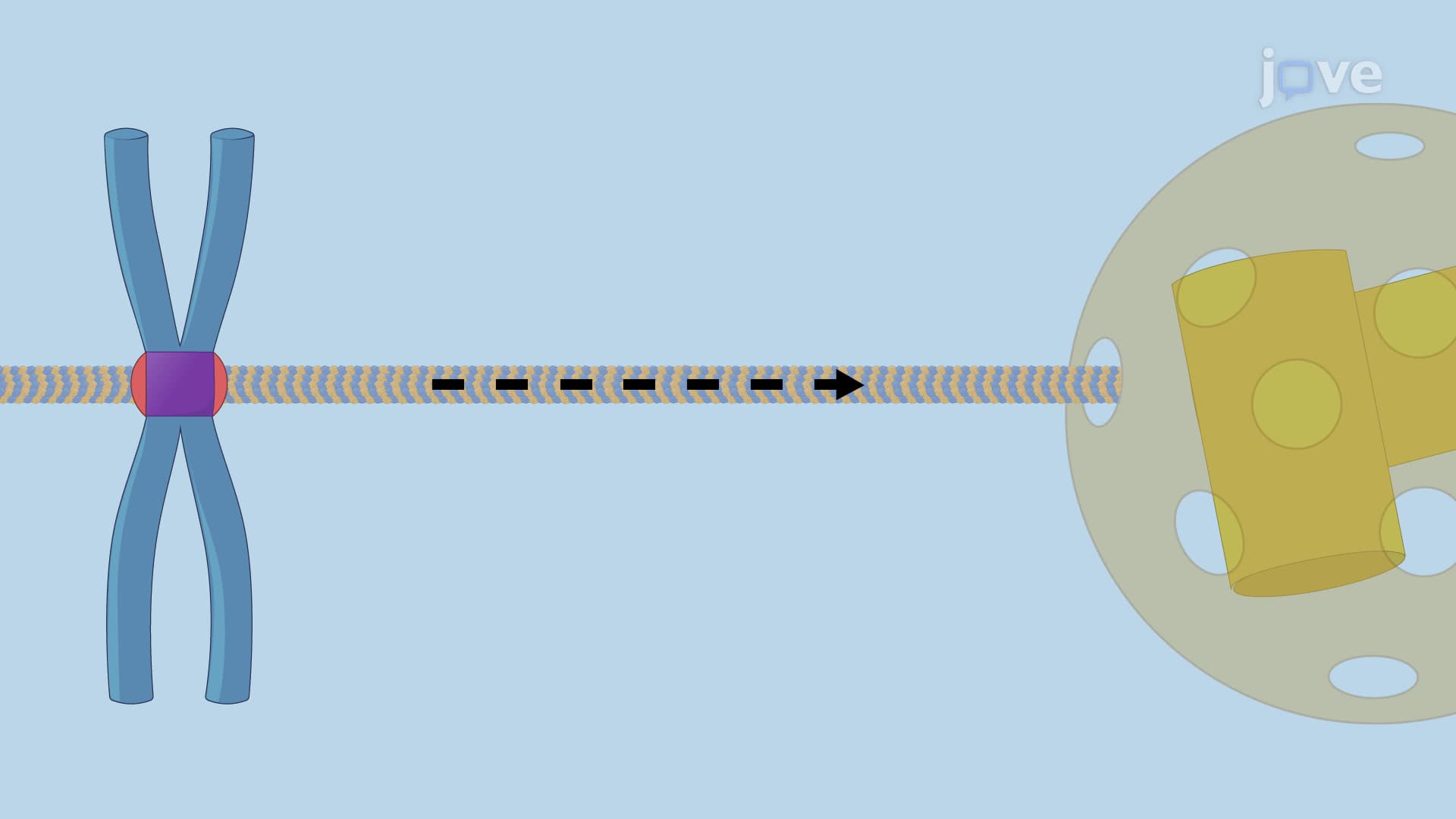
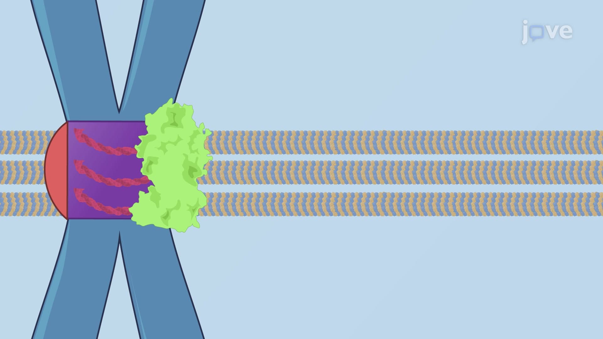
5. Chromosome Attachment and Bi-orientation Kinetochores are multi-protein complexes assembled at centromeres that link chromosomes to spindle microtubules. The NDC80 complex forms the critical connection between kinetochores and microtubule plus-ends, allowing continued attachment during microtubule dynamics. Proper bi-orientation occurs when sister chromatids attach to opposite spindle poles, creating tension that stabilizes attachments. Aurora B kinase acts as a tension sensor, destabilizing incorrect attachments until proper bi-orientation is achieved. These mechanisms are studied extensively in US research laboratories developing targeted cancer therapeutics.
6. Spindle Assembly Checkpoint Control The spindle assembly checkpoint prevents anaphase onset until all chromosomes achieve proper bi-orientation on the spindle. Unattached kinetochores recruit checkpoint proteins including Mad2, forming the mitotic checkpoint complex (MCC) that inhibits the anaphase-promoting complex (APC/C). Only when all chromosomes are correctly attached does the checkpoint signal disappear, allowing APC/C activation and anaphase progression. Checkpoint defects contribute to aneuploidy in cancer cells. Major pharmaceutical companies in the United States are developing drugs that exploit checkpoint weaknesses in cancer therapy.
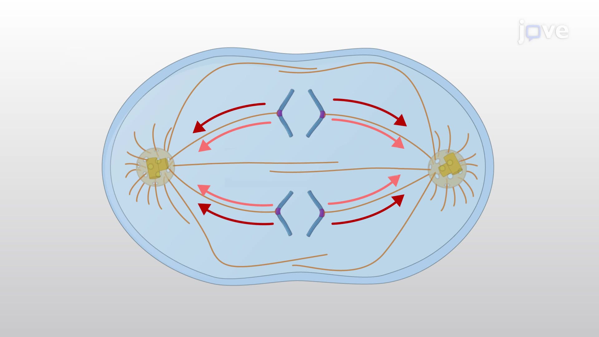
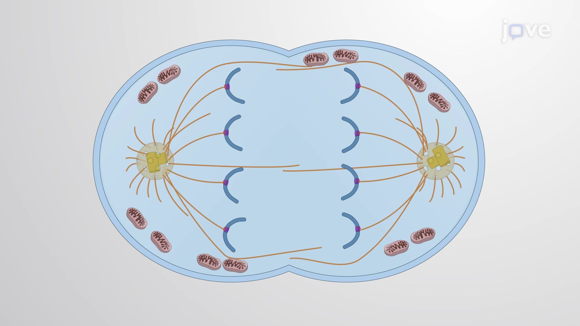
7. Anaphase Progression and Chromosome Segregation Anaphase consists of two overlapping processes: Anaphase A involves chromosome movement toward spindle poles through kinetochore microtubule shortening, while Anaphase B involves spindle pole separation through interpolar microtubule sliding. The anaphase-promoting complex triggers sister chromatid separation by degrading securin protein, activating separase protease to cleave cohesin rings. Multiple forces including microtubule depolymerization, microtubule flux, and motor protein activity drive chromosome segregation. Understanding these mechanisms helps explain chromosomal abnormalities observed in genetic disorders diagnosed in US medical facilities.
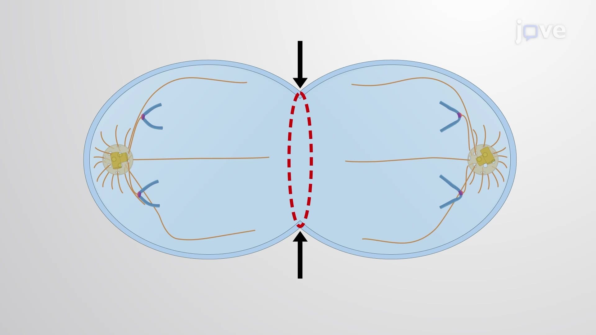
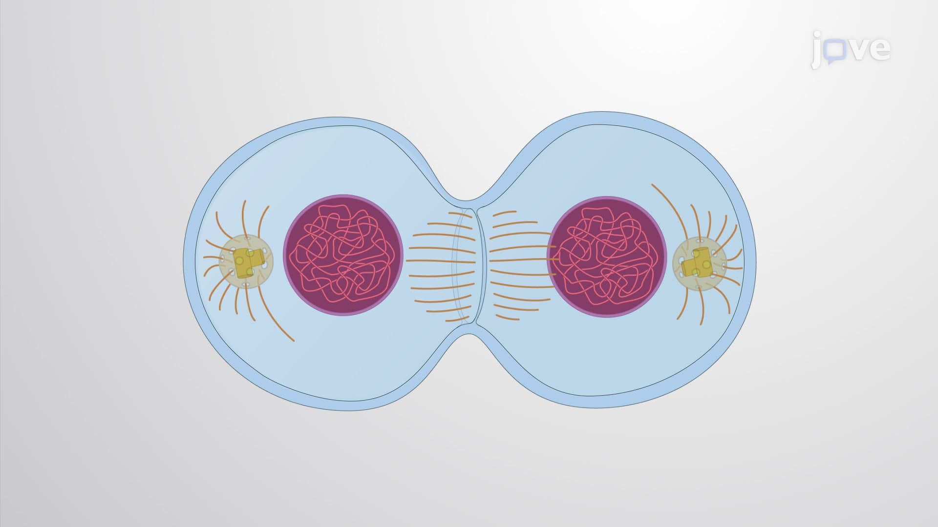
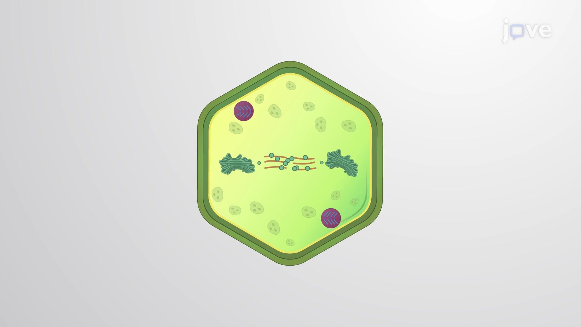
8. Cytokinesis and Contractile Ring Formation Cytokinesis physically divides the cytoplasm through contractile ring formation and function. RhoA GTPase regulates contractile ring assembly at the cell equator, promoting actin filament polymerization and myosin II motor recruitment. The contractile ring generates force through myosin-driven actin filament sliding, creating the cleavage furrow that deepens until cell division is complete. Cytokinesis defects can lead to multinucleated cells and genomic instability. Research at institutions like the National Institutes of Health investigates how cytokinesis failures contribute to cancer progression and therapeutic resistance.