Loading subject page…

- Molecular Biology
- Cell Division
- Microtubule Instability
Micro-course: Cell Division
101,001 views
Learn key concepts and earn your certificate.



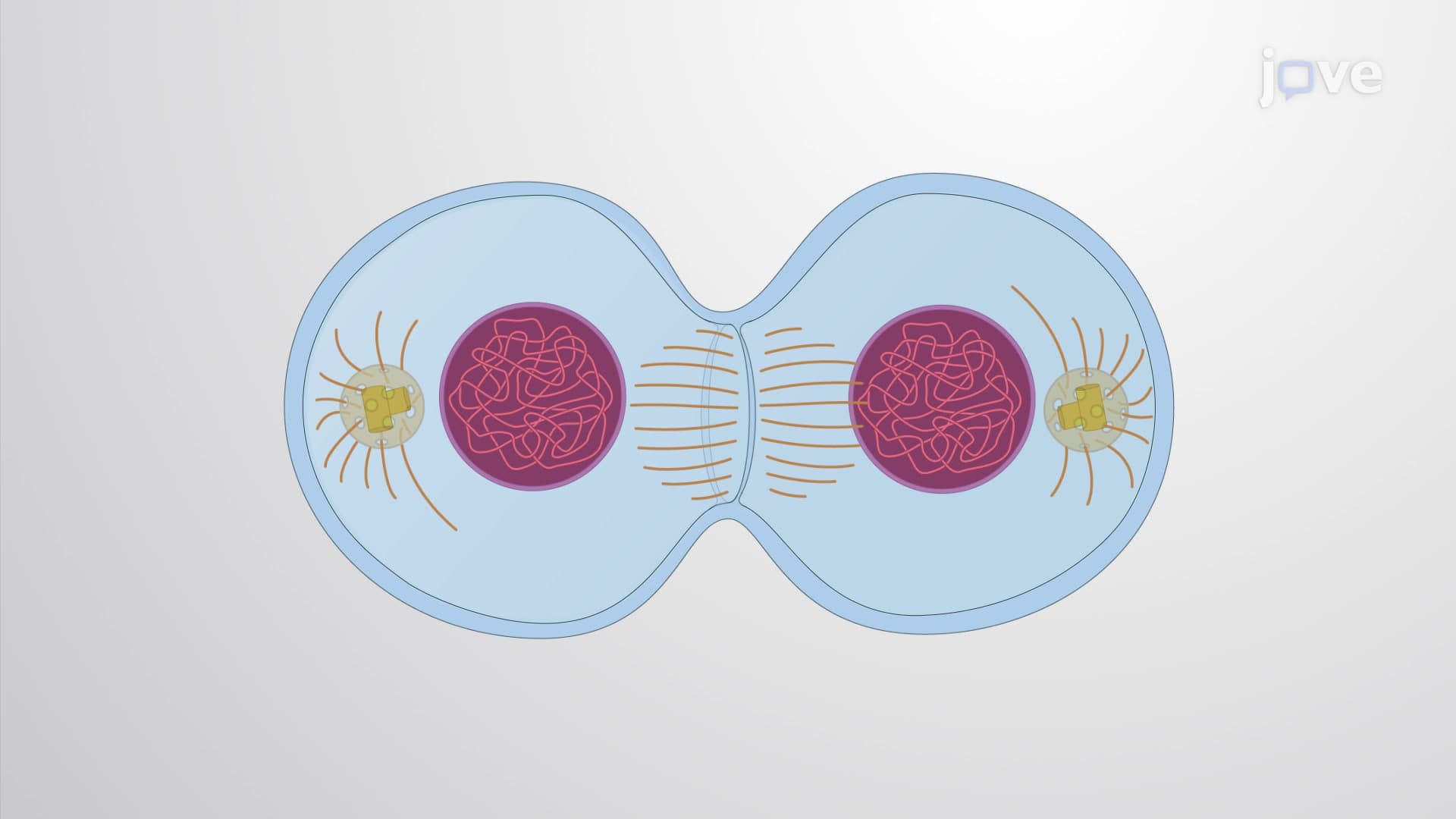
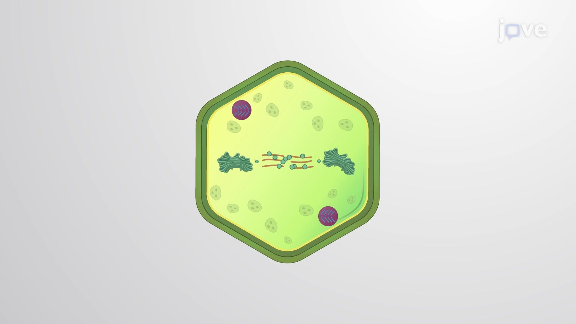
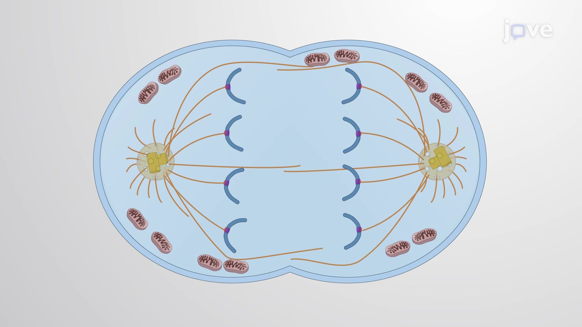

Microtubule Instability

00:00
02:17
Video Summary: What Is Microtubule Instability
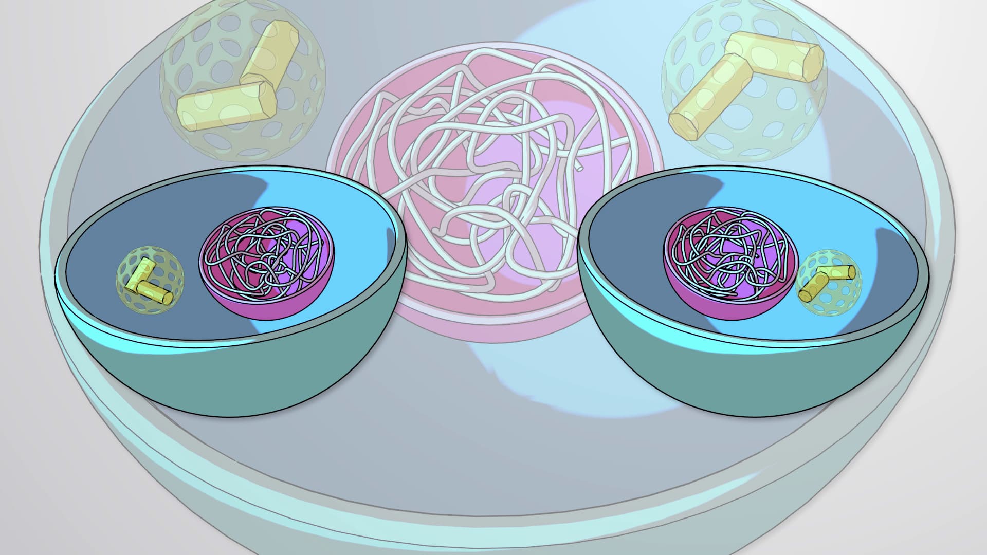
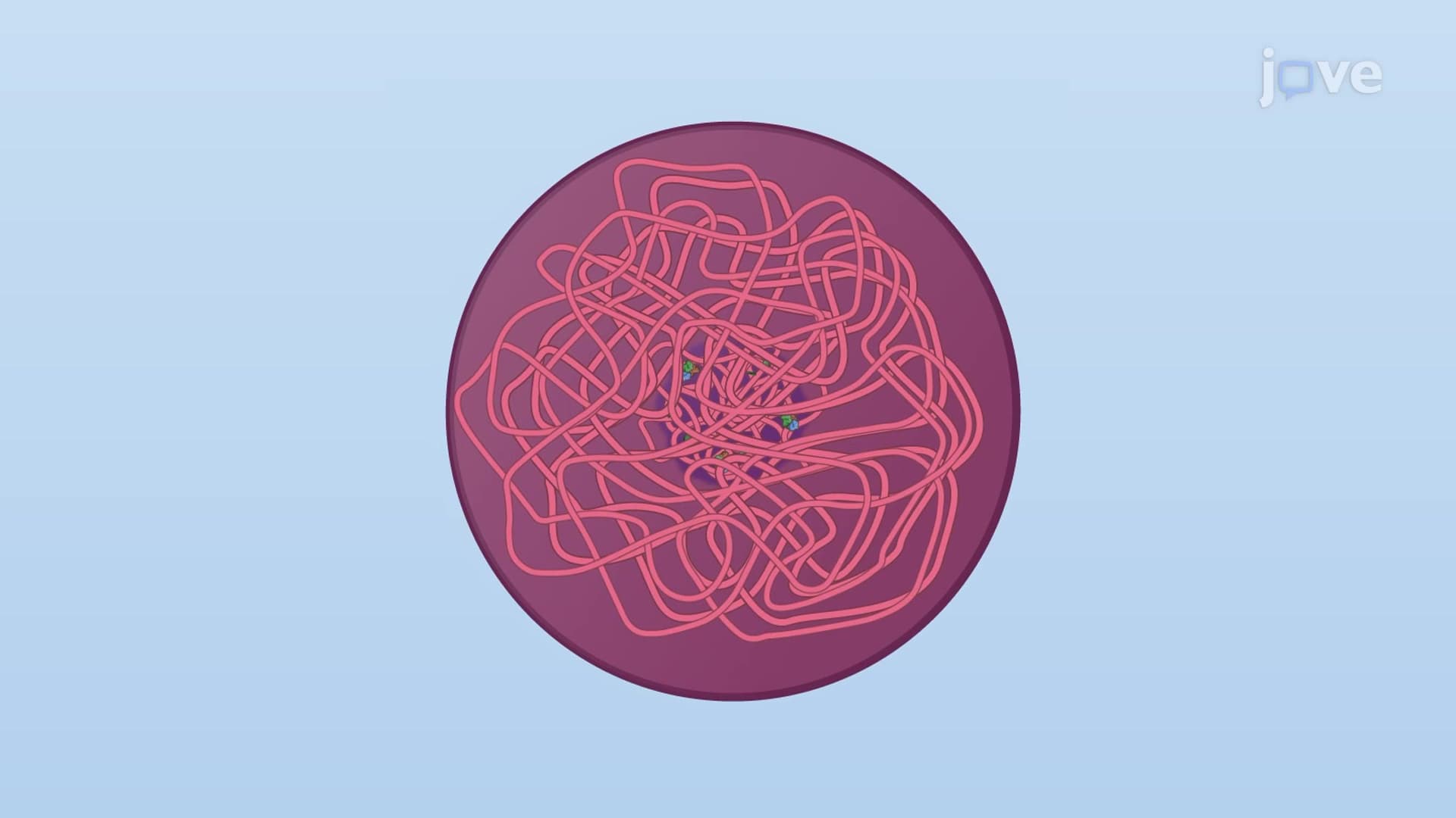
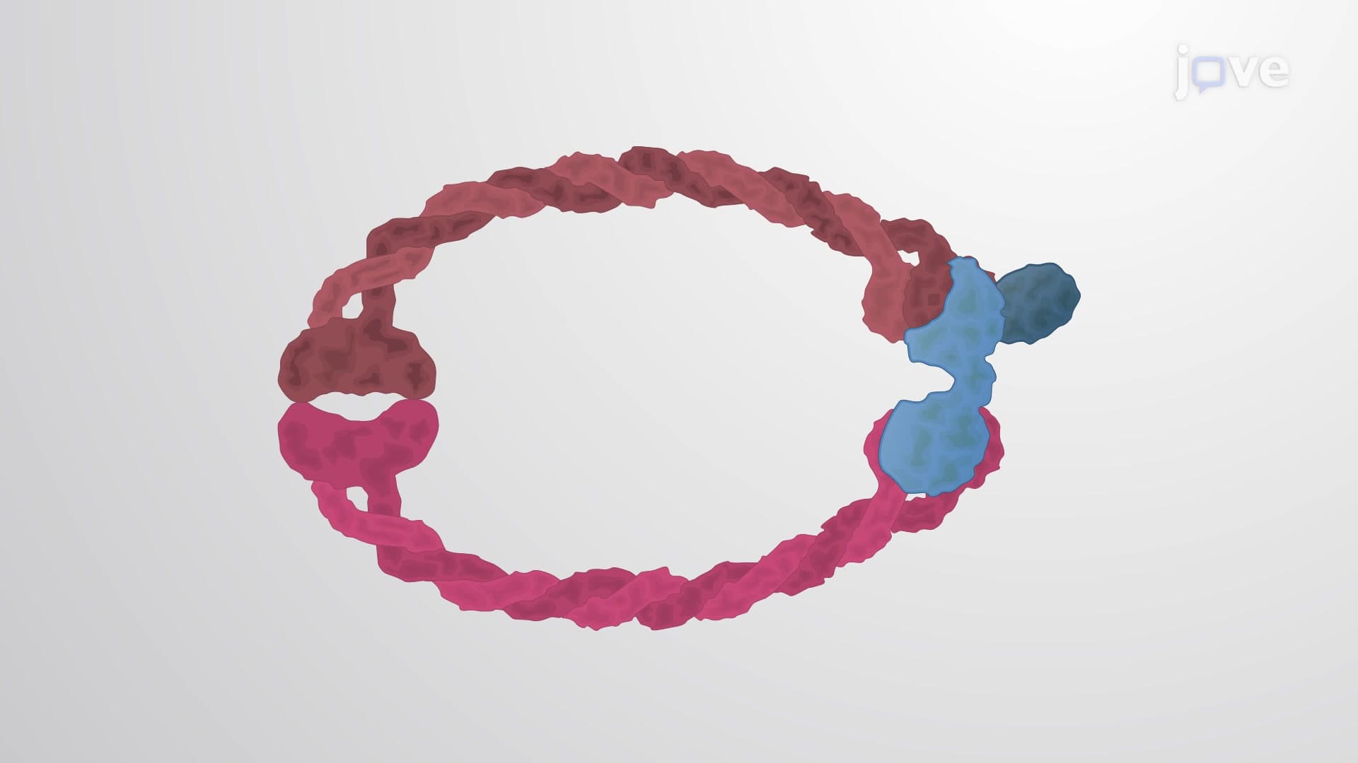
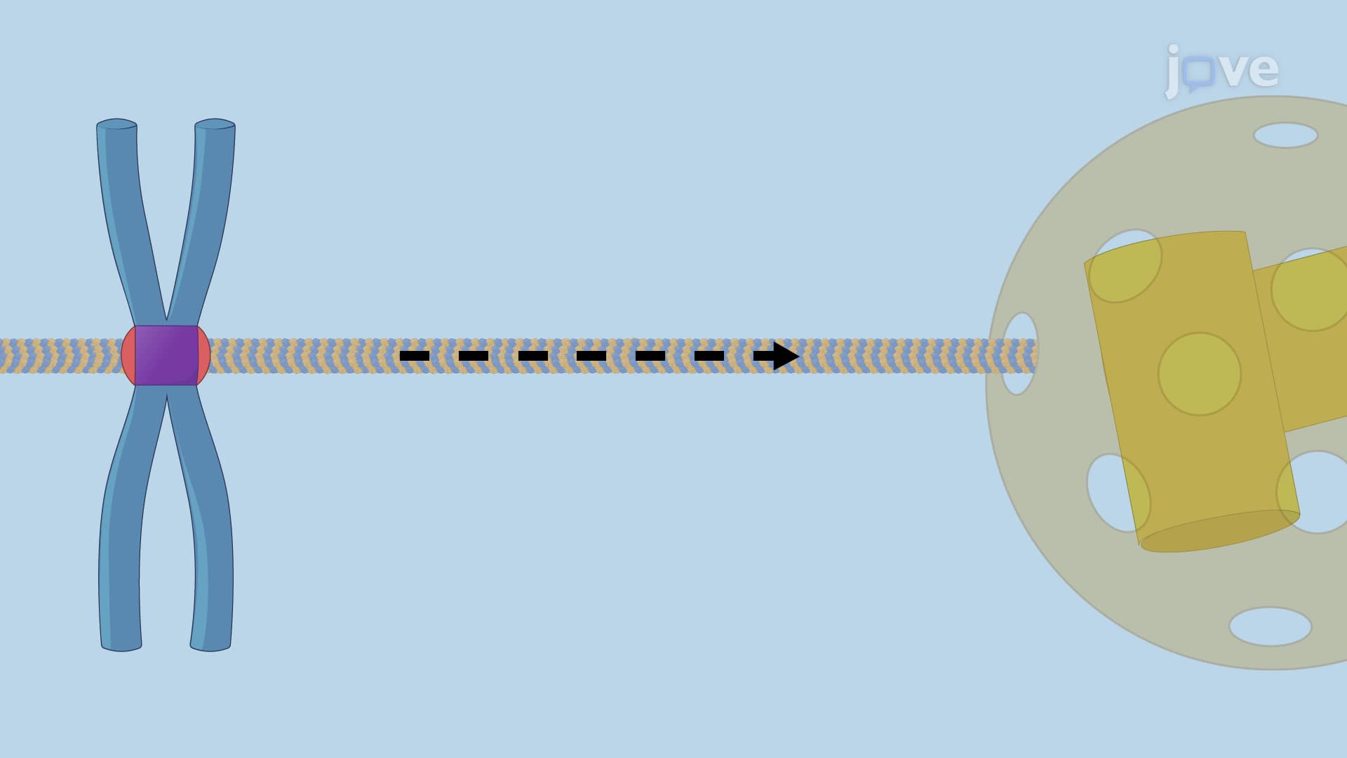
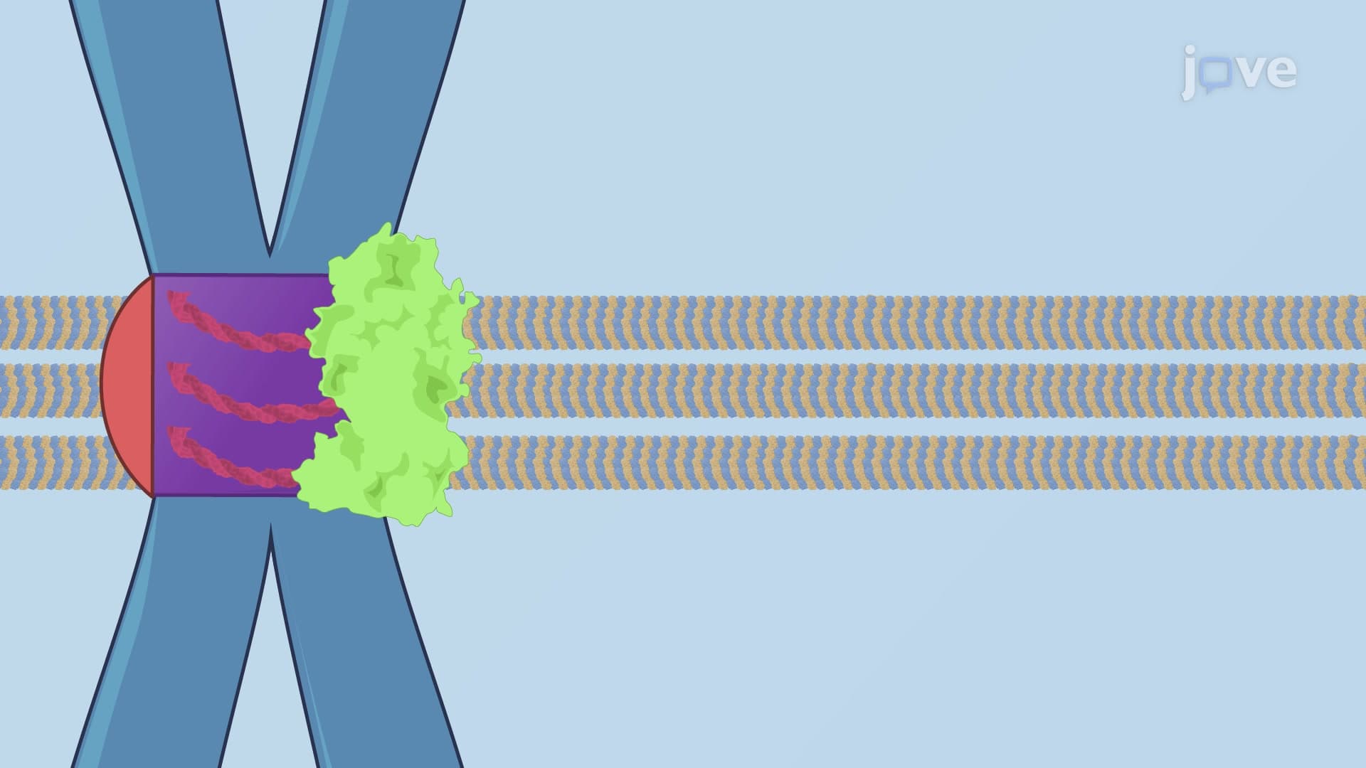
Ever wonder how your cells know exactly when and where to divide? Microtubule instability is the dynamic process that allows cellular structures to rapidly grow and shrink, enabling precise chromosome separation during cell division. Picture the protein scaffolding inside a NASA clean room facility being constantly assembled and disassembled as technicians prepare spacecraft components, that's similar to how microtubules behave in your cells. This cellular phenomenon involves continuous switching between growth phases called "rescue" and breakdown phases called "catastrophe." Watch the full video on JoVE Coach to master this concept with expert-led visuals and step-by-step explanations.
- Understand what is microtubule instability and its fundamental role in cellular processes
- Identify the key phases of dynamic instability: catastrophe (growth to shrinkage) and rescue (shrinkage to growth)
- Analyze how GTP hydrolysis in tubulin subunits drives microtubule depolymerization
- Compare the regulatory roles of microtubule-associated proteins (MAPs) versus catastrophe factors
- Evaluate how cells modify microtubule dynamics throughout different cell cycle phases
- Apply knowledge of tubulin heterodimer structure to explain polymerization mechanisms
- Recognize the connection between increased instability and mitotic spindle formation
- Explore real-world implications of microtubule dysfunction in disease states
Understanding Microtubule Instability Fundamentals
Microtubule instability represents one of the most fascinating examples of controlled chaos in cell biology. Unlike static structural elements, microtubules exist in a constant state of dynamic equilibrium, rapidly transitioning between growth and shrinkage phases. This phenomenon enables cells to quickly reorganize their internal architecture in response to changing needs, particularly during cell division.
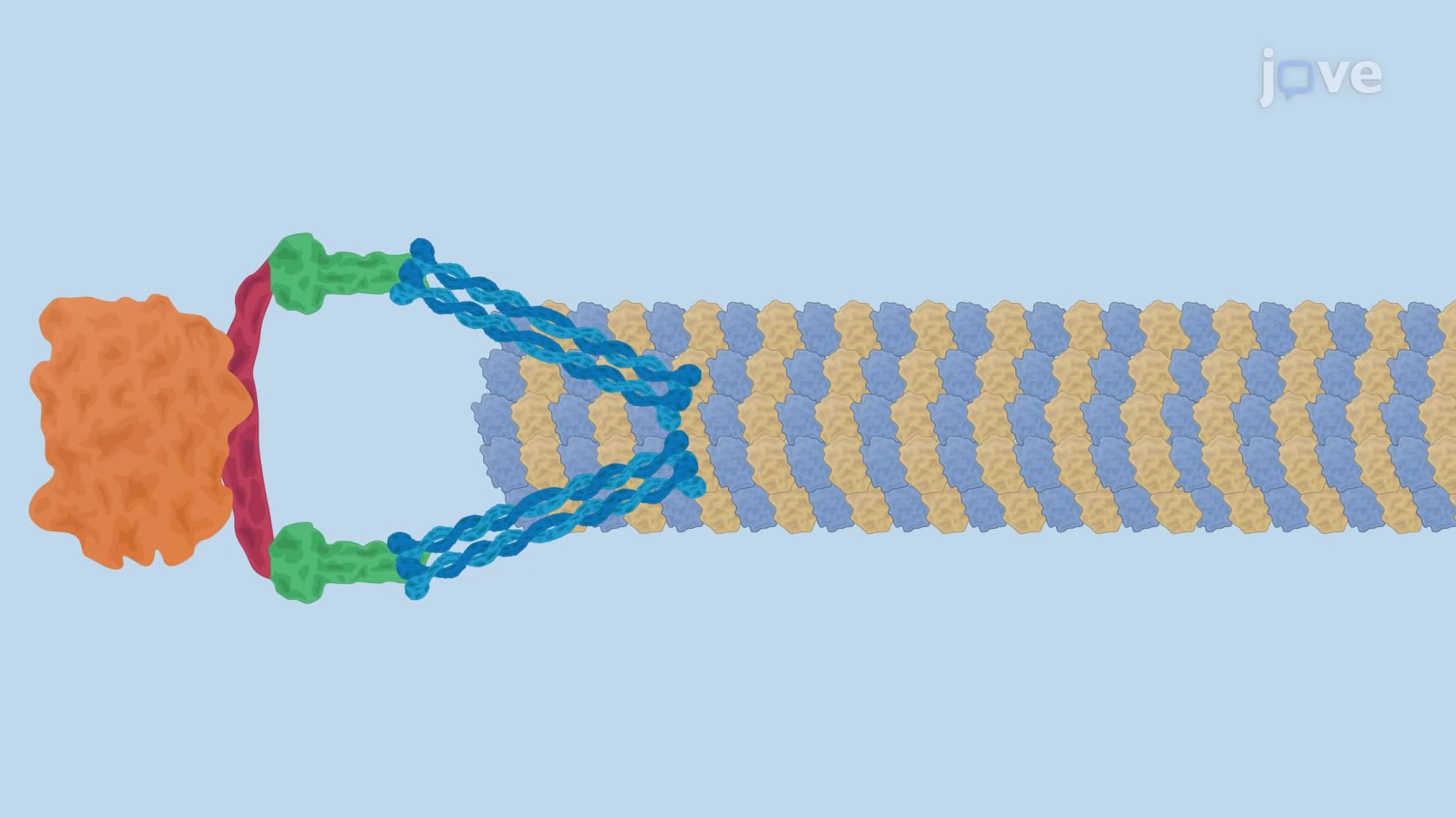
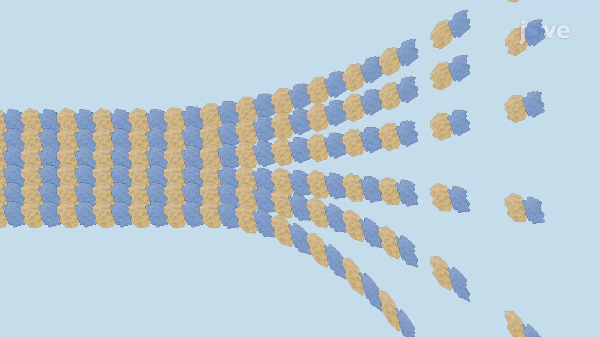
The molecular basis of this instability lies in the unique properties of tubulin, the protein building block of microtubules. Each tubulin heterodimer consists of alpha and beta subunits, with the beta subunit carrying a crucial GTP molecule. When GTP hydrolyzes to GDP, it dramatically destabilizes the microtubule structure, causing the characteristic "splaying" at the microtubule tip that propagates depolymerization throughout the structure.
Dynamic Phases: Catastrophe and Rescue Events
The unpredictable switching between growth and shrinkage defines microtubule dynamic instability. "Catastrophe" events mark the sudden transition from growth to rapid shrinkage, while "rescue" events represent the opposite shift. These transitions occur stochastically, meaning they cannot be predicted for individual microtubules, yet they follow statistical patterns that cells can regulate.
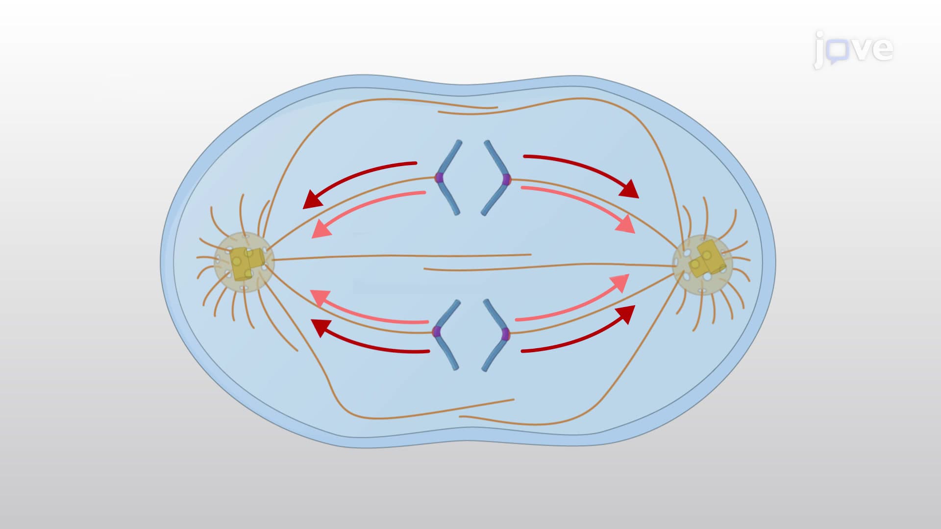
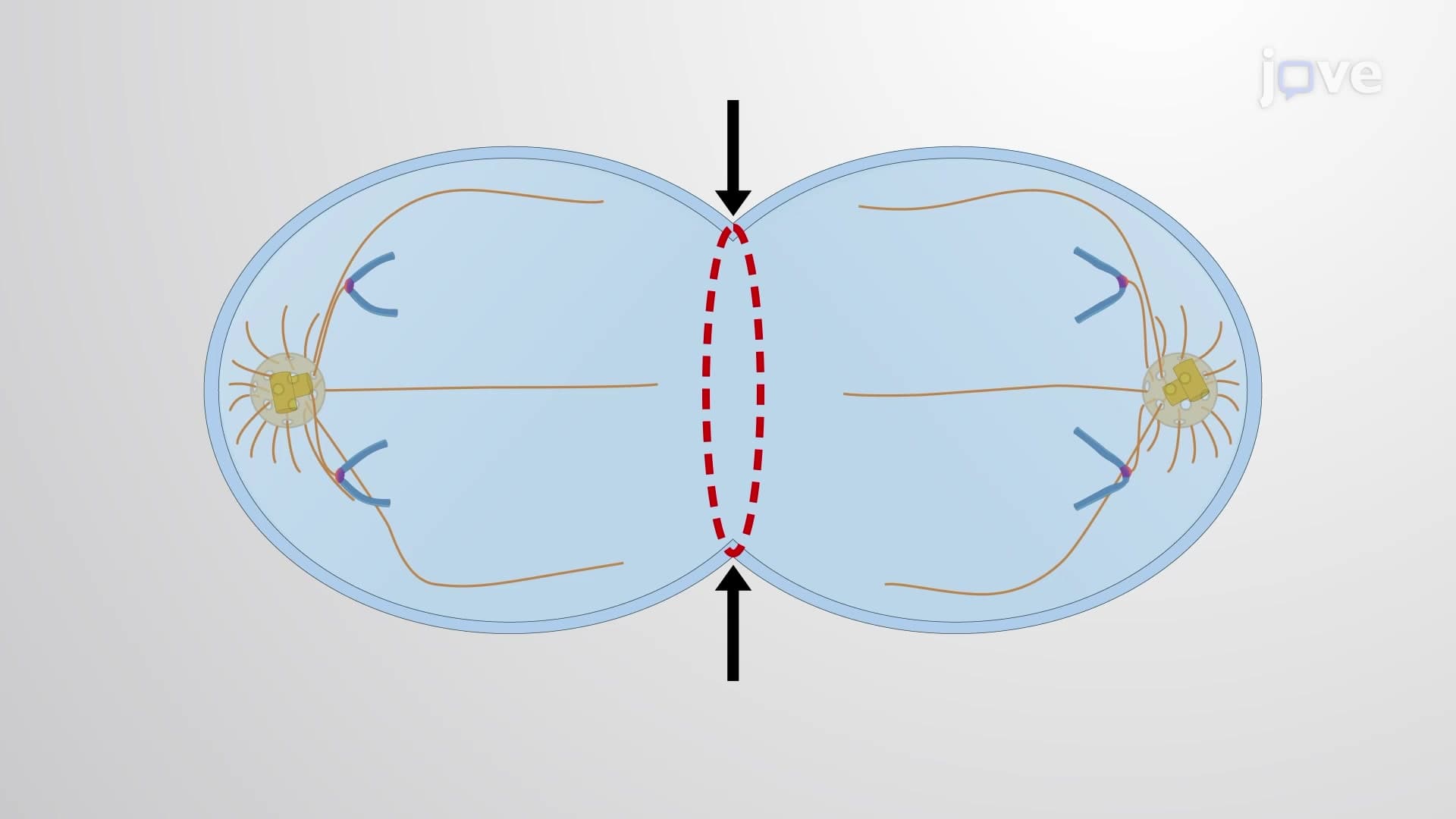
Research at institutions like Harvard Medical School and MIT has revealed that catastrophe frequencies increase dramatically during mitosis, when cells need to disassemble their interphase microtubule networks and build mitotic spindles. This regulatory mechanism ensures proper chromosome segregation, a process critical for preventing cancer-causing chromosomal abnormalities.
Regulatory Control Mechanisms
Cells don't leave microtubule dynamics to chance. Microtubule-associated proteins (MAPs) act as stabilizing factors, binding along microtubule shafts to promote growth and prevent catastrophe. Conversely, catastrophe factors like the kinesin-13 family actively promote microtubule breakdown by removing tubulin subunits from microtubule ends.
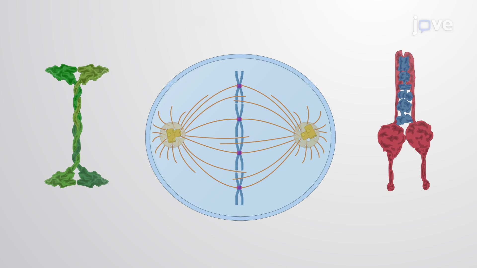
This regulatory system becomes particularly important during the cell cycle. During interphase, cells maintain long, stable microtubules radiating from centrosomes to support intracellular transport. As cells enter mitosis, increased catastrophe factor activity and decreased MAP activity create the short, highly dynamic microtubules necessary for spindle formation.
Clinical and Educational Relevance
Understanding microtubule instability proves essential for students preparing for the MCAT, AP Biology exams, or college-level cell biology courses. Many anti-cancer drugs, including taxanes used in chemotherapy at centers like MD Anderson Cancer Center, work by disrupting microtubule dynamics. Students frequently encounter questions about these mechanisms on standardized tests, making this concept both academically and medically relevant.
Frequently Asked Questions
Microtubule instability is the rapid, unpredictable switching between growth and shrinkage phases in cellular protein structures called microtubules. This dynamic behavior allows cells to quickly reorganize their internal framework, especially during cell division when chromosomes must be precisely separated into daughter cells.
MCAT questions often focus on the molecular mechanisms of GTP hydrolysis and drug interactions with microtubules. AP Biology exams typically test understanding of cell cycle regulation and how microtubule dynamics facilitate chromosome separation during mitosis. Students should know the terms "catastrophe" and "rescue" and their roles in spindle formation.
Catastrophe occurs when GTP-bound tubulin hydrolyzes to GDP, destabilizing the microtubule structure and causing rapid depolymerization. Rescue events happen when new GTP-tubulin subunits are added faster than hydrolysis occurs, stabilizing the growing end. Regulatory proteins also influence these transitions by promoting or inhibiting either process.
Many chemotherapy drugs used in American cancer centers target microtubule instability. Taxanes like paclitaxel stabilize microtubules, preventing the dynamic instability needed for cancer cell division. Conversely, vinca alkaloids destabilize microtubules, both approaches ultimately blocking tumor cell proliferation by disrupting normal mitotic processes.
While the molecular details can seem complex, the basic concept is quite accessible. Think of microtubules like construction scaffolding that's constantly being built up and torn down as needed. The key is understanding that this apparent chaos is actually precisely controlled and essential for proper cell function.
Focus on drawing diagrams that show the relationship between GTP/GDP states and microtubule stability. Practice explaining how regulatory proteins affect dynamics, and memorize the definitions of catastrophe and rescue. Create concept maps linking microtubule instability to cell cycle phases and specific examples of drugs that target these processes.
Cells use spatial and temporal regulation through localized proteins. Centrosomes concentrate nucleation factors, while specific MAPs and catastrophe factors are activated or inhibited based on cell cycle phase. Post-translational modifications of tubulin also create "microtubule codes" that recruit different regulatory proteins to different microtubule populations.
Explore related topics like kinetochore structure and function, spindle checkpoint mechanisms, and cytoskeletal motor proteins like dynein and kinesin. Understanding how microtubule instability integrates with these systems provides a complete picture of cellular division and organization processes.
Related Micro-courses