Loading subject page…

- Molecular Biology
- Cell Division
- The Mitotic Spindle
Micro-course: Cell Division
101,001 views
Learn key concepts and earn your certificate.

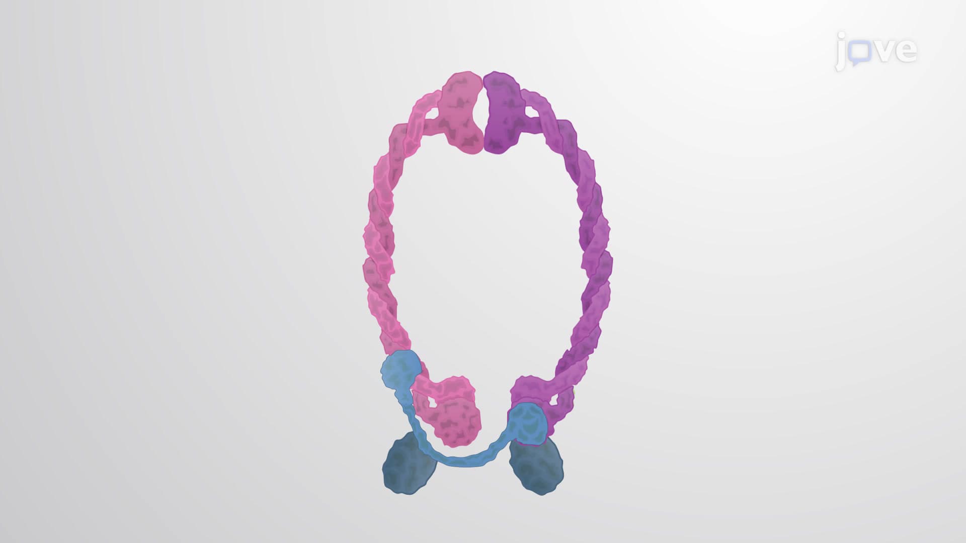
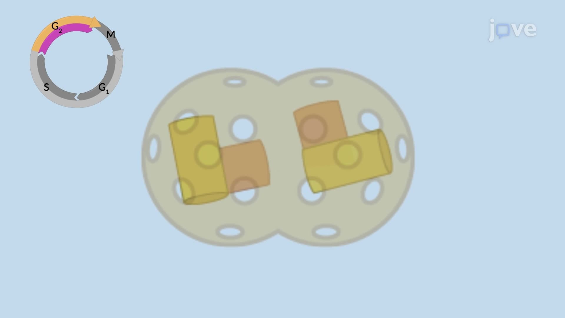
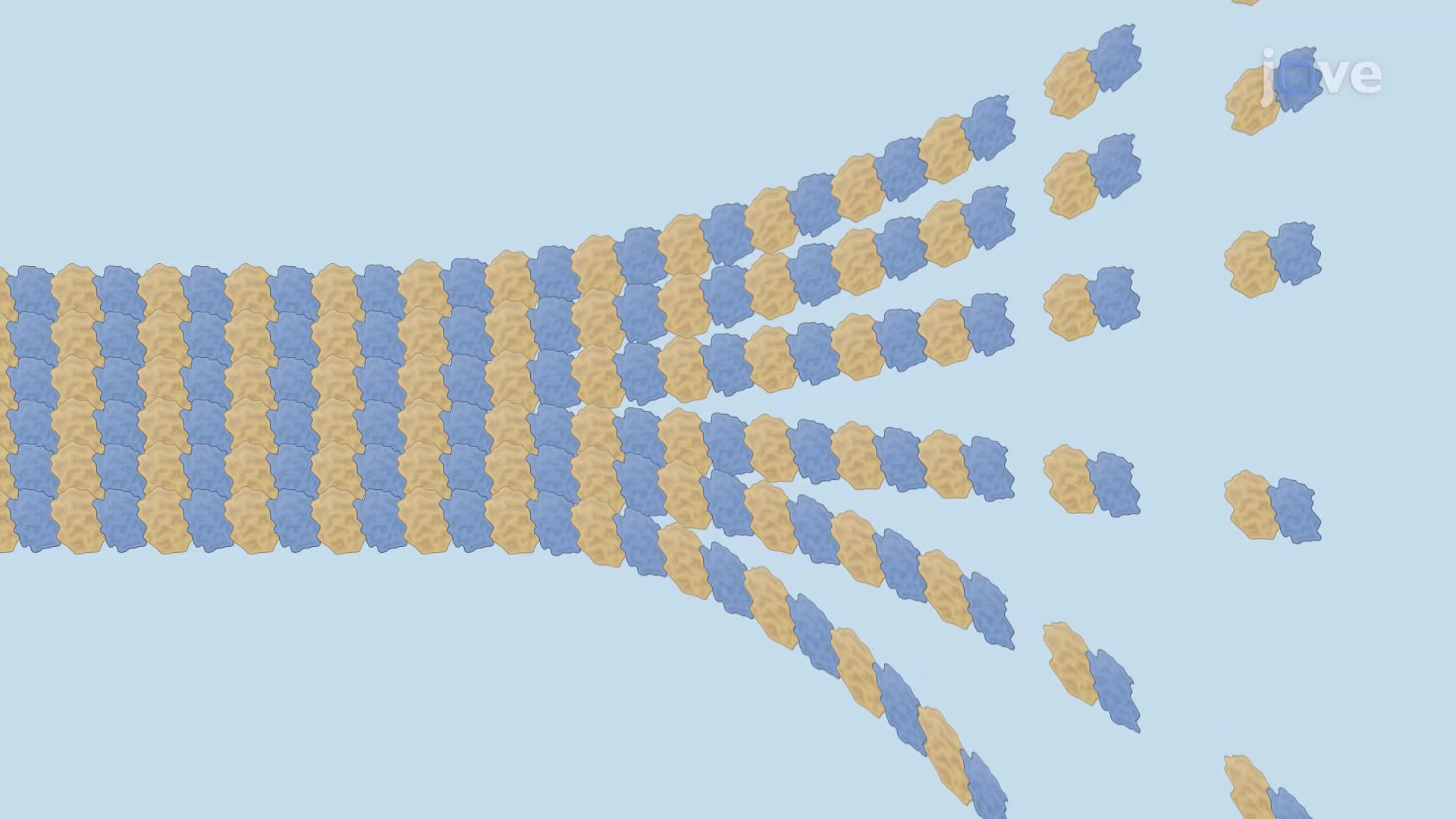

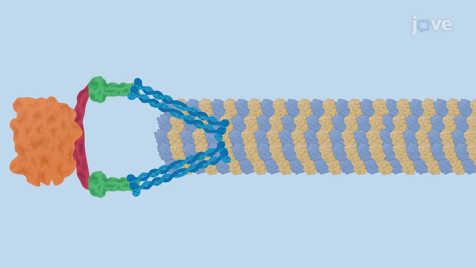


The Mitotic Spindle

00:00
02:27
Video Summary: What Is the Mitotic Spindle
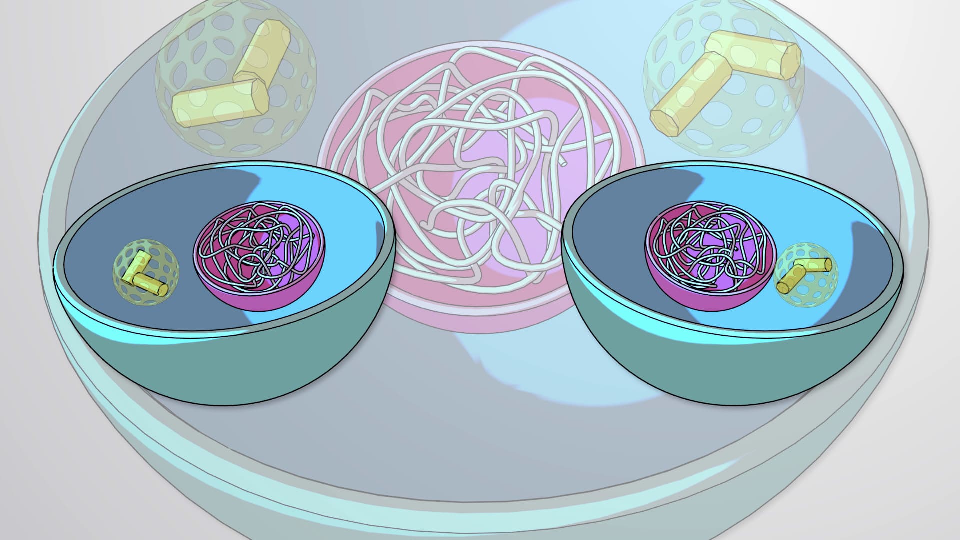
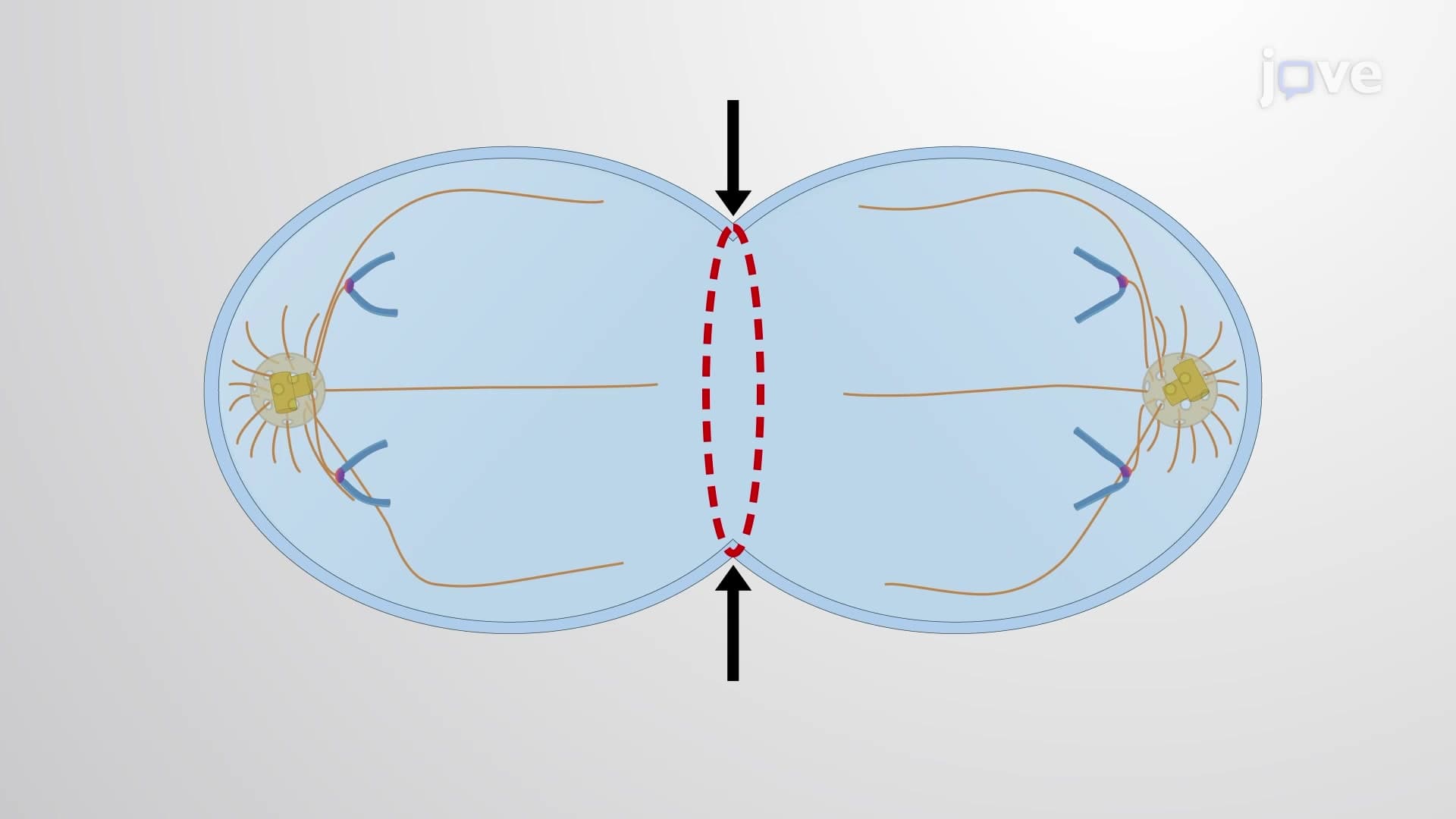
Every second, approximately 25 million cells in your body divide to replace old or damaged tissue. The mitotic spindle is the cellular machinery that ensures each new cell receives exactly the right number of chromosomes during this division process. Like the precisely choreographed assembly line at a Ford manufacturing plant in Michigan, this microscopic structure coordinates the separation and distribution of genetic material with remarkable accuracy. The mitotic spindle consists of protein filaments called microtubules that form a bipolar structure, pulling sister chromatids apart during cell division. Watch the full video on JoVE Coach to master this concept with expert-led visuals and step-by-step explanations.
- Understand how the mitotic spindle functions as the primary mechanism for chromosome separation during cell division
- Identify the three main types of microtubules that compose the spindle: kinetochore, interpolar, and astral microtubules
- Analyze the role of centrosomes in organizing and anchoring microtubules at each spindle pole
- Compare the functions of kinesin and dynein motor proteins in spindle assembly and chromosome movement
- Evaluate how the polarity of microtubules (minus and plus ends) facilitates directional chromosome transport
- Apply knowledge of kinetochore structure and function to explain how chromosomes attach to the spindle
- Recognize the importance of spindle checkpoint mechanisms in preventing chromosome missegregation
- Explore how spindle defects contribute to genetic disorders and cancer development
Understanding the Mitotic Spindle Architecture
The mitotic spindle represents one of biology's most sophisticated molecular machines, orchestrating the precise distribution of genetic material during cell division. This bipolar structure emerges during prophase and reaches full maturity by metaphase, consisting of hundreds of dynamic protein filaments working in perfect coordination.
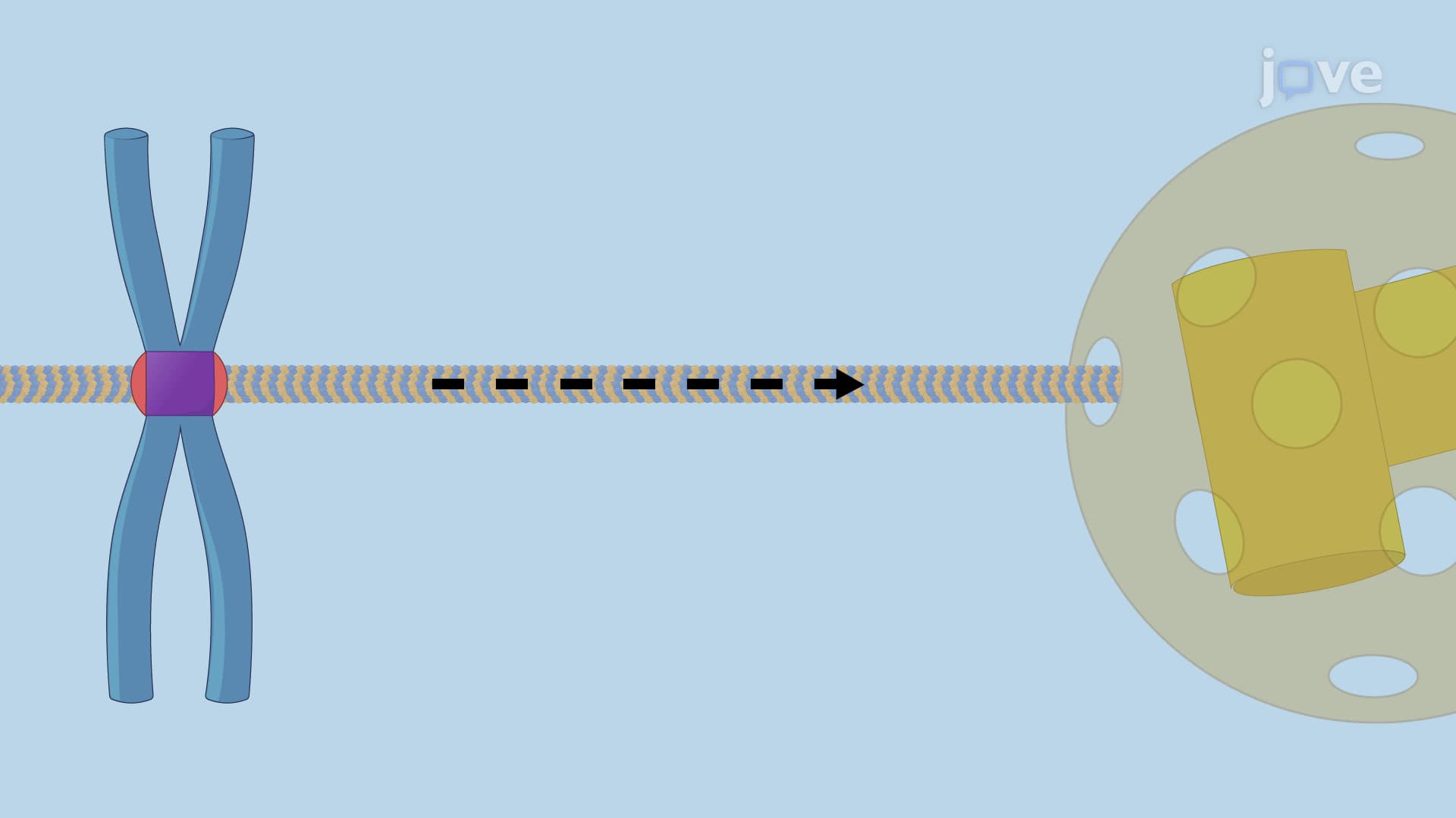
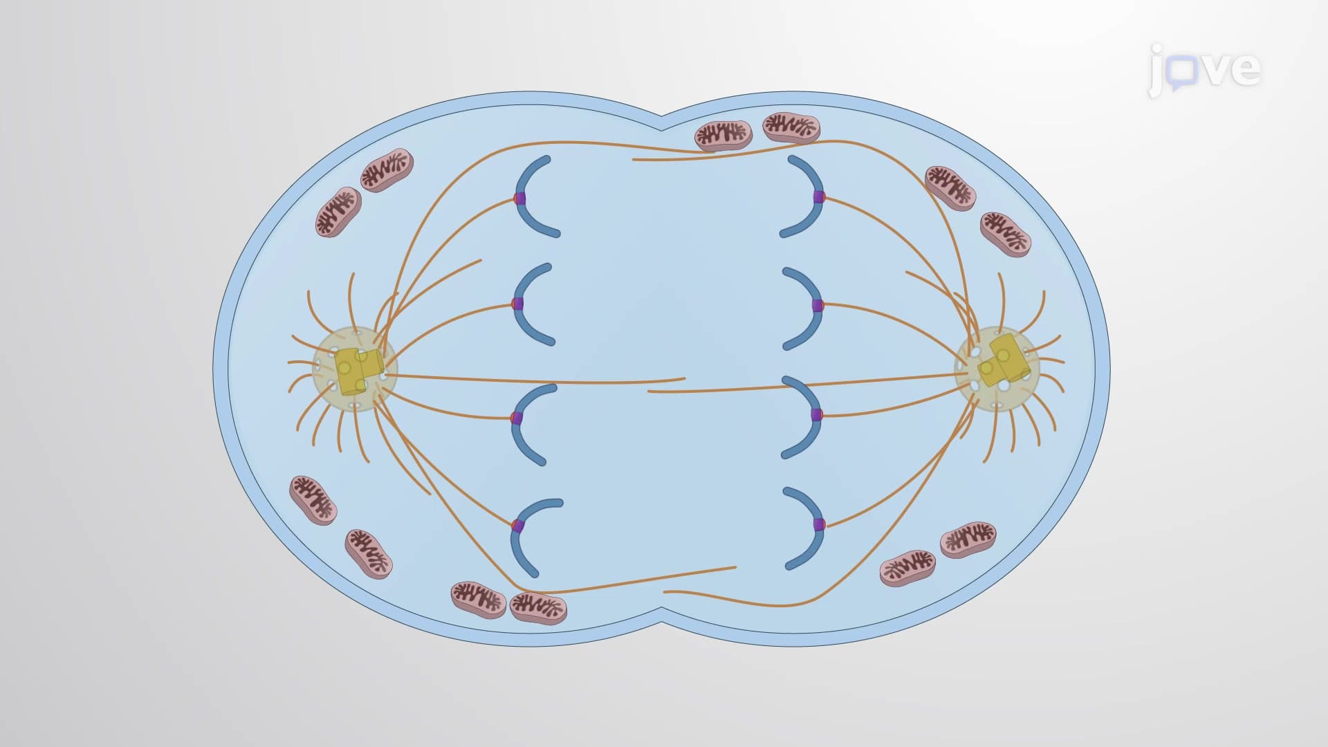
At its core, the spindle is built from microtubules-hollow cylindrical structures composed of tubulin proteins. These microtubules exhibit inherent polarity, with structurally distinct minus ends that anchor at spindle poles and plus ends that extend outward. This polarity is crucial for the directional movement of chromosomes and motor proteins.
Microtubule Organization and Centrosome Function
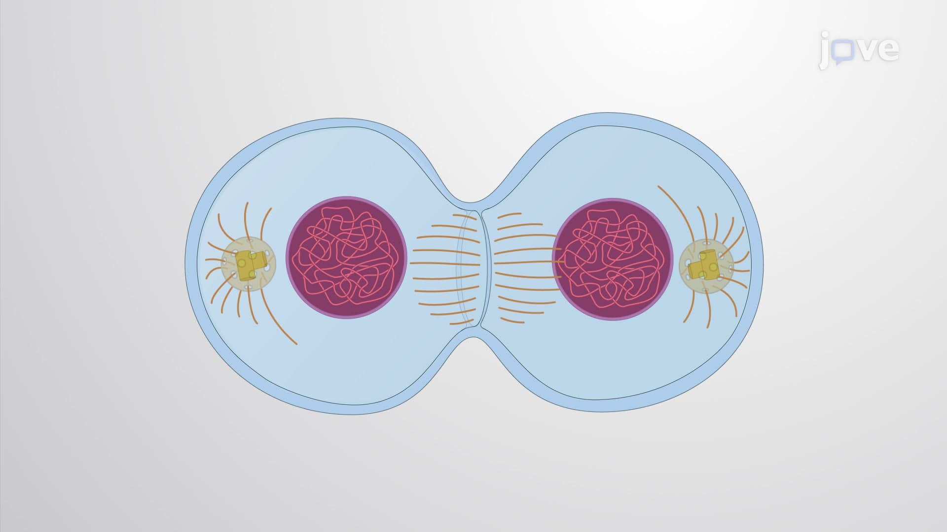
In animal cells, two centrosomes serve as the organizing centers for microtubule nucleation and spindle pole formation. Each centrosome contains a pair of centrioles embedded within the pericentriolar matrix, a protein-rich region that serves as the microtubule organizing center. During cell division, centrosomes duplicate and migrate to opposite sides of the cell, establishing the bipolar spindle framework.
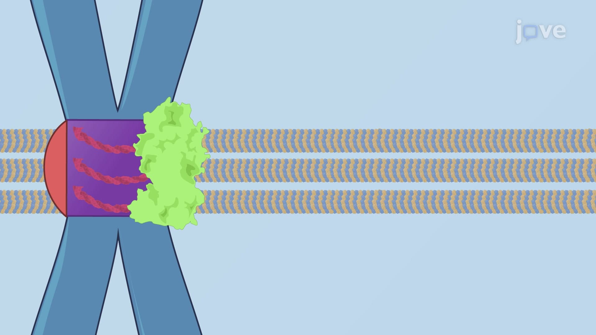
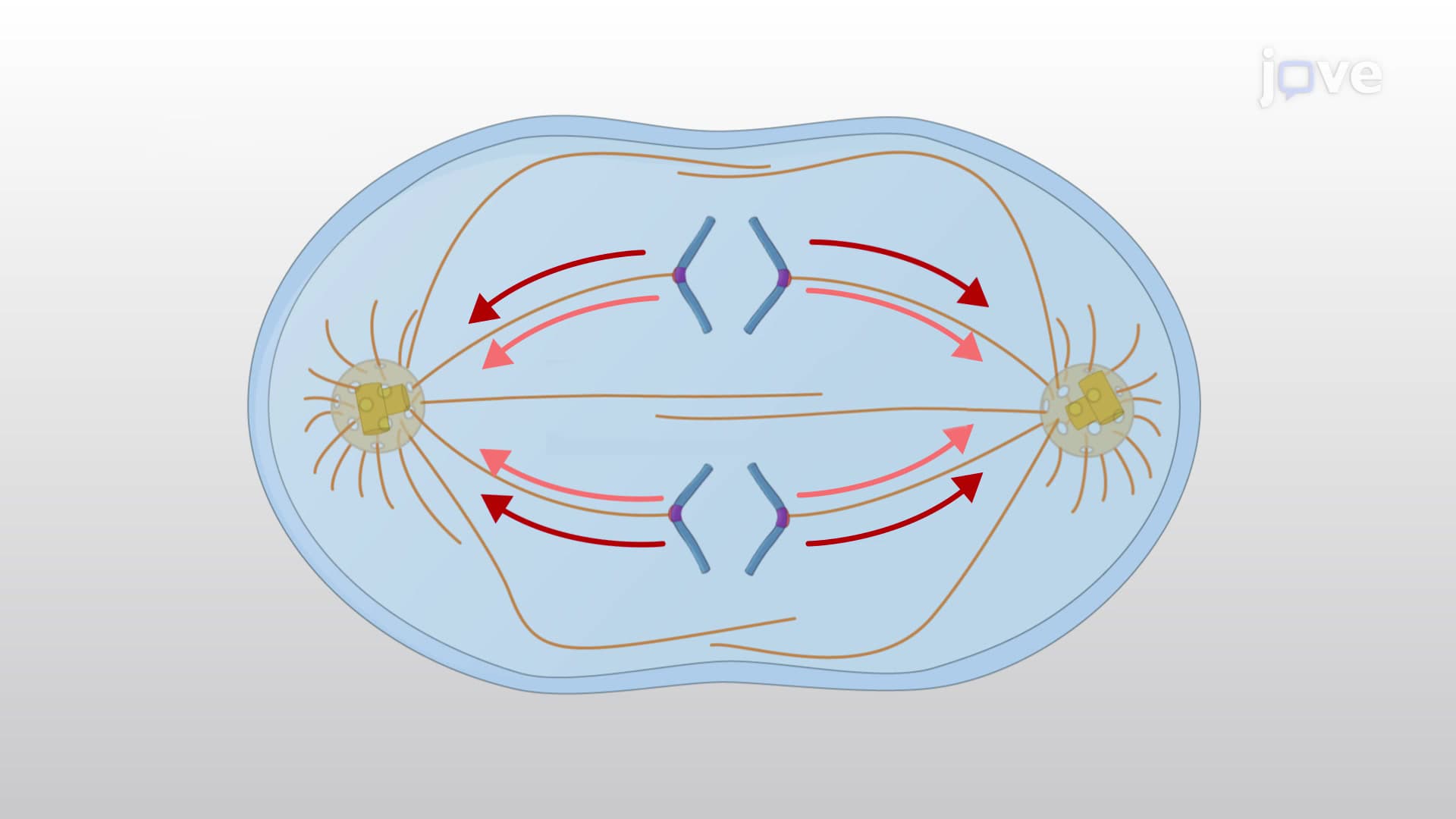
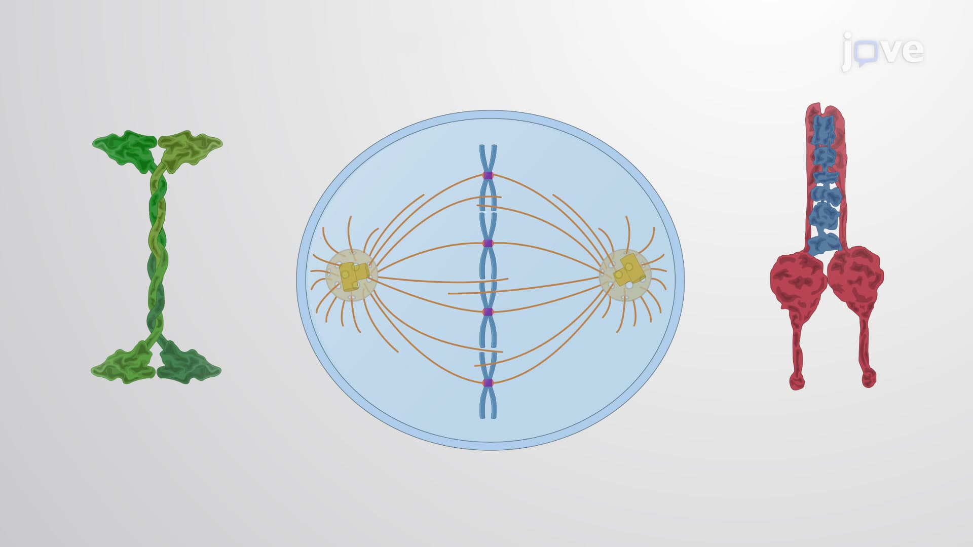
The three distinct classes of spindle microtubules each serve specific functions. Kinetochore microtubules directly connect chromosomes to spindle poles through protein complexes called kinetochores, which assemble at chromosome centromeres. Interpolar microtubules from opposite poles overlap at the spindle midzone, providing structural integrity and generating forces for spindle elongation. Astral microtubules extend toward the cell cortex, anchoring the spindle and helping position it within the cell.
Motor Proteins and Chromosome Movement
Two families of motor proteins power spindle dynamics and chromosome movement. Kinesin proteins typically move toward microtubule plus ends, driving processes like chromosome alignment and spindle pole separation. Dynein motors move toward minus ends, contributing to spindle pole focusing and chromosome oscillations. These opposing forces create the mechanical framework necessary for accurate chromosome segregation.
Students preparing for the AP Biology exam should understand how spindle defects can lead to aneuploidy-cells with abnormal chromosome numbers. The MCAT frequently tests spindle function in the context of cell cycle regulation and cancer biology. Medical students studying for the USMLE will encounter spindle-targeting drugs like taxanes and vinca alkaloids, which disrupt microtubule dynamics in cancer chemotherapy.
Clinical and Research Applications
Understanding spindle biology has direct implications for human health and disease. Spindle checkpoint defects contribute to chromosomal instability in many cancers, while certain genetic disorders result from mutations in spindle-associated proteins. Research institutions like the National Cancer Institute and major universities including Harvard Medical School and Johns Hopkins continue advancing our knowledge of spindle mechanisms for therapeutic development.
Frequently Asked Questions
The mitotic spindle is a dynamic protein structure that separates chromosomes during cell division, ensuring each daughter cell receives the correct genetic material. It consists of microtubules organized around centrosomes that capture and move chromosomes through precisely coordinated molecular mechanisms. Without a functional spindle, cells cannot divide properly, leading to genetic instability or cell death.
MCAT questions often focus on spindle function in cancer biology, drug mechanisms, and cell cycle checkpoints, requiring you to analyze experimental data or predict outcomes of spindle disruption. AP Biology exams emphasize the relationship between spindle structure and chromosome movement, frequently asking students to explain how microtubule polarity enables directional transport. Both exams may include questions about centrosome function and motor protein roles.
College exams typically test deeper mechanistic understanding, requiring you to explain molecular interactions between kinetochores, microtubules, and motor proteins. Focus on understanding the three microtubule types, their specific functions, and how their organization changes throughout mitosis. Many professors emphasize the connection between spindle defects and human disease, particularly cancer.
Major cancer centers like MD Anderson in Houston and Memorial Sloan Kettering in New York use spindle-targeting chemotherapy drugs such as paclitaxel and vincristine to treat various cancers. These medications disrupt microtubule dynamics, preventing cancer cells from completing division and ultimately causing cell death. Understanding spindle biology helps oncologists optimize treatment protocols and develop new therapeutic strategies.
The mitotic spindle builds on basic cell biology concepts you already know, like protein structure and organelle function, making it very approachable for motivated high school students. Start by understanding that it's essentially a molecular machine made of protein tubes that pull chromosomes apart. Most students find the visual aspects particularly helpful-think of it like a microscopic crane system moving genetic cargo.
Create detailed diagrams showing the three microtubule types and their specific attachments, then practice explaining each component's function out loud. Focus on understanding the cause-and-effect relationships: how centrosome positioning determines spindle orientation, how kinetochore attachment enables chromosome capture, and how motor protein activity drives chromosome movement. Many students find success connecting spindle function to familiar mechanical analogies.
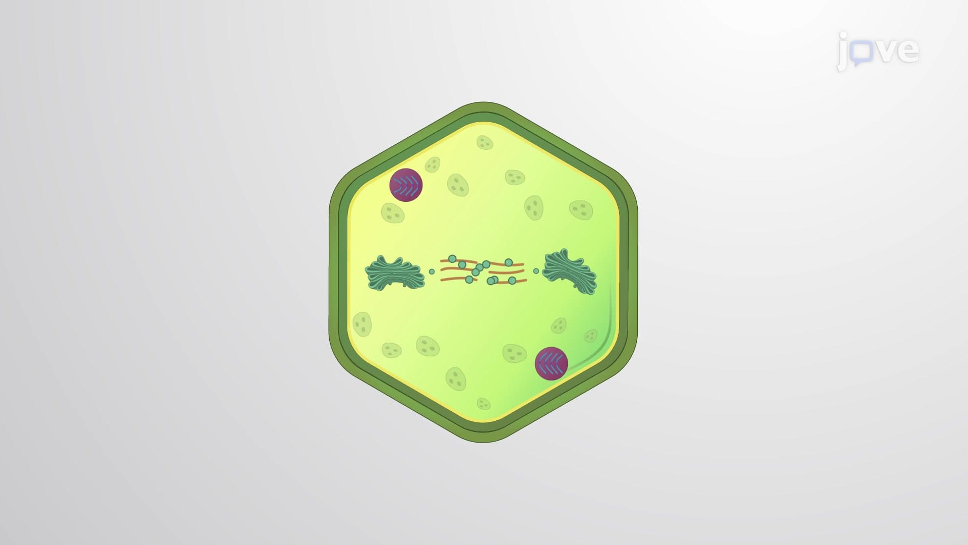
Consider studying spindle checkpoint mechanisms that ensure proper chromosome attachment before division proceeds, or explore how plant cells accomplish mitosis without centrosomes. Advanced students often investigate the role of spindle defects in genetic disorders or research how different organisms have evolved unique solutions to the chromosome segregation problem.
Related Micro-courses