Loading subject page…


14 Concepts
11 Concepts
29 Concepts
19 Concepts
24 Concepts
20 Concepts
13 Concepts
10 Concepts
12 Concepts
12 Concepts
7 Concepts
6 Concepts
8 Concepts
6 Concepts
8 Concepts
8 Concepts
8 Concepts
6 Concepts
9 Concepts
8 Concepts
8 Concepts
10 Concepts
7 Concepts
5 Concepts
16 Concepts
 12 Concepts
 21.2K Views
 Popular topics


Cardiovascular drugs: antiarrhythmic and heart failure drugs represent critical therapeutic interventions for managing cardiac rhythm disorders and compromised heart function. This comprehensive course explores the electrophysiology of normal cardiac rhythm, mechanisms underlying arrhythmias, and the four classes of antiarrhythmic agents. Additionally, it covers heart failure pathophysiology and therapeutic approaches including inotropic agents, diuretics, and RAAS inhibitors, essential knowledge for US healthcare professionals and students preparing for medical examinations with JoVE Coach.
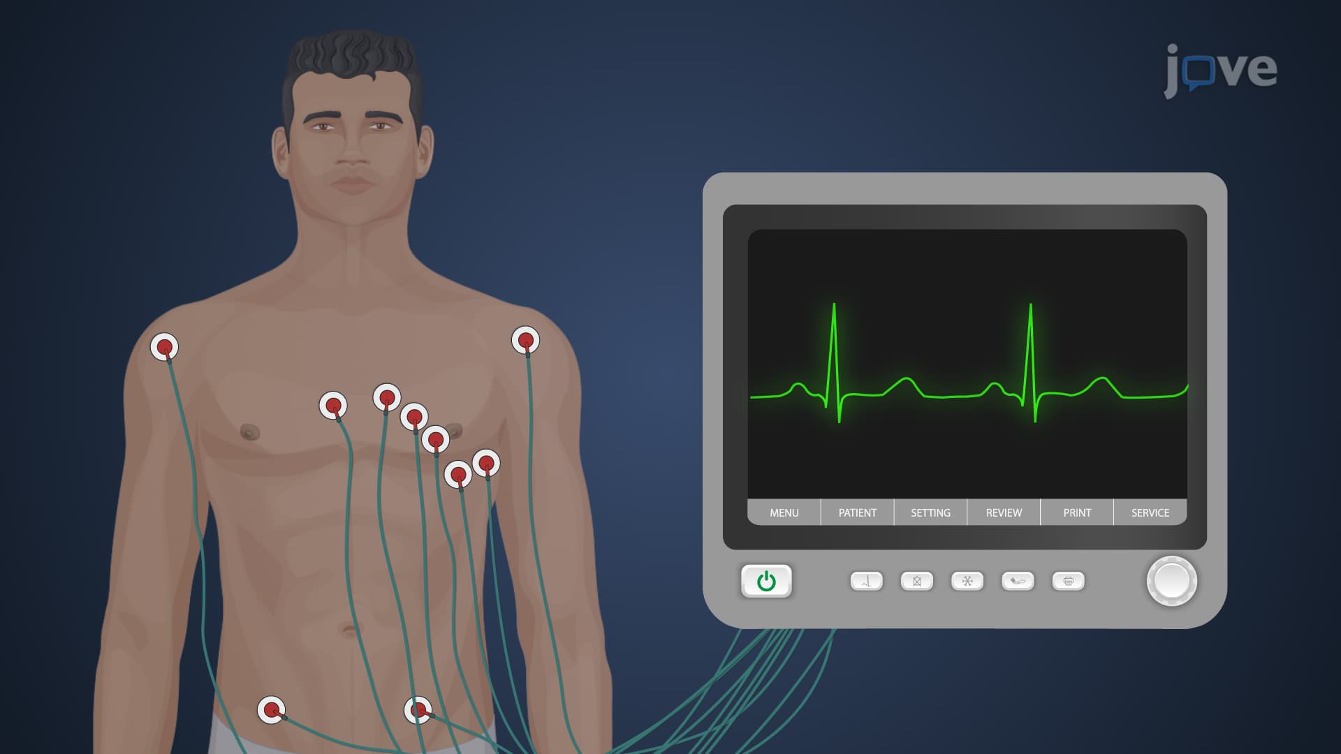
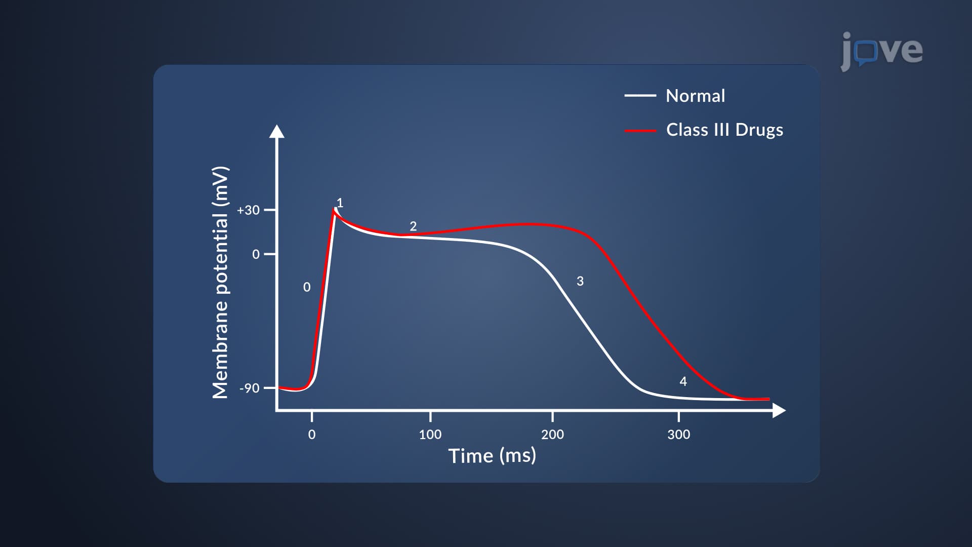
1. Cardiac Electrophysiology and Normal Rhythm: The heart's electrical system begins with the sinoatrial (SA) node, the natural pacemaker containing specialized non-contractile cells. Electrical impulses travel through the atrioventricular (AV) node to coordinate ventricular contraction. Cardiac muscle cells generate a five-phase action potential: Phase 0 involves rapid sodium influx causing depolarization, Phase 1 shows partial repolarization, Phase 2 creates a calcium-mediated plateau, Phase 3 involves potassium efflux for repolarization, and Phase 4 represents the resting potential. This coordinated electrical activity ensures effective blood circulation throughout the body, detectable through electrocardiogram (ECG) monitoring in clinical settings.
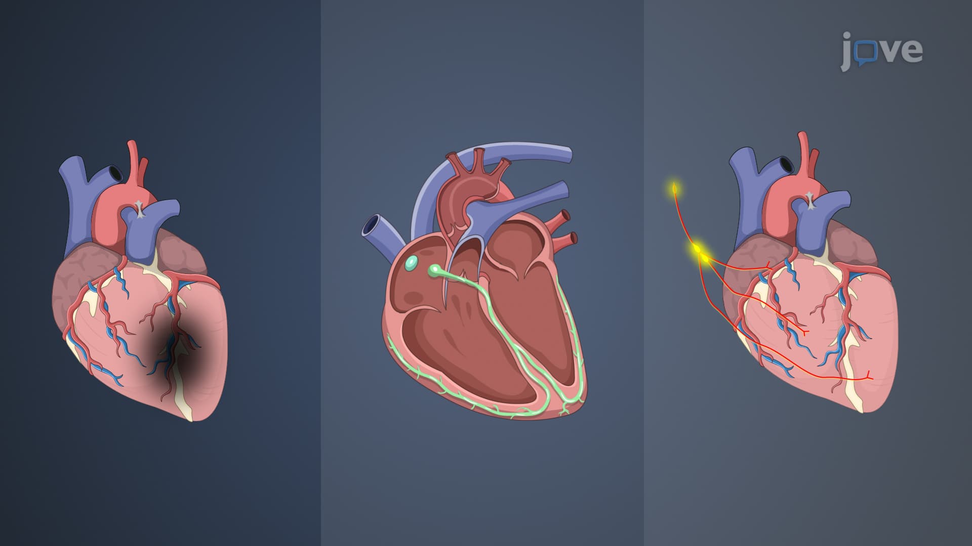
2. Mechanisms of Cardiac Arrhythmias: Arrhythmias result from abnormal impulse formation or defective cardiac conduction, manifesting as bradyarrhythmias (slow heart rate) or tachyarrhythmias (rapid heart rate). Common mechanisms include ectopic pacemaker activity where impulses originate from sites other than the SA node, enhanced automaticity due to elevated intracellular calcium levels, and reentry circuits caused by myocardial damage altering normal conduction pathways. These conditions can lead to symptoms like palpitations, dizziness, and syncope. In US emergency departments, ECG analysis remains the primary diagnostic tool for identifying arrhythmic patterns and guiding appropriate treatment strategies.
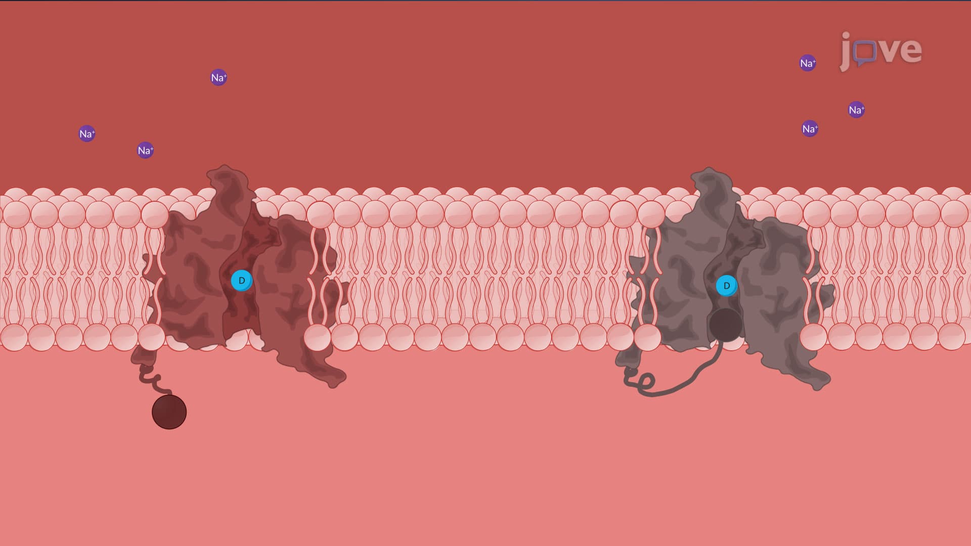
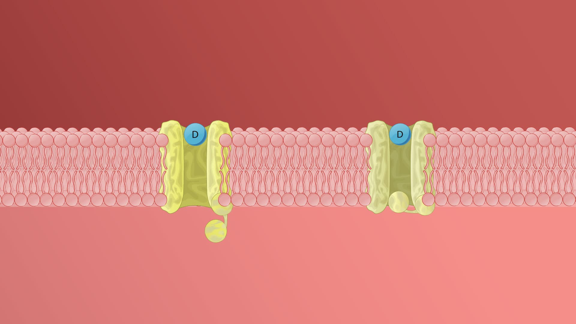
3. Class I Antiarrhythmic Drugs - Sodium Channel Blockers: Class I agents block voltage-sensitive sodium channels in continuously depolarizing tissues, preventing auto-excitation and hindering action potential propagation. They are subdivided based on dissociation kinetics: Class IA drugs (like quinidine) have intermediate kinetics and prolong action potential duration; Class IB drugs (like lidocaine) have fast dissociation and shorten action potential duration; Class IC drugs (like flecainide) have slow dissociation without affecting duration. All Class I drugs carry proarrhythmic risks and can cause delayed conduction, negative inotropy, and potential tachycardia. US physicians must carefully monitor patients for these adverse effects during treatment.
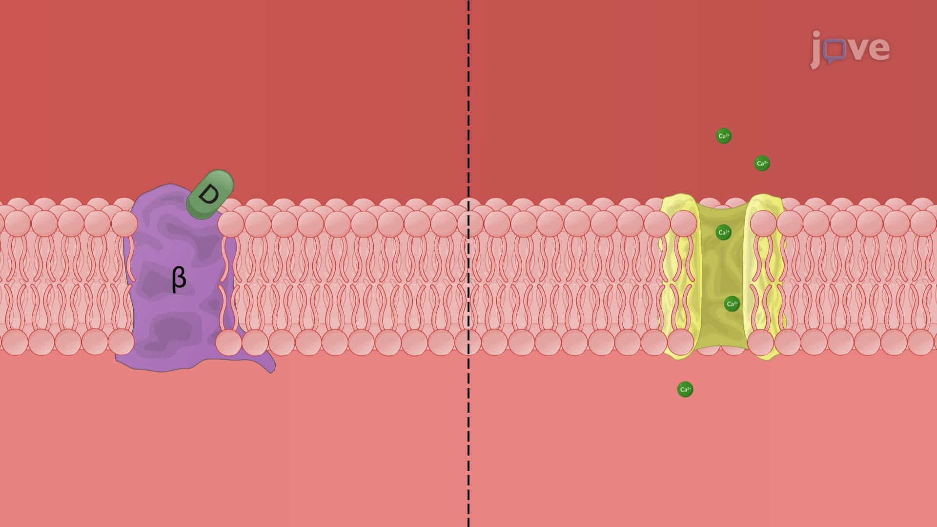
4. Class II Antiarrhythmic Drugs - β-Adrenergic Blockers: Class II drugs are β-adrenoceptor antagonists that indirectly block calcium channels by counteracting sympathetic stimulation. They primarily depress Phase 4 depolarization, reducing automaticity, prolonging AV conduction, and decreasing heart rate and contractility. Examples include propranolol (non-selective), metoprolol (β1-selective), esmolol (short-acting IV), and acebutolol (for ventricular ectopic beats). These medications vary in selectivity and intrinsic sympathomimetic activity. Common adverse effects include fatigue, bradycardia, bronchospasm, and hypotension. US healthcare providers must avoid abrupt discontinuation to prevent dangerous rebound arrhythmias, particularly in patients with underlying coronary artery disease.
5. Class III and IV Antiarrhythmic Drugs: Class III agents primarily block potassium channels, prolonging action potential and refractory periods without affecting resting membrane potential. Amiodarone, the most widely used Class III drug, has multiple mechanisms but causes significant toxicity including pulmonary fibrosis and thyroid dysfunction due to its iodine content. Class IV drugs block L-type calcium channels in a use-dependent manner, slowing SA and AV node conduction. Verapamil and diltiazem are primary examples, commonly used for supraventricular tachycardia and rate control in atrial fibrillation. US cardiologists often prefer these agents for specific arrhythmias while monitoring for bradycardia and hypotension.
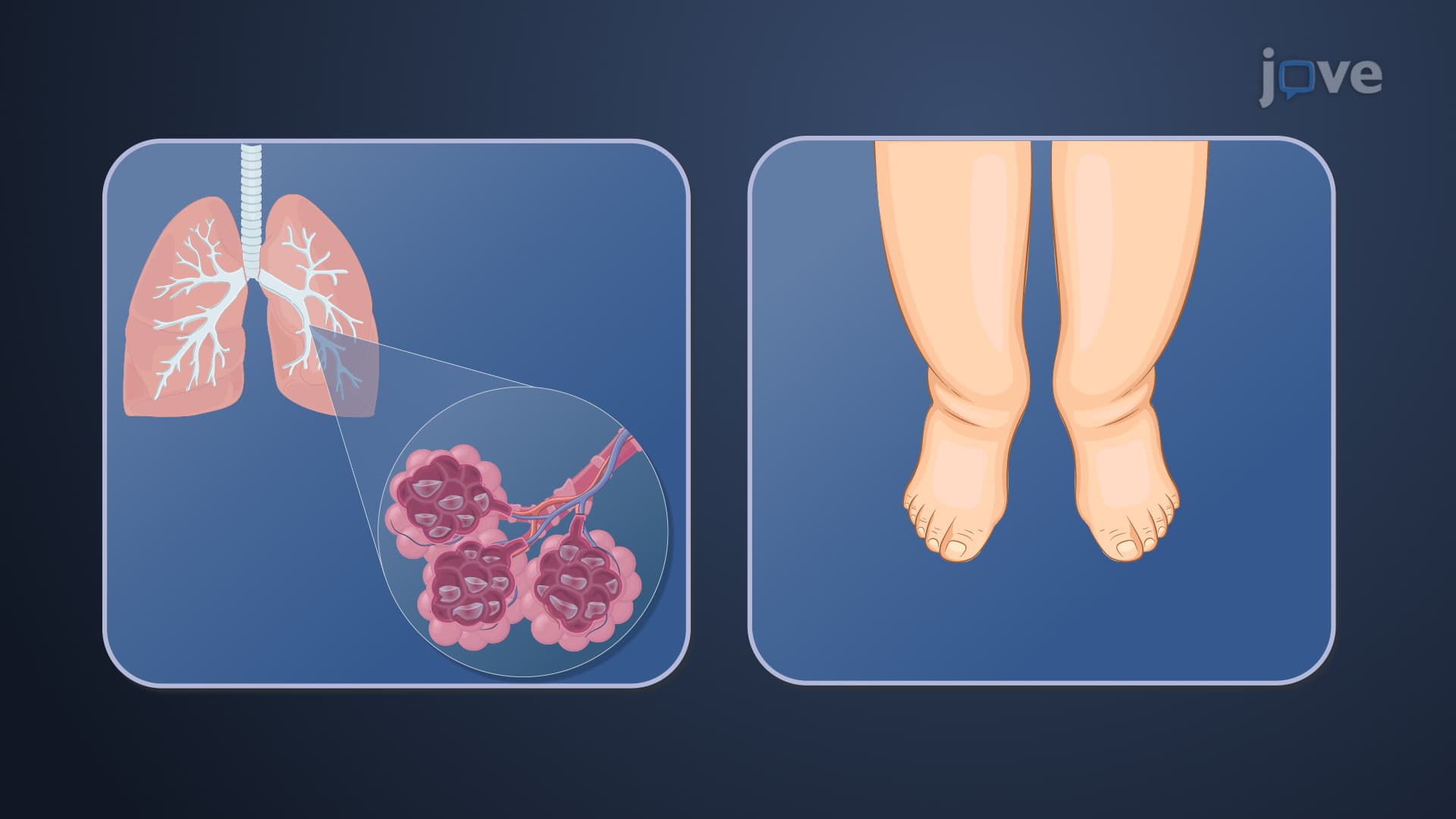
6. Heart Failure Pathophysiology and Compensatory Mechanisms: Heart failure involves the heart's inability to pump blood effectively to meet metabolic demands. The pathophysiology includes decreased cardiac output triggering compensatory cardiac remodeling with myocyte hypertrophy, fibroblast proliferation, and collagen deposition. These changes paradoxically worsen heart function through apoptosis and necrosis. Neurohumoral activation of the sympathetic nervous system and renin-angiotensin-aldosterone system (RAAS) initially compensates but ultimately creates a vicious cycle. In the US, heart failure affects over 6 million adults, with hospitalization rates exceeding 1 million annually, making understanding these mechanisms crucial for healthcare providers.
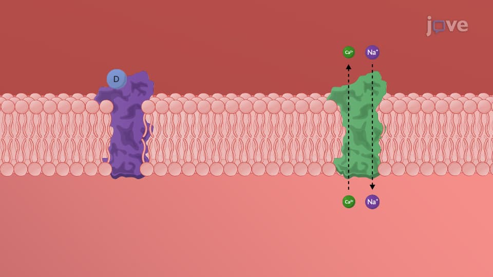
7. Inotropic Agents and Diuretics in Heart Failure: Positive inotropic agents like cardiac glycosides (digoxin) increase cardiac contractility by inhibiting the Na+/K+ pump, raising intracellular calcium levels. These drugs improve cardiac output and renal perfusion but have a narrow therapeutic window requiring careful monitoring of serum levels and electrolytes. Diuretics treat fluid overload symptoms by increasing sodium excretion: loop diuretics (furosemide) are most potent, thiazide diuretics provide moderate effects, and potassium-sparing diuretics prevent hypokalemia. US heart failure guidelines recommend loop diuretics as first-line therapy for volume management, with combination therapy often needed in refractory cases.
8. RAAS Inhibitors and β-Blockers in Heart Failure Management: ACE inhibitors block angiotensin II formation, reducing cardiac afterload and preventing harmful remodeling. ARBs provide similar benefits by blocking AT1 receptors and are used when patients cannot tolerate ACE inhibitors due to cough. Mineralocorticoid receptor antagonists (spironolactone, eplerenone) provide additional mortality benefits by blocking aldosterone effects. β-blockers like carvedilol and metoprolol succinate counteract harmful sympathetic overstimulation, reducing heart rate and myocardial oxygen demand. US heart failure guidelines recommend these evidence-based therapies as standard of care, with careful dose titration required to optimize benefits while minimizing adverse effects in clinical practice.