Loading video…


 29 Concepts
 135.5K views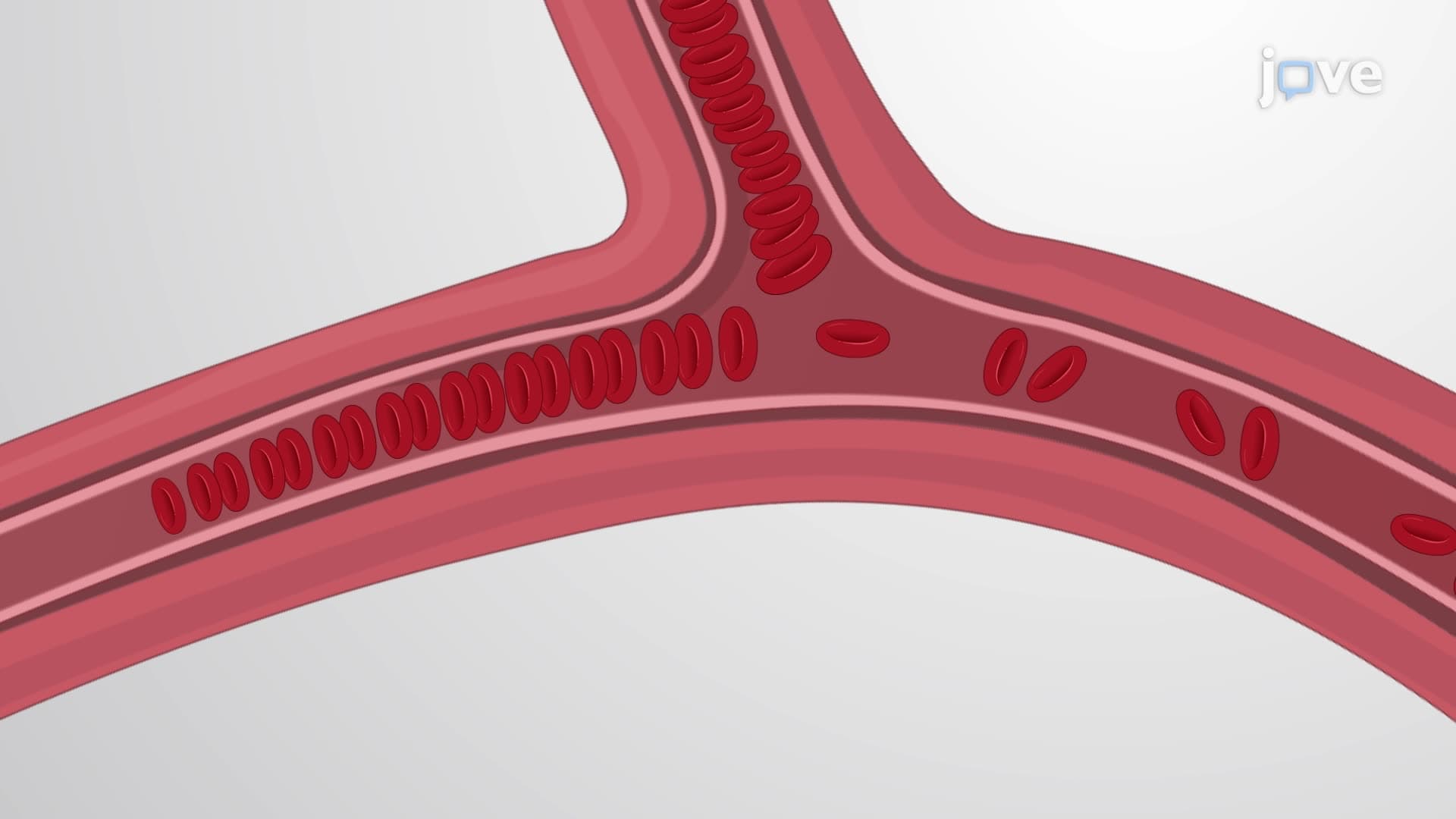

Video Summary: Structure and Function of Erythrocytes Explained
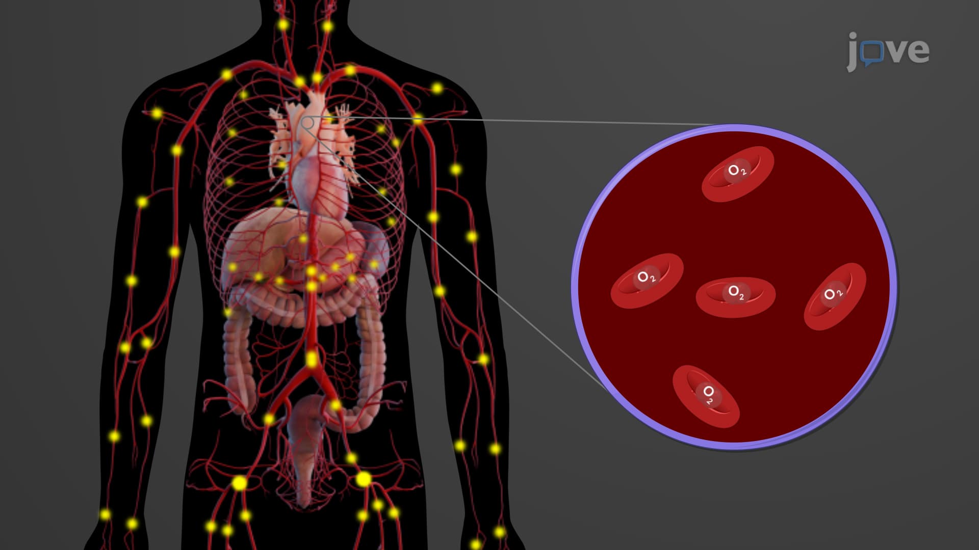
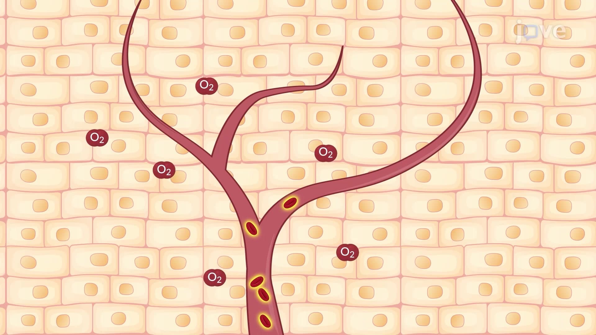
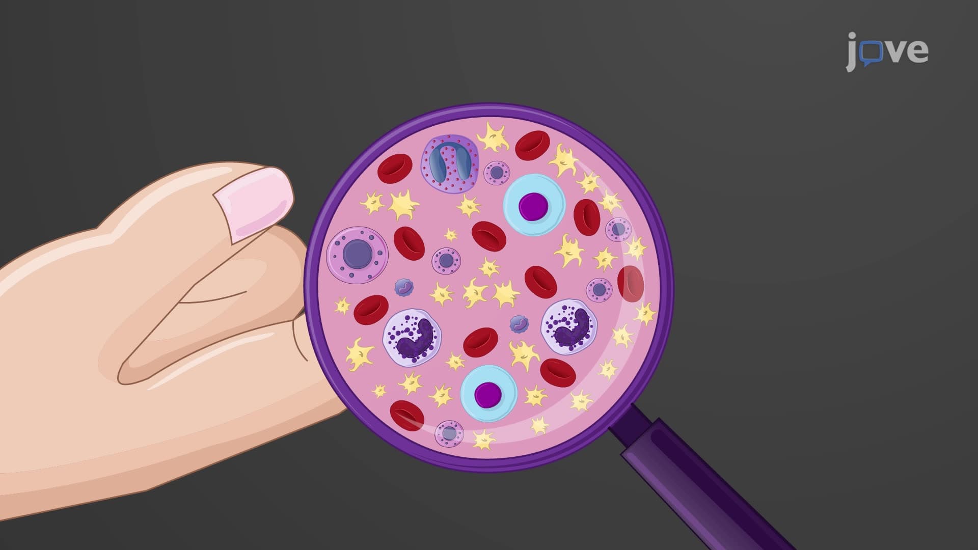
Every drop of your blood contains millions of specialized cells working as oxygen delivery trucks throughout your body. Understanding erythrocyte structure function biology reveals how these remarkable red blood cells achieve 99% efficiency in oxygen transport. During a typical blood donation at American Red Cross centers, approximately 450 mL of blood contains over 2 trillion erythrocytes, each perfectly designed for gas exchange. Their unique biconcave shape and hemoglobin-packed interior make them nature's most efficient oxygen carriers. Watch the full video on JoVE Coach to master this concept with expert-led visuals and step-by-step explanations.
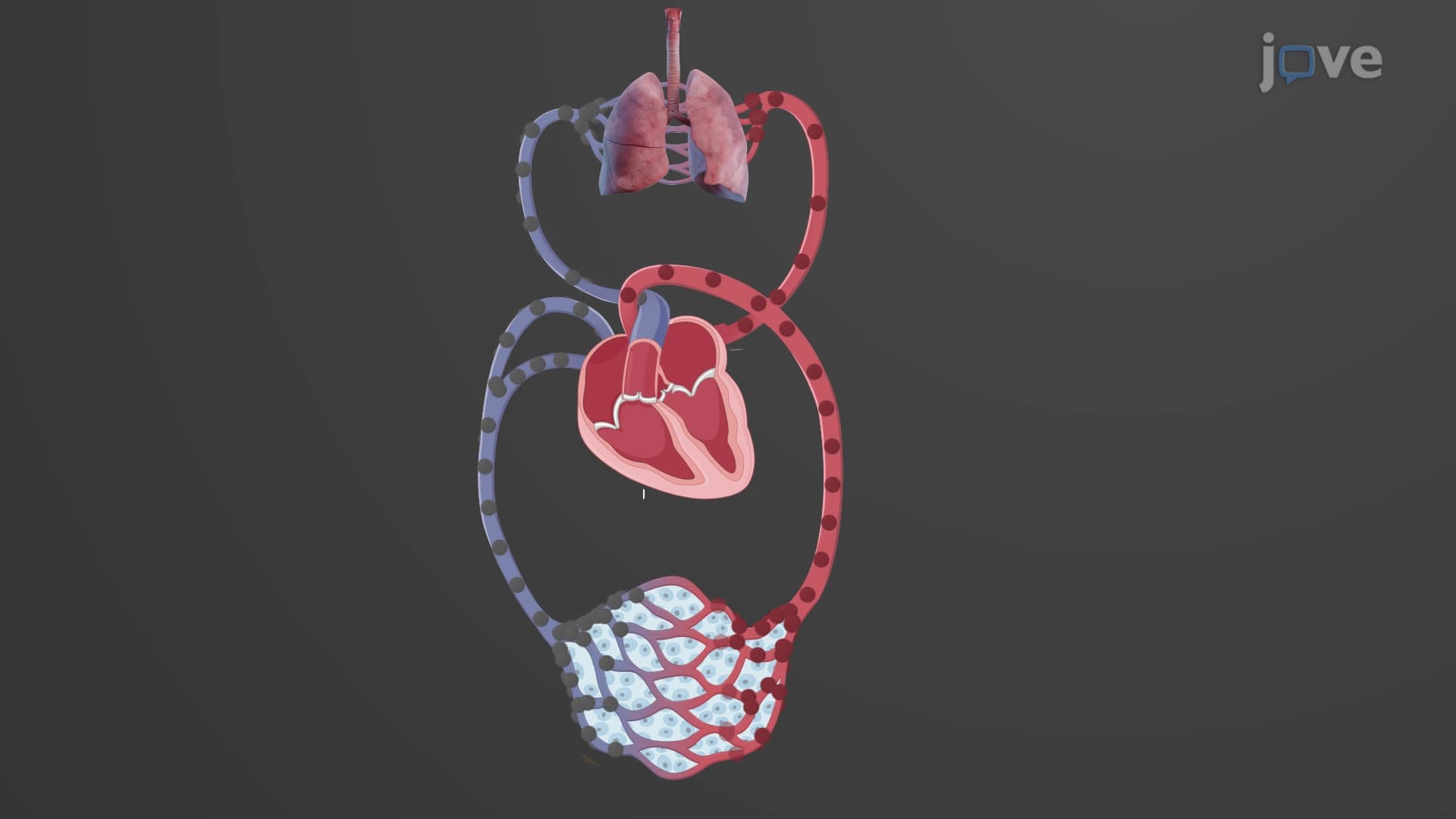
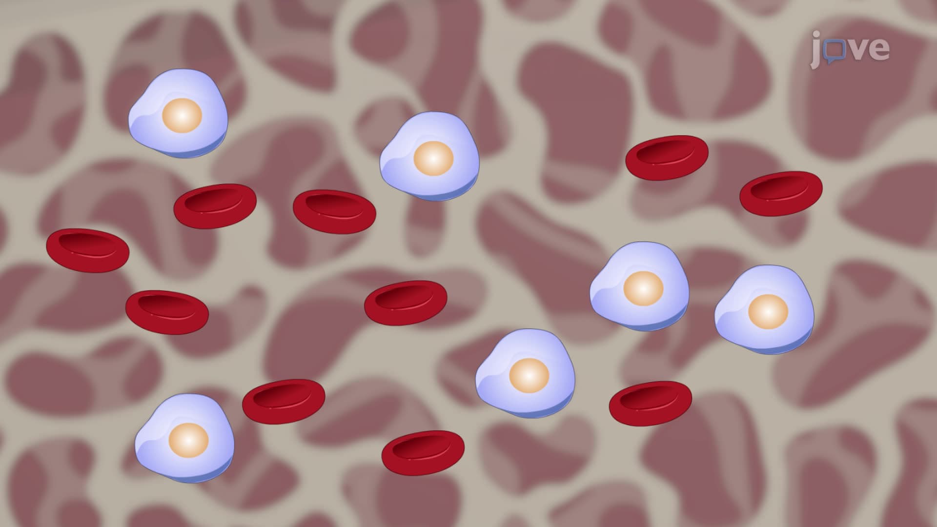
Red blood cells represent one of biology's most specialized cellular designs. With 4.2-6 million erythrocytes per microliter, these cells must function flawlessly to maintain tissue oxygenation. Their evolution reflects millions of years of optimization for a single critical task: efficient gas transport throughout the circulatory system.
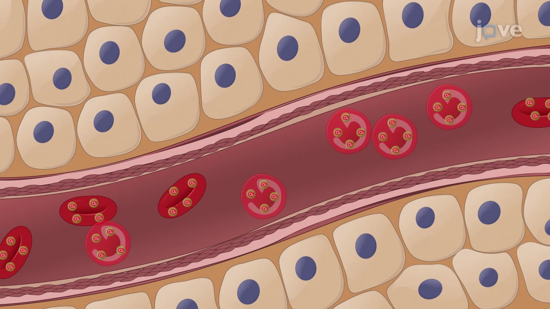
The distinctive biconcave disc shape isn't merely aesthetic-it's functional engineering. This flattened, donut-like structure without a central hole increases surface area by approximately 30% compared to spherical cells of equivalent volume. For students preparing for AP Biology or college physiology exams, this concept frequently appears in questions about structure-function relationships. The increased surface area positions hemoglobin molecules closer to the cell membrane, reducing diffusion distance for oxygen and carbon dioxide exchange.
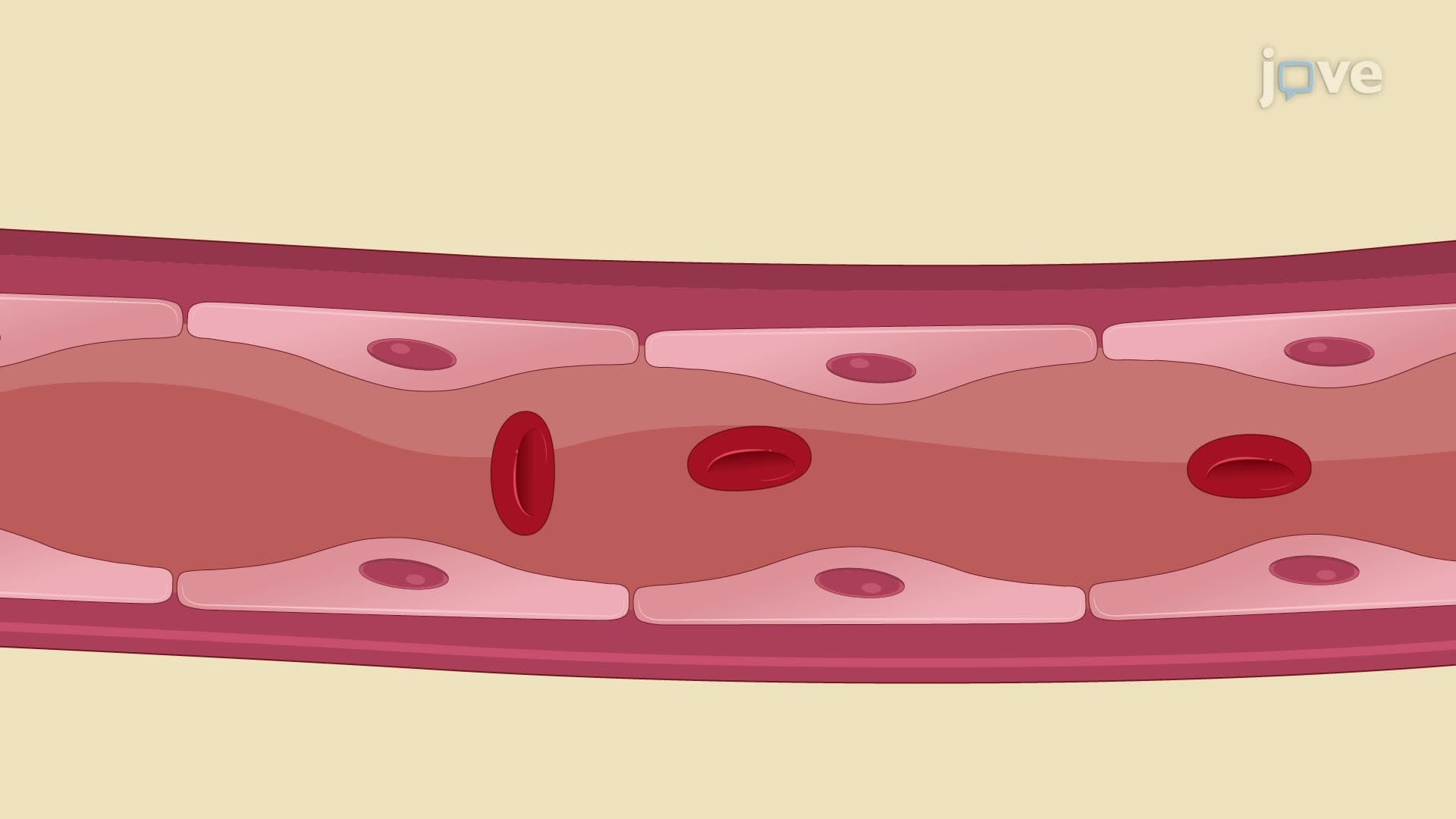
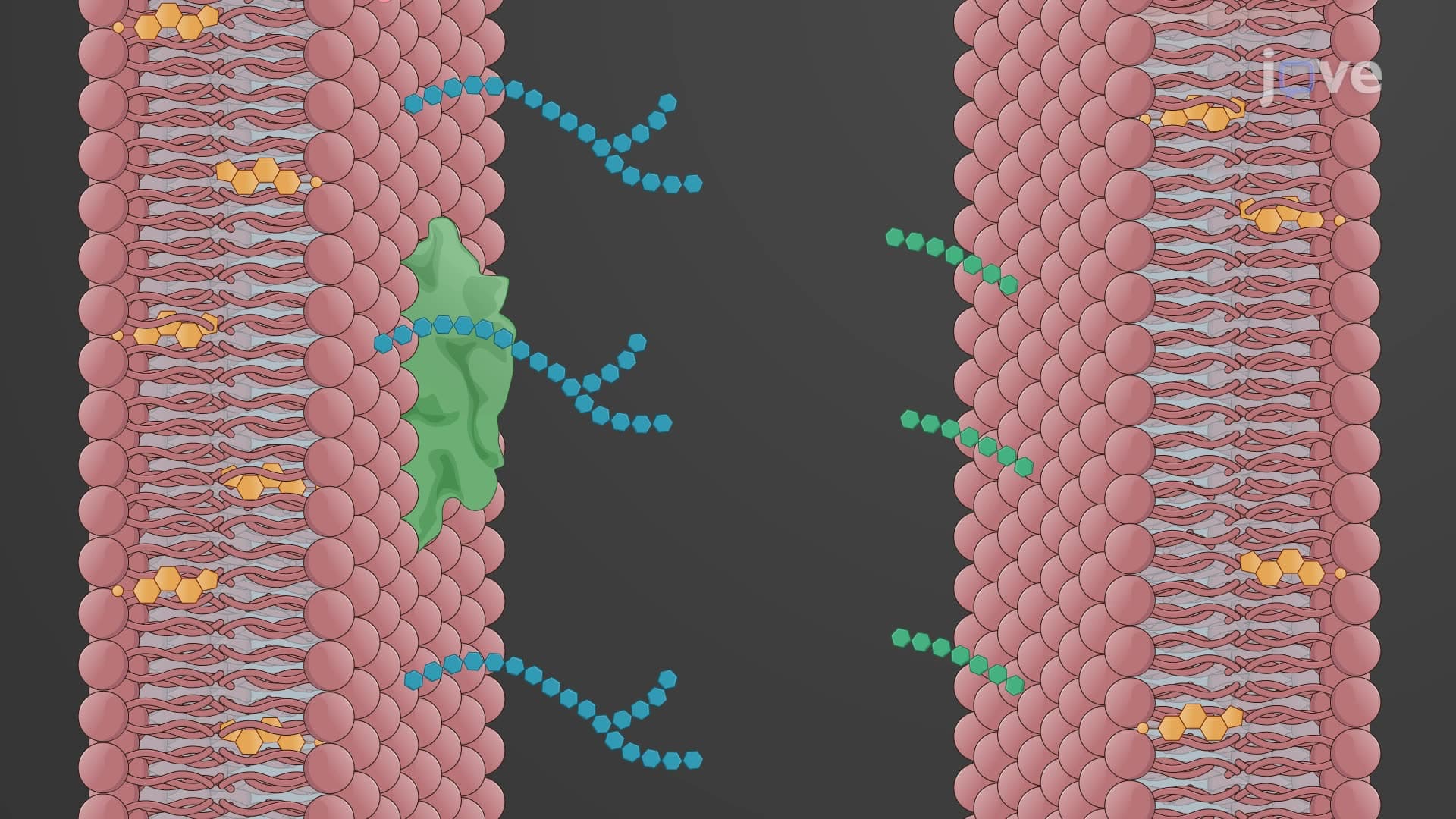
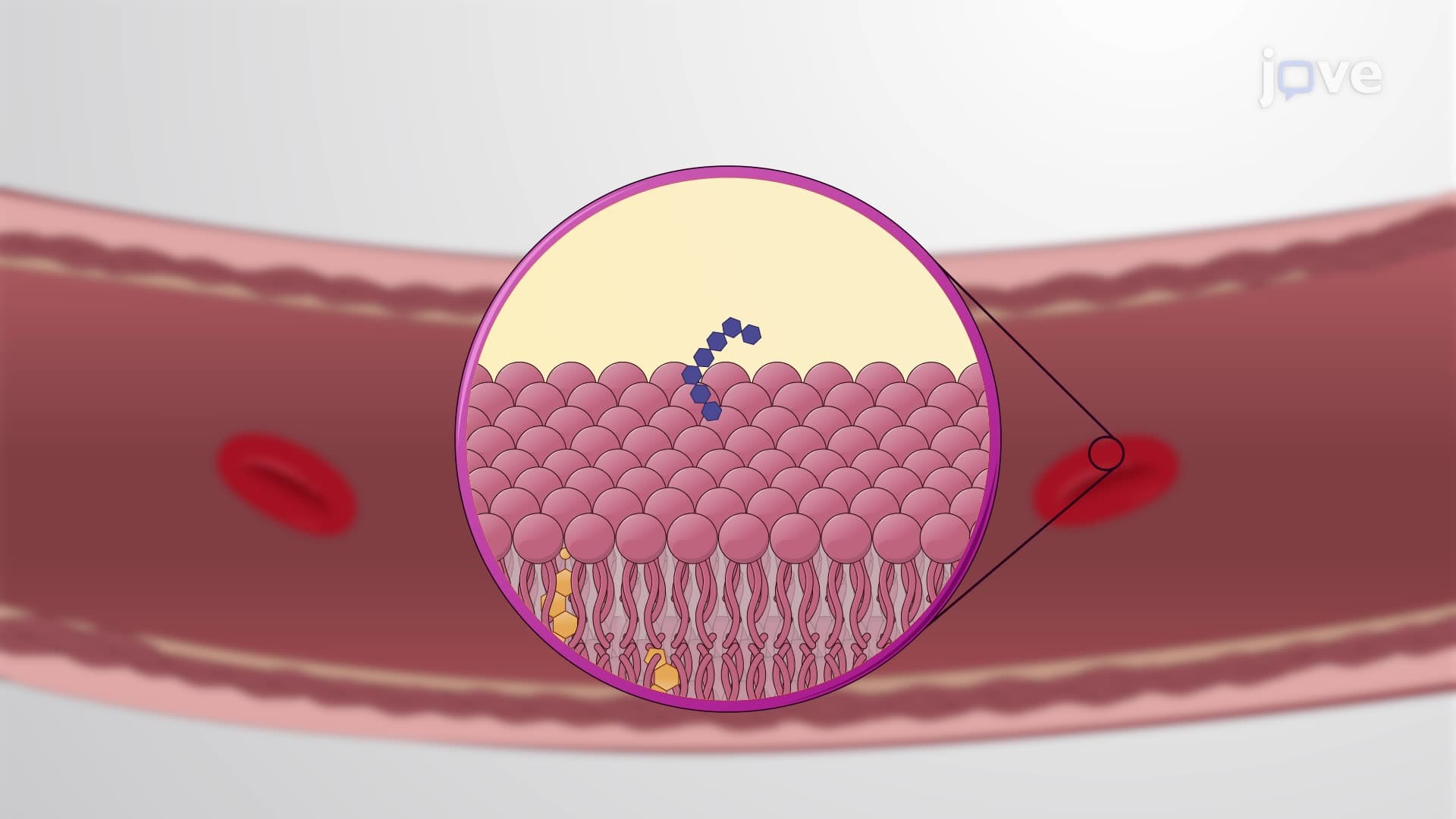
Erythrocyte membranes contain specialized proteins, particularly spectrin, forming a flexible cytoskeletal network beneath the lipid bilayer. This protein meshwork allows red blood cells to deform dramatically-squeezing through capillaries only 2-3 micrometers in diameter while maintaining structural integrity. Medical students studying for the MCAT often encounter questions about this flexibility, especially regarding conditions like hereditary spherocytosis where spectrin defects cause membrane rigidity.
Unlike most human cells, mature erythrocytes lack nuclei and organelles-a strategic trade-off maximizing internal space for hemoglobin. This design creates room for approximately 280 million hemoglobin molecules per cell, constituting 97% of cytosolic proteins. Without mitochondria, erythrocytes rely entirely on anaerobic glycolysis for ATP production, consuming no oxygen themselves. This metabolic strategy appears frequently on USMLE Step 1 examinations, particularly in questions about cellular respiration and oxygen transport efficiency.
The absence of organelles also means erythrocytes cannot synthesize new proteins or repair themselves, limiting their lifespan to approximately 120 days. Students studying hematology for nursing entrance exams like HESI A2 should understand this concept when learning about erythrocyte turnover and anemia pathophysiology.
Related Micro-courses