Loading subject page…


135,527 views

Video Summary: Structure and Function of Platelets Explained
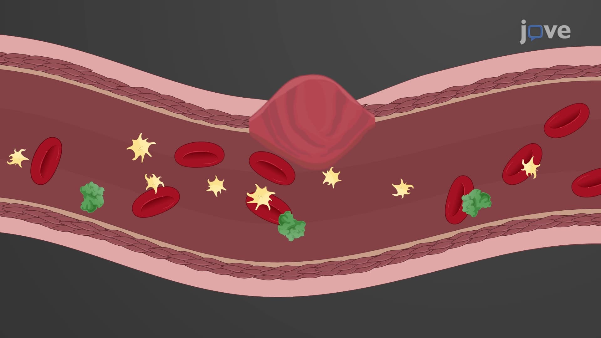
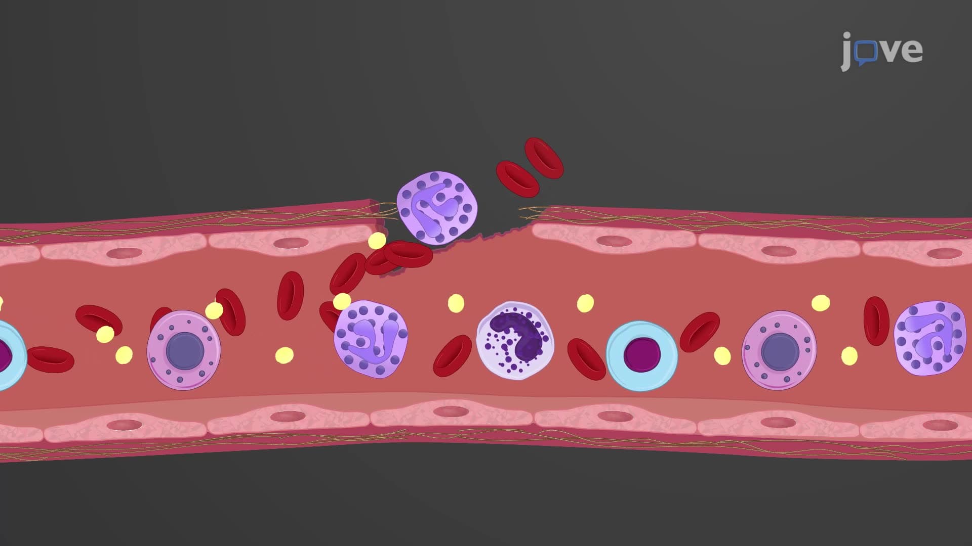
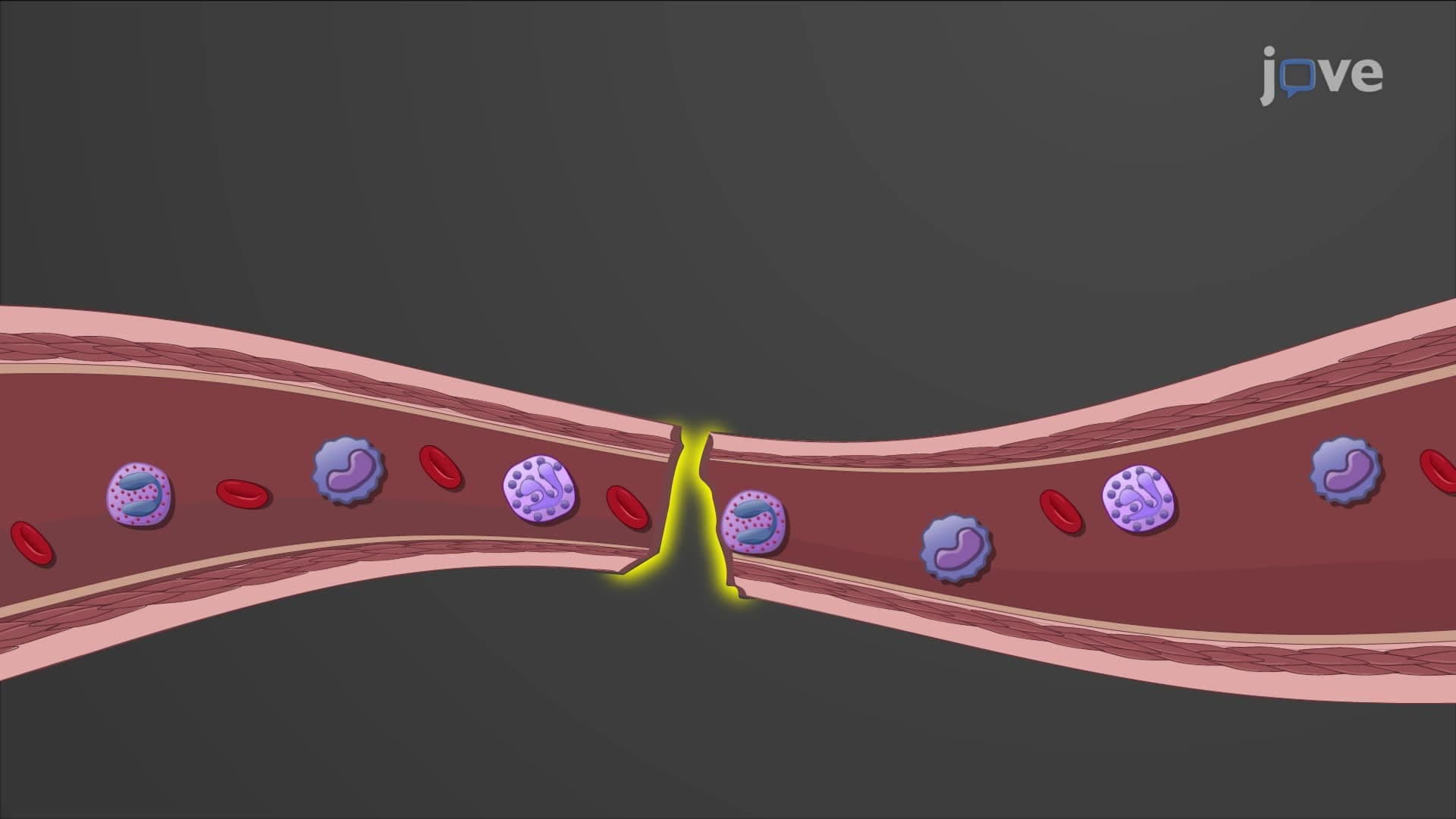
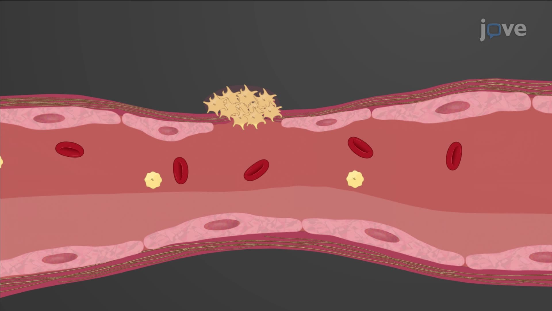
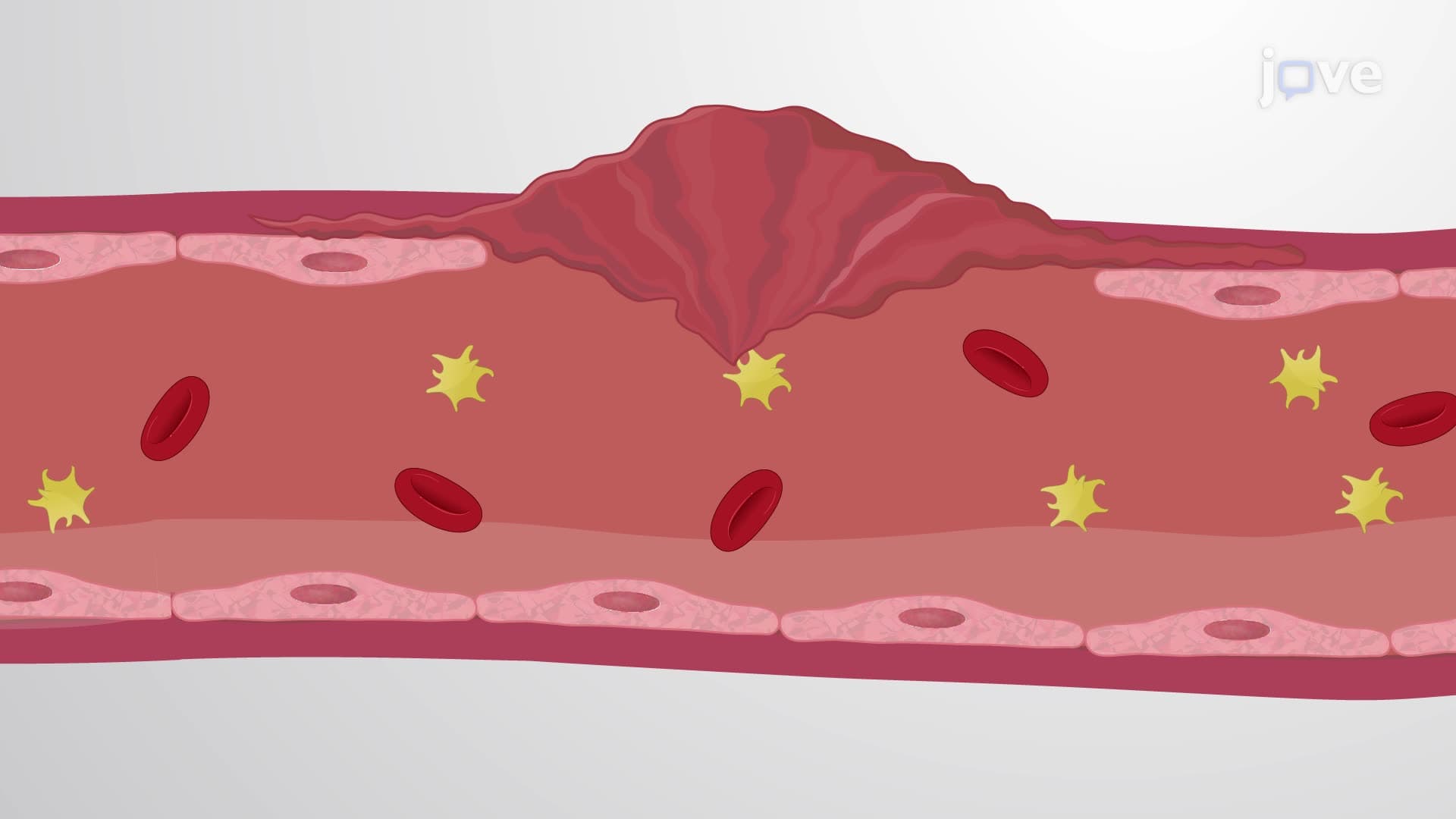
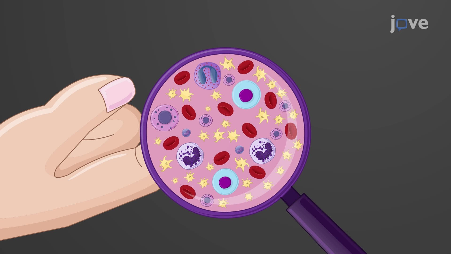
Every time you get a paper cut, millions of tiny cellular fragments called platelets rush to seal the wound within seconds-this remarkable platelet structure function biology showcases one of your body's most critical survival mechanisms. These disc-shaped cell fragments, measuring just 2-4 micrometers, originate from massive bone marrow cells and contain specialized granules packed with clotting factors. Understanding the Structure And Function of Platelets Explained reveals how these nucleus-free fragments can rapidly form life-saving plugs at injury sites. Watch the full video on JoVE Coach to master this concept with expert-led visuals and step-by-step explanations.
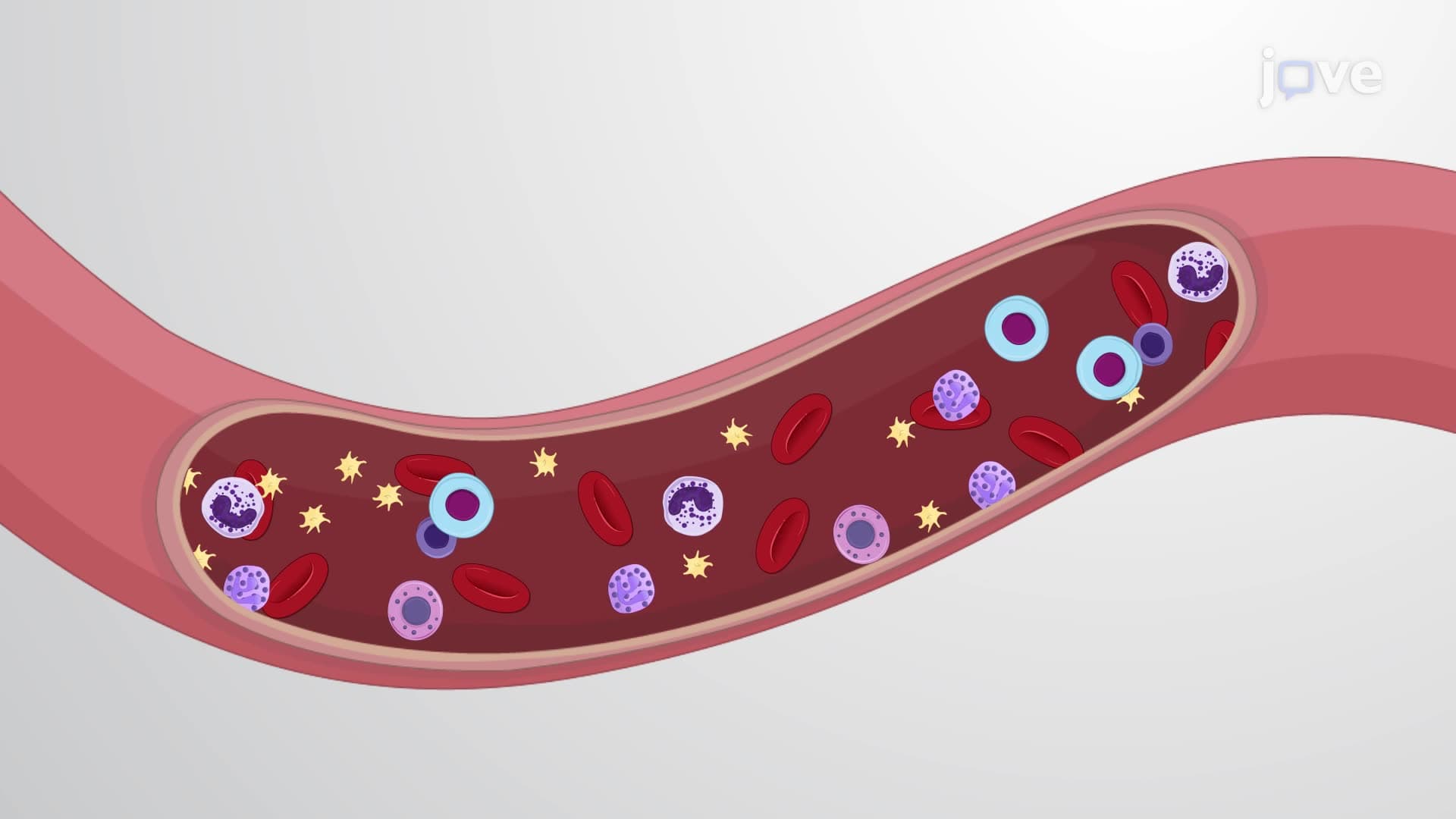
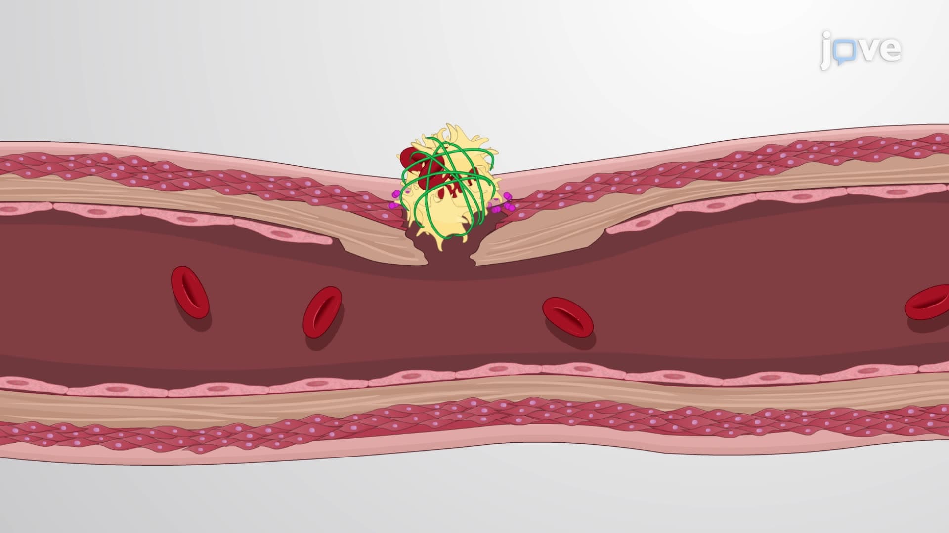
The Structure And Function of Platelets Explained begins with recognizing these remarkable cellular fragments as essential components of hemostasis. Unlike typical cells, platelets lack nuclei but compensate with highly organized cytoplasmic contents. Their small size (2-4 micrometers) allows rapid circulation through even the smallest capillaries, while their disc shape maximizes surface area for adhesion during vascular injury.
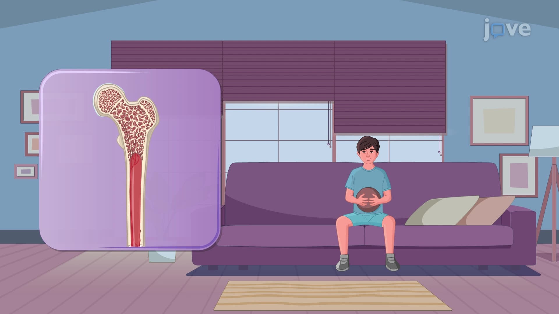
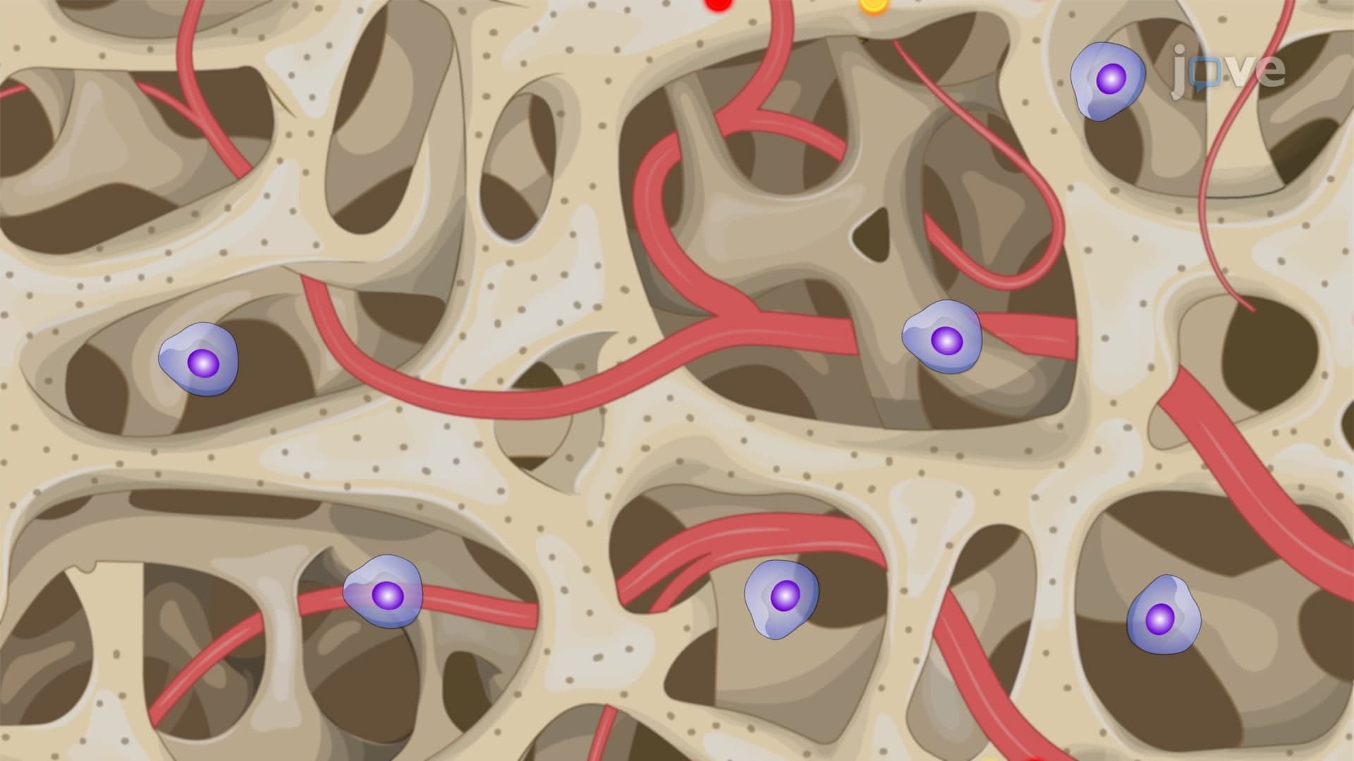
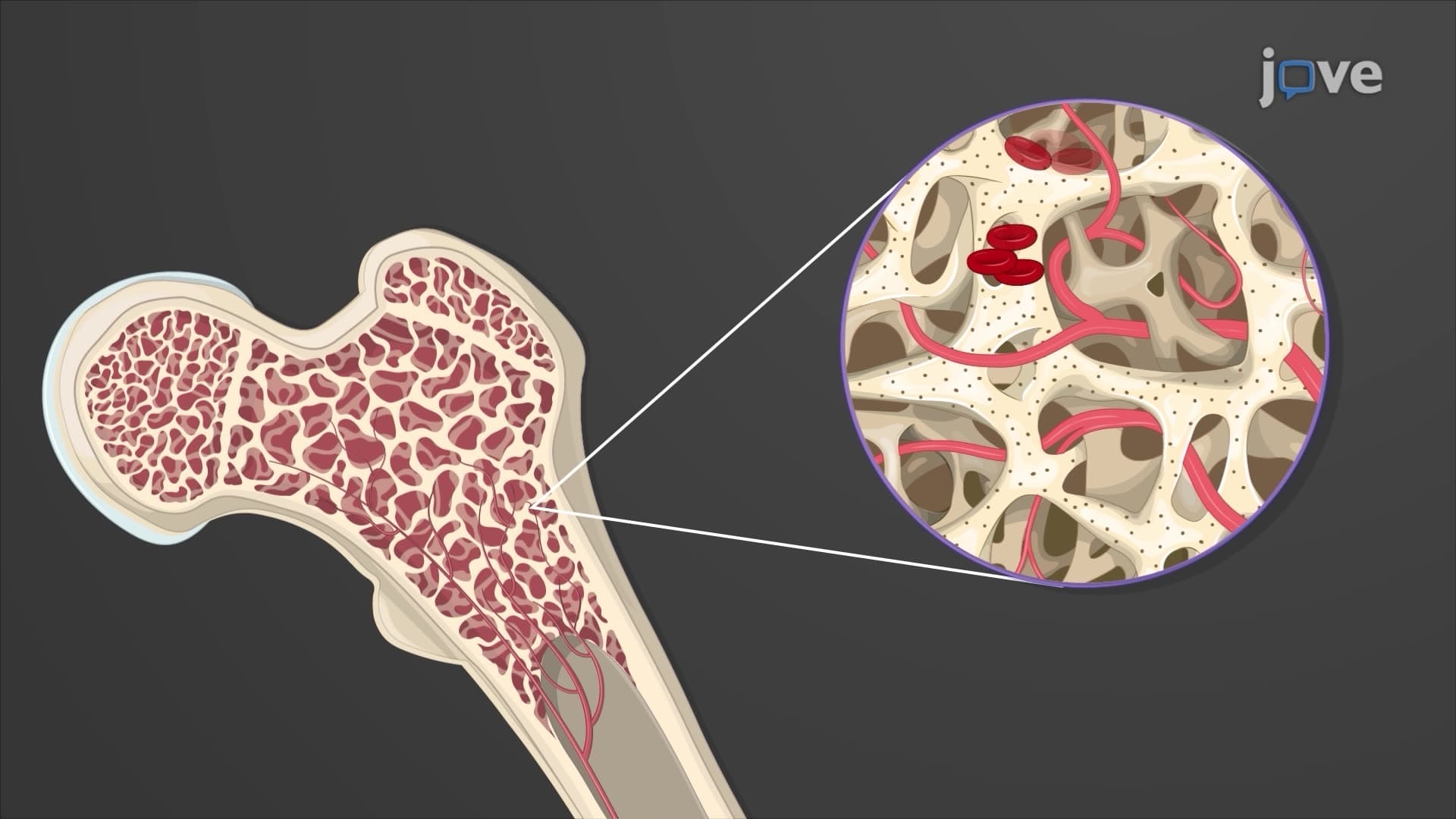
Blood platelet anatomy explained starts in the bone marrow, where myeloid stem cells differentiate into megakaryoblasts under hormonal influence. Thrombopoietin, primarily produced by the liver and kidneys, serves as the key regulatory hormone driving this process. Interleukin-6 provides additional stimulation during inflammatory states. The megakaryoblast undergoes endomitosis-DNA replication without cell division-creating polyploid megakaryocytes that can reach 100 micrometers in diameter. These giant cells extend cytoplasmic projections through sinusoid walls, eventually fragmenting to release 1,000-3,000 platelets per megakaryocyte.
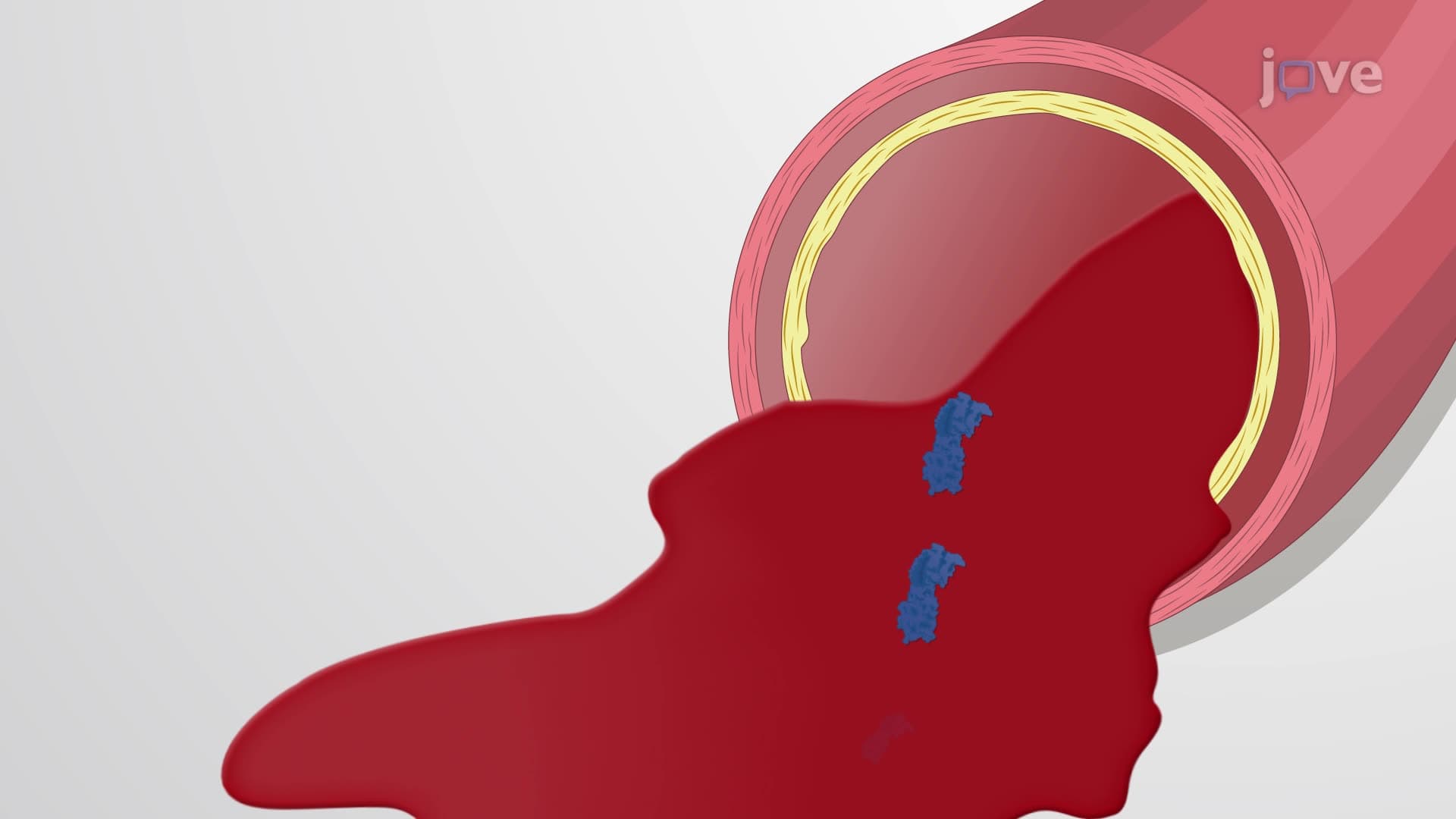
Platelet granule secretion involves three distinct granule types, each serving specific hemostatic functions. Alpha granules contain fibrinogen, von Willebrand factor, platelet factor 4, and platelet-derived growth factor. Dense granules store ADP, ATP, serotonin, and calcium ions. Lysosomes house hydrolytic enzymes for clot remodeling. During activation, these granules undergo exocytosis, releasing their contents to amplify the coagulation cascade and recruit additional platelets.
Understanding what is the structure and function of platelets proves crucial for MCAT preparation, particularly in biological sciences sections covering cardiovascular physiology. AP Biology students encounter platelet function when studying cell structure and intercellular communication. In clinical settings, platelet disorders like thrombocytopenia affect over 200,000 Americans annually, making this knowledge essential for healthcare professionals. Medical students preparing for USMLE Step 1 must master platelet plug formation mechanisms to answer hematology questions effectively.
Related Micro-courses