Loading video…


 29 Concepts
 135.5K views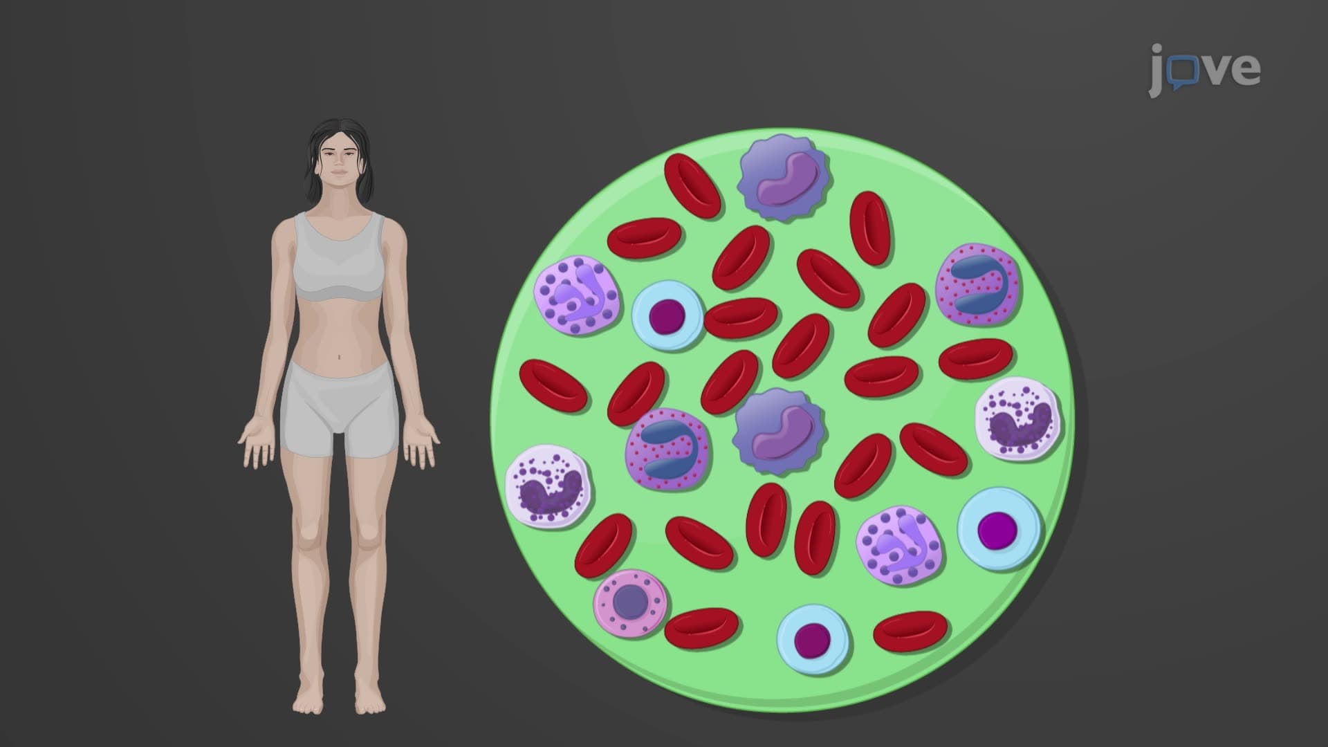

Video Summary: Structure and Function of Leukocytes Explained
Did you know that when you get a cut or infection, your white blood cell count can double within hours? This remarkable response showcases the critical leukocyte structure function biology that keeps us healthy. During the 2020 pandemic, healthcare workers regularly monitored COVID-19 patients' white blood cell counts to assess immune system response. The Structure And Function of Leukocytes Explained reveals how these cellular defenders patrol our bodies, squeeze through blood vessel walls, and eliminate threats through specialized mechanisms. Watch the full video on JoVE Coach to master this concept with expert-led visuals and step-by-step explanations.
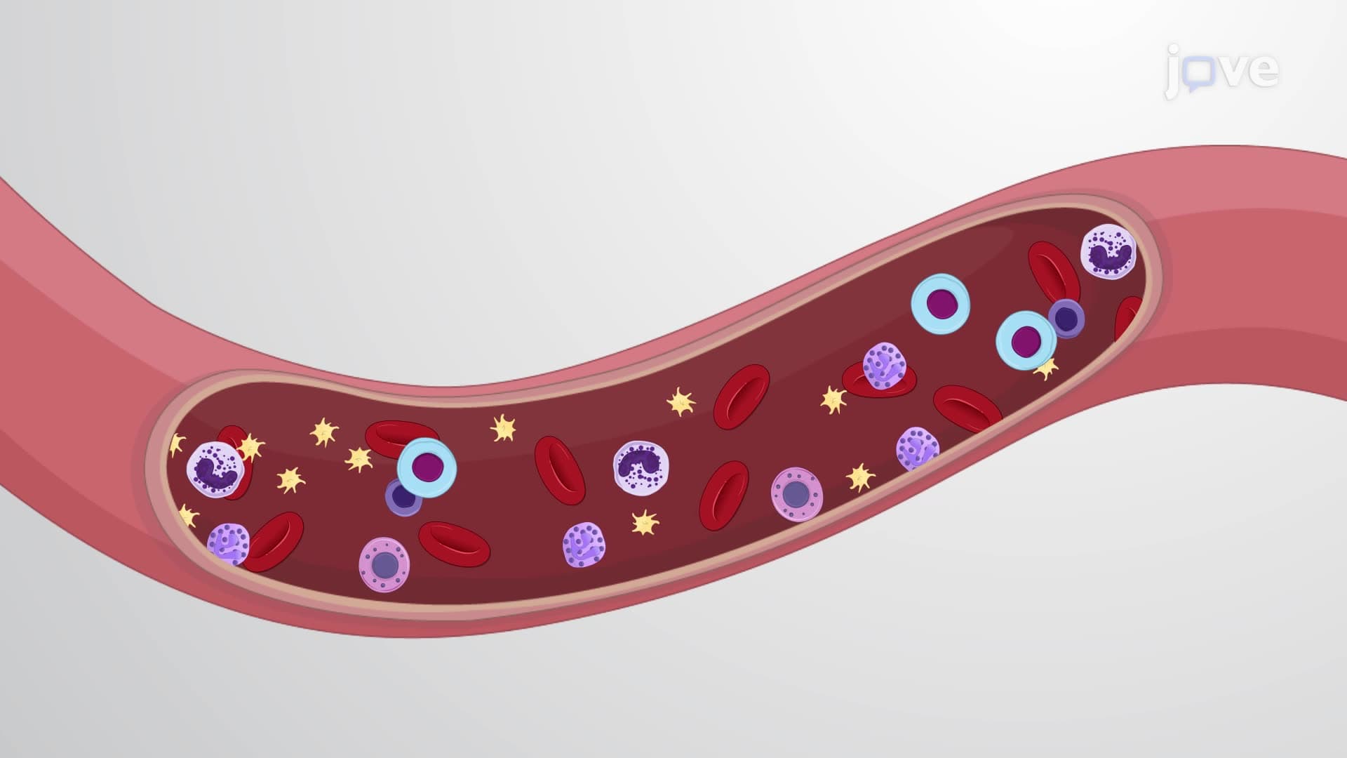
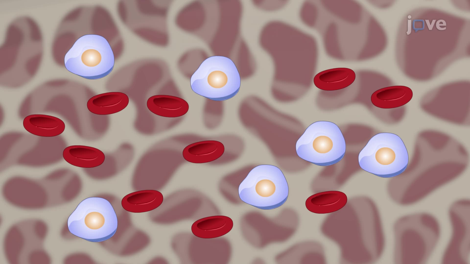
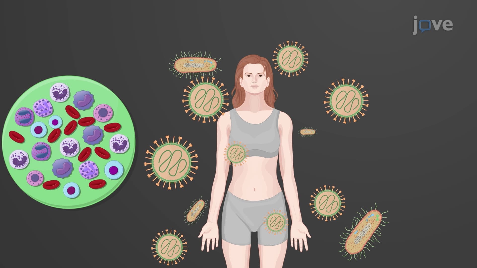
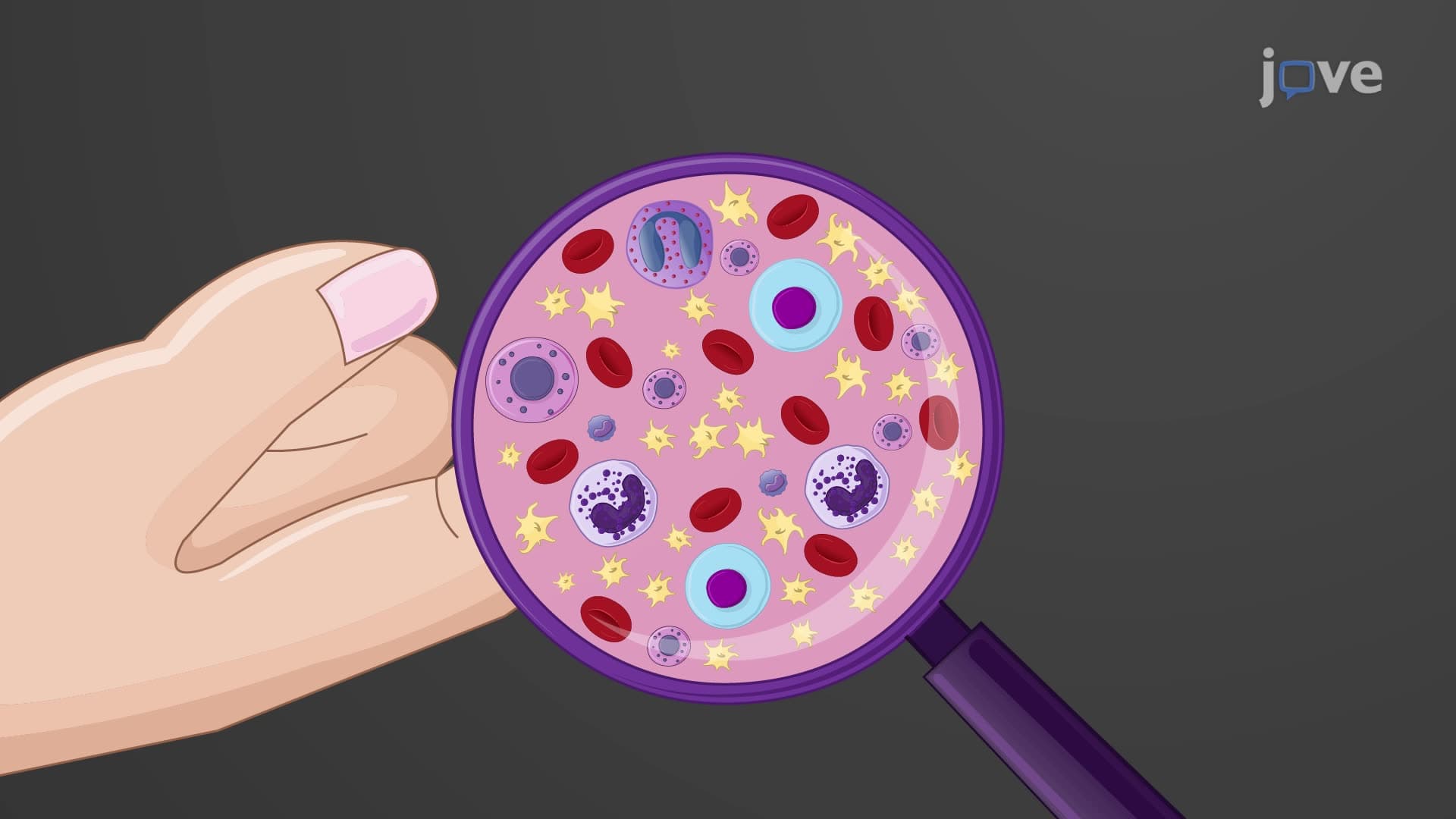
White blood cell structure function represents one of the most sophisticated defense mechanisms in human physiology. Unlike red blood cells, leukocytes are complete cells containing nuclei and full organelle complements, enabling them to perform complex immune functions. These cellular guardians comprise only 1% of blood volume yet orchestrate our entire immune response system.
The normal range of 4,500 to 11,000 leukocytes per microliter serves as a critical diagnostic marker. When physicians at Johns Hopkins Hospital evaluate infection severity, they monitor what is the structure and function of leukocytes by tracking these counts. Elevated levels often indicate bacterial infections, while decreased counts may suggest viral infections or immunocompromised states.
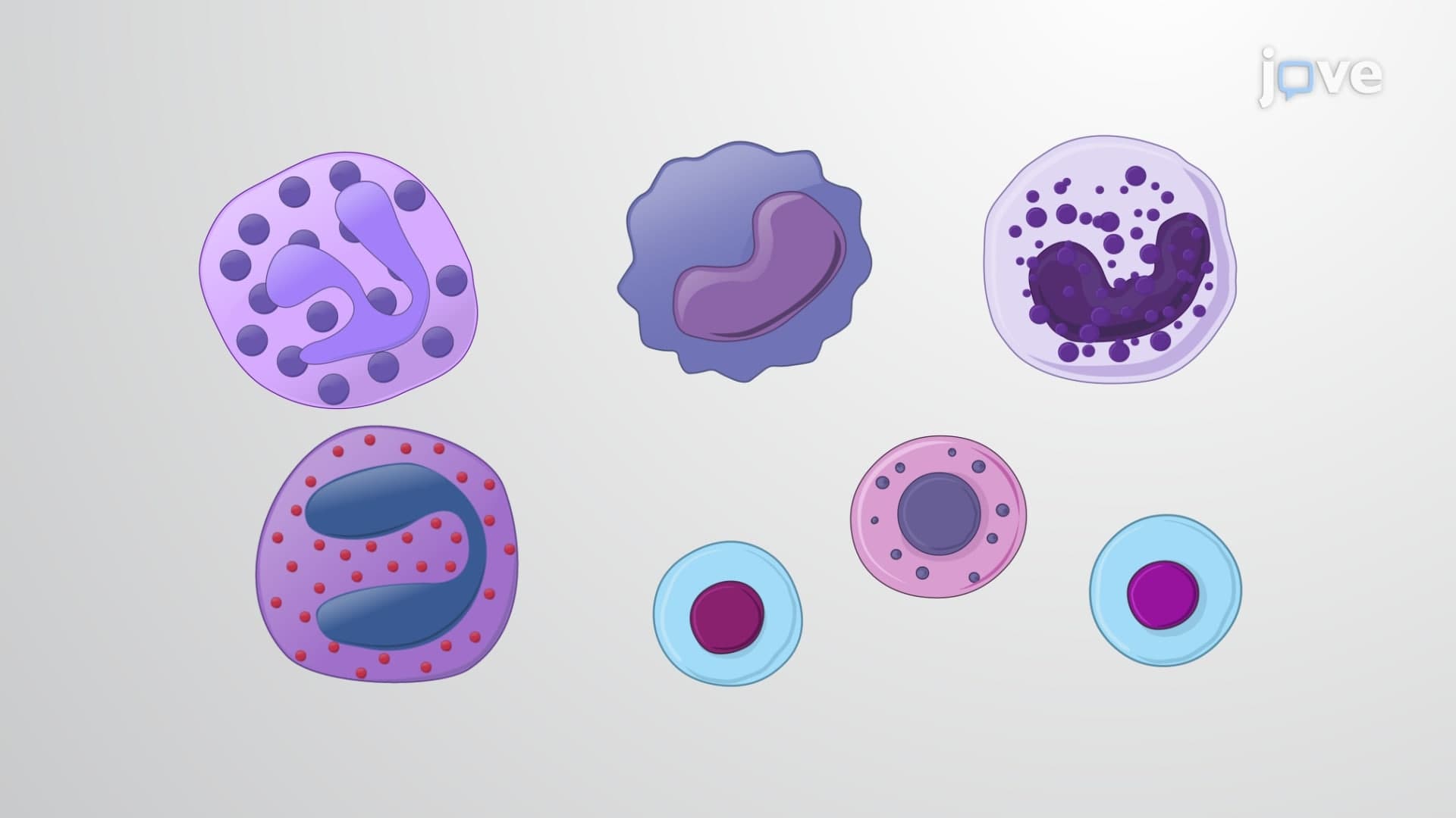
Granulocyte agranulocyte leukocyte distinctions form the foundation of hematology studies. Granulocytes include neutrophil eosinophil basophil cells, each with specialized granules containing different enzymes and chemical mediators. Neutrophils dominate bacterial responses, eosinophils target parasites and allergic reactions, while basophils release histamine during inflammatory responses.
Agranulocytes encompass lymphocyte monocyte function categories. Lymphocytes include T-cells, B-cells, and natural killer cells that provide adaptive immunity. Monocytes differentiate into macrophages and dendritic cells, serving as tissue-resident immune sentinels.
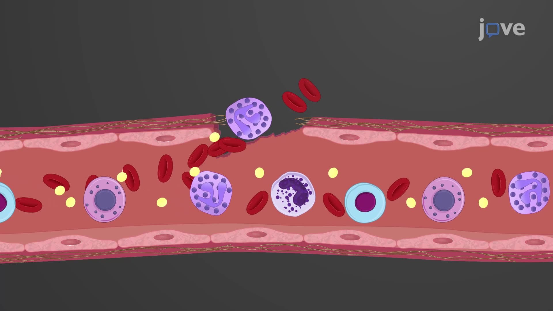
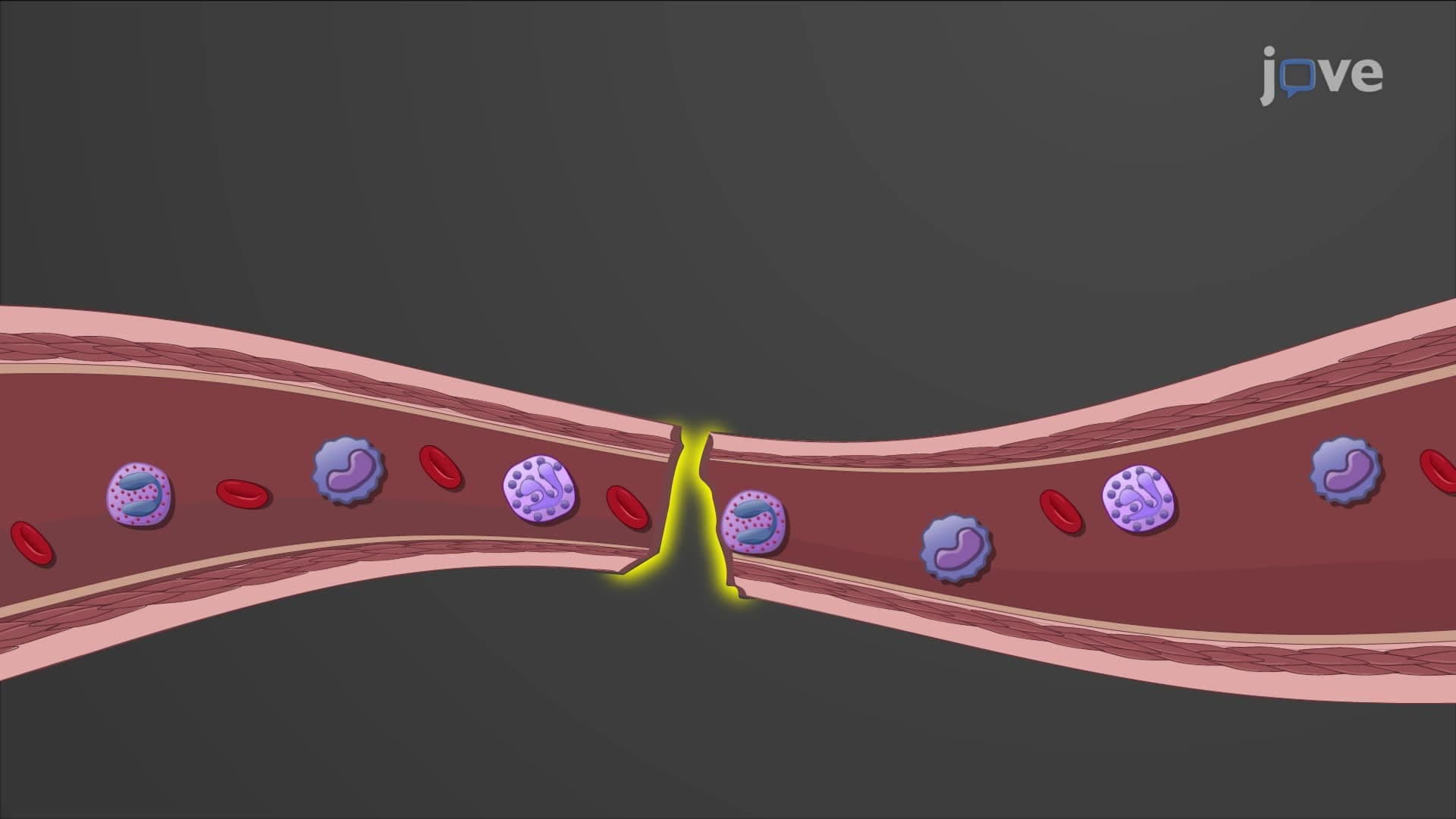
The WBC immune defense function operates through multiple coordinated processes. During tissue invasion, damaged cells release cytokines and chemokines that attract circulating leukocytes. This chemical gradient guides white blood cells to infection sites through a process called chemotaxis.
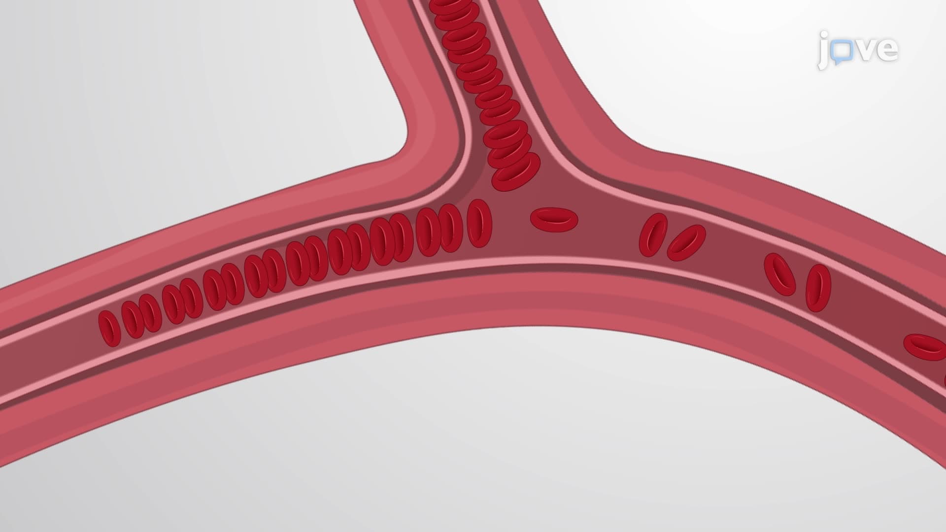
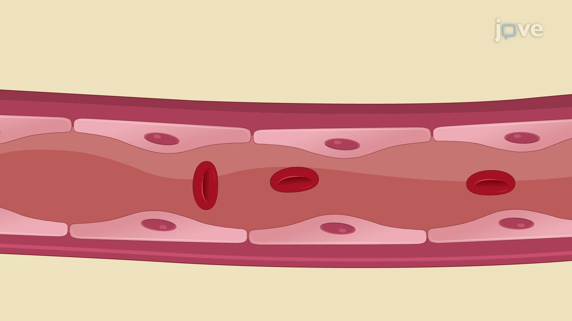
Leukocyte adhesion represents a critical step where white blood cells bind to endothelial cells lining blood vessels. Rolling, firm adhesion, and transmigration allow these cells to exit circulation and enter infected tissues. This process, studied extensively at medical schools like Harvard and Stanford, demonstrates how leukocyte type anatomy determines specific immune functions.
Understanding leukocyte biology proves essential for MCAT success, particularly in biological sciences sections covering immune system function. AP Biology students encounter these concepts when studying homeostasis and immune responses. College anatomy and physiology courses at institutions like UCLA and University of Michigan emphasize leukocyte mechanisms in their curricula.
Healthcare professionals rely on complete blood counts (CBCs) to assess leukocyte populations. During medical training at institutions like Mayo Clinic, students learn to interpret differential counts that reveal specific leukocyte subtype proportions, providing diagnostic insights into various pathological conditions.
Related Micro-courses