Loading video…


 20 Concepts
 68.3K views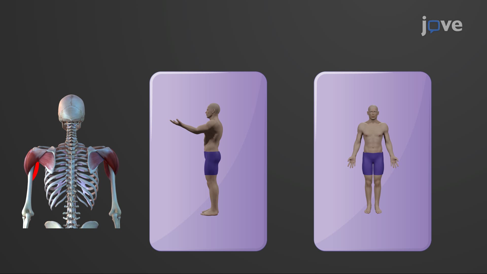

Video Summary: Muscles That Move the Arm Explained
Did you know that throwing a baseball at 90 mph requires precise coordination of at least nine different muscle groups? Understanding arm movement muscles anatomy is crucial for anyone studying human physiology, from aspiring physical therapists at the University of California to high school students preparing for AP Biology. The complex interplay between axial and scapular muscles enables everything from a surgeon's delicate movements to a basketball player's powerful slam dunk. Muscles That Move The Arm Explained breaks down this intricate system into digestible components. Watch the full video on JoVE Coach to master this concept with expert-led visuals and step-by-step explanations.
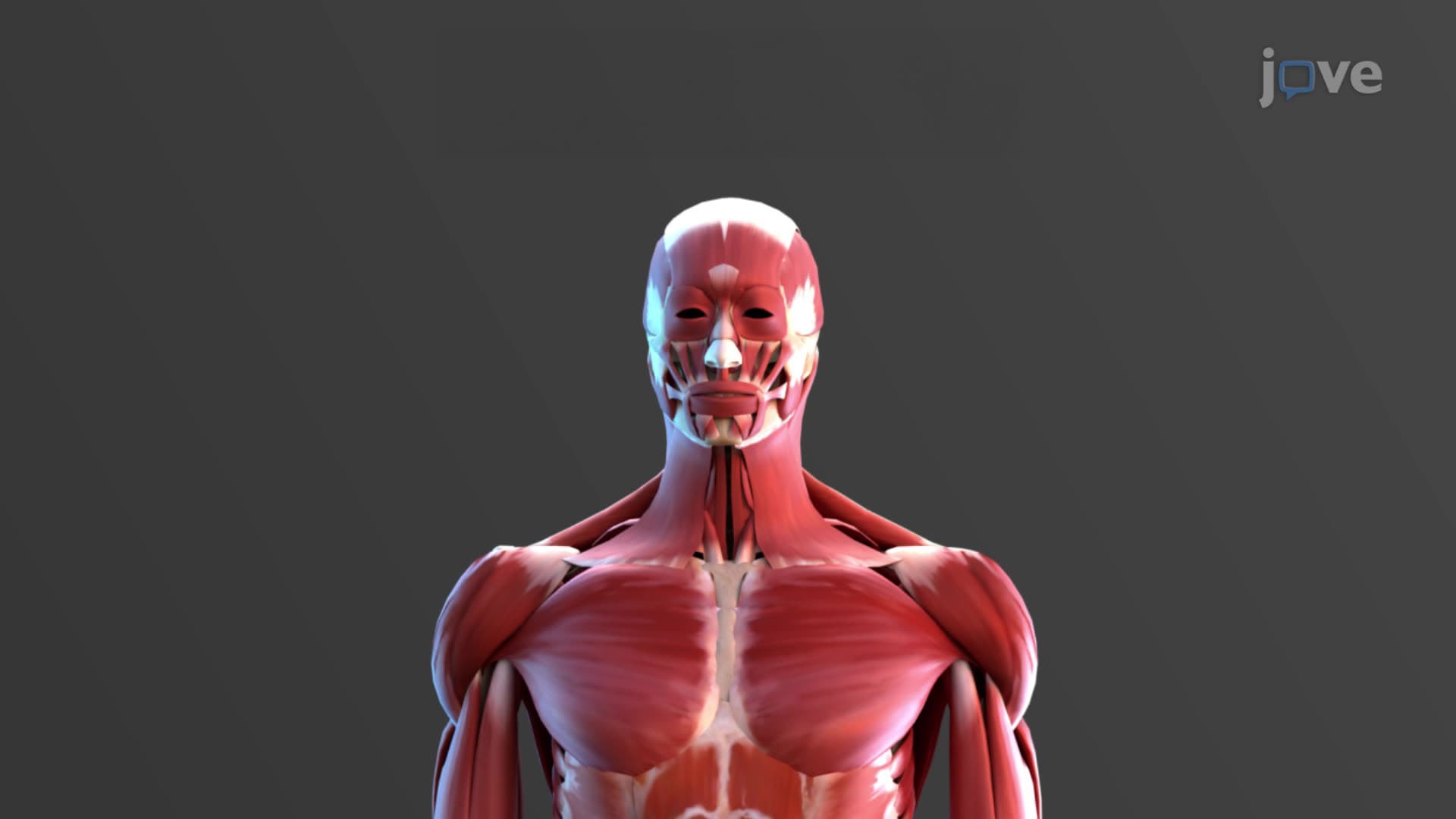
The human arm's remarkable mobility stems from a sophisticated network of muscles working in precise coordination. These muscles responsible for arm movement fall into two primary categories: axial muscles (originating from the trunk) and scapular muscles (originating from the shoulder blade). This organization reflects evolutionary adaptations that allow humans to perform complex manipulative tasks, from a surgeon's precise incisions at Johns Hopkins Hospital to a pitcher's curveball at Yankee Stadium.
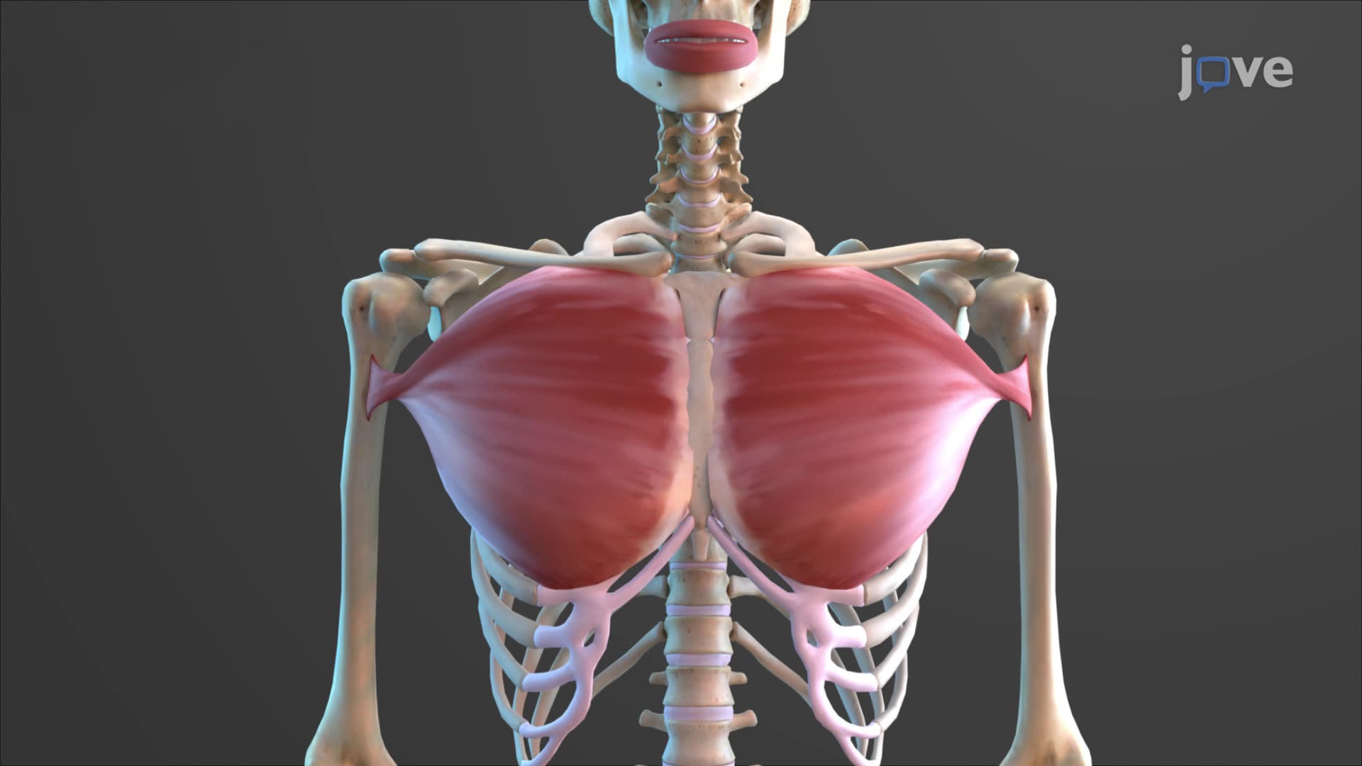
The pectoralis major and latissimus dorsi represent the body's primary arm movement muscle group originating from the central axis. The pectoralis major, with its distinctive clavicular and sternocostal heads, demonstrates functional specialization rarely seen in anatomy. The clavicular head primarily flexes the arm (raising it forward), while the sternocostal head extends it (pulling it backward from a raised position). This dual functionality appears frequently on MCAT questions and AP Biology exams, where students must distinguish between these actions.
The latissimus dorsi, often called the "swimmer's muscle," creates the characteristic V-shape of trained athletes. Its primary function-arm extension, adduction, and medial rotation-makes it crucial for activities like rock climbing and swimming. Physical therapy programs at universities like USC emphasize this muscle's role in shoulder rehabilitation following sports injuries.
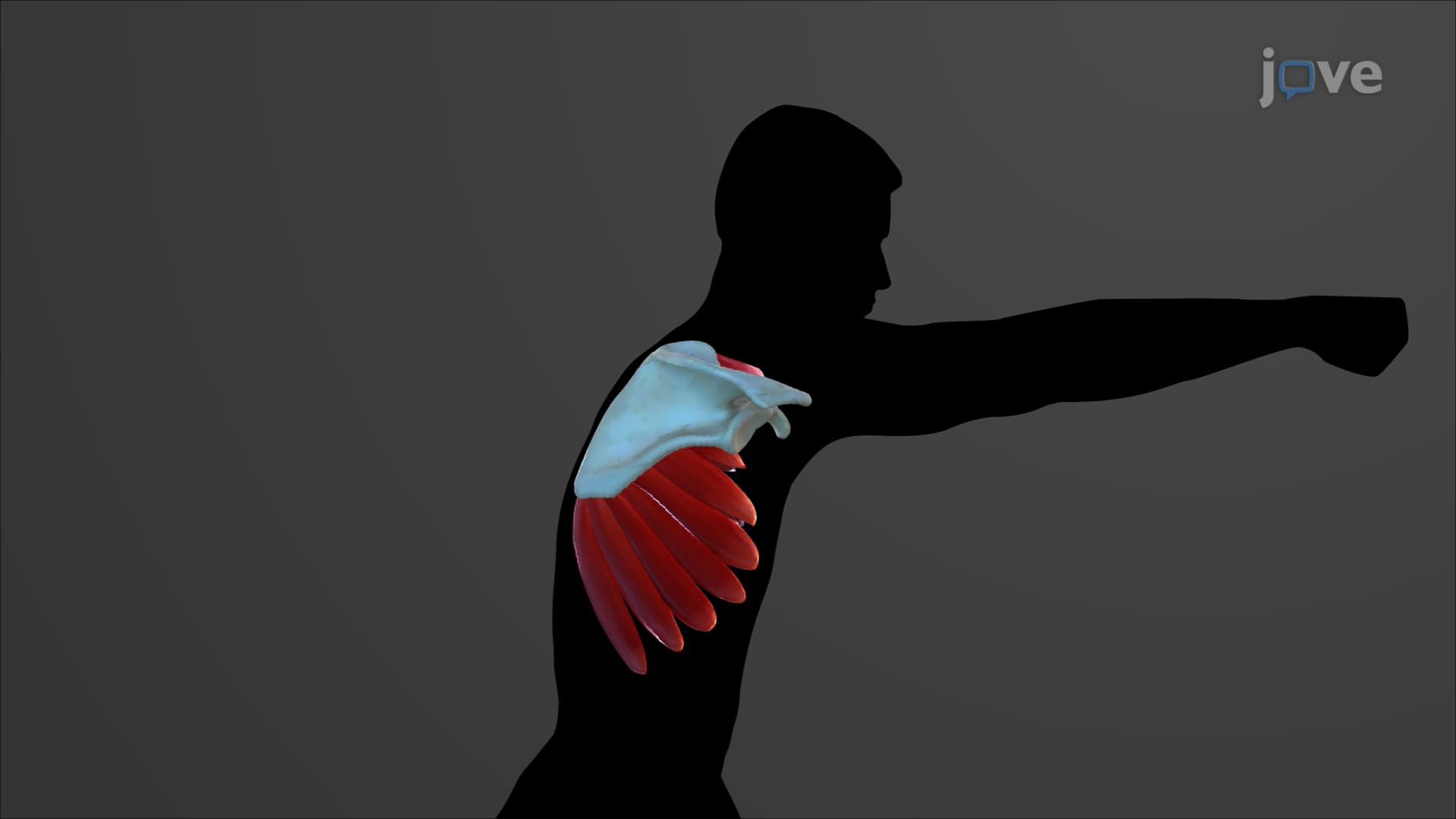
The seven scapular muscles include the functionally vital rotator cuff group: supraspinatus, infraspinatus, teres minor, and subscapularis. Medical students learning for the USMLE Step 1 often use the mnemonic "SITS" to remember these muscles. The rotator cuff's primary role involves stabilizing the glenohumeral joint while allowing the deltoid to produce powerful movements.
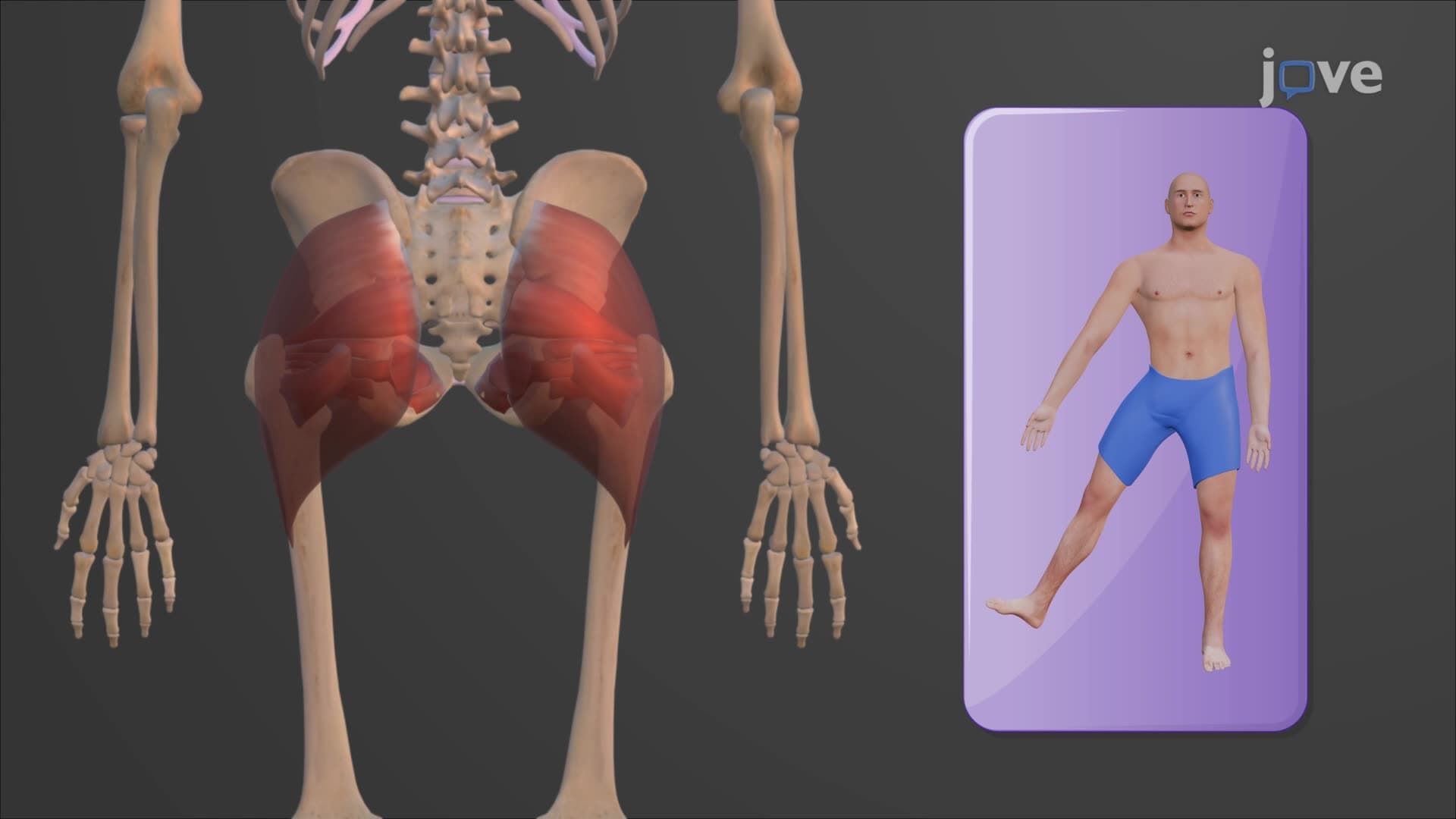
What muscles are responsible for moving the arm in abduction? The deltoid, covering the shoulder like a cap, works with the supraspinatus to lift the arm laterally. This coordinated action becomes compromised in rotator cuff injuries, explaining why patients with supraspinatus tears struggle to initiate arm abduction-a clinical finding nursing students encounter during NCLEX preparation.
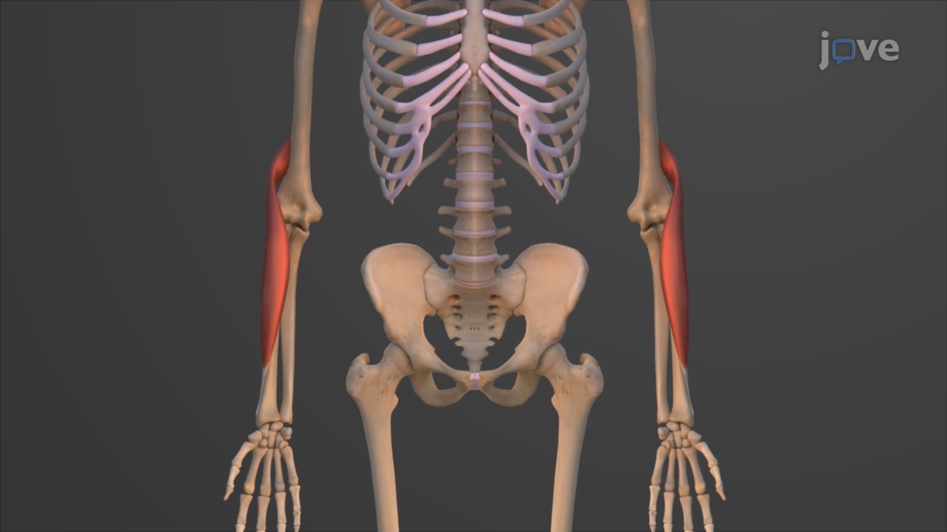
Understanding these muscle relationships proves essential for healthcare professionals. The biceps brachii flexion arm action, while not detailed in the transcript, works synergistically with muscles like the coracobrachialis to produce smooth, controlled movement. Physical therapy students studying at institutions like Northwestern University learn to assess these muscle interactions when developing rehabilitation protocols for athletes and injury patients.
Related Micro-courses