Loading video…


 11 Concepts
 1.2M views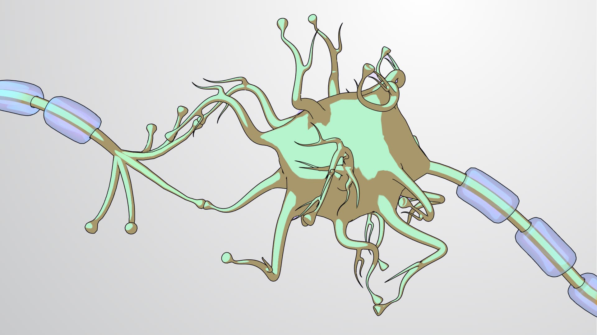

Video Summary: What Is Neuron Structure
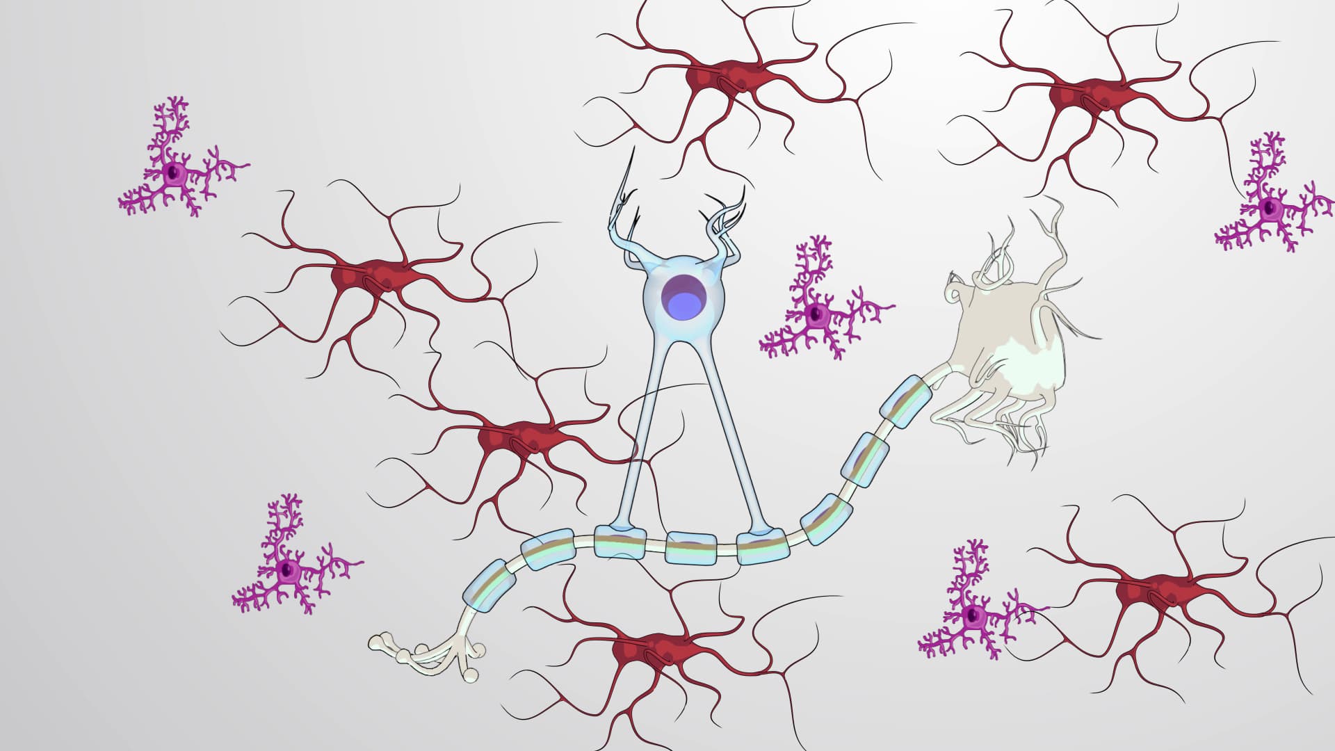
Every time you blink, think, or move your finger, billions of specialized cells called neurons fire electrical signals at lightning speed through your body. Neuron structure forms the foundation of all nervous system function, from reflexes that protect NFL players from concussions to the complex neural networks that help medical students at Johns Hopkins memorize anatomy. These remarkable cells share common structural components-cell bodies, dendrites, axons, and synaptic terminals-that work together like a biological electrical circuit. Understanding what is neuron structure reveals how your brain processes everything from solving calculus problems to recognizing your best friend's voice. Watch the full video on JoVE Coach to master this concept with expert-led visuals and step-by-step explanations.
Neuron structure represents one of biology's most elegant examples of form following function. These specialized cells have evolved precise architectural features that enable rapid, long-distance electrical communication throughout the human body.
The soma, or cell body, serves as the neuron's metabolic headquarters. Unlike other cells, the neuronal cell body must support extremely long cellular extensions-some motor neurons in tall individuals have axons extending over three feet from spinal cord to toe muscles. The nucleus within the soma contains the DNA blueprint for producing specialized proteins essential for neural function, including ion channels, neurotransmitter receptors, and synaptic machinery. Students preparing for the AP Biology exam should remember that the cell body's extensive rough endoplasmic reticulum, called Nissl bodies, produces the massive amounts of protein needed to maintain these far-reaching cellular extensions.
Dendrites function as the neuron's receiving equipment, their branched, tree-like structure maximizing surface area for synaptic connections. A single cortical neuron may receive input from thousands of other neurons across its dendritic tree. This neuron structure biology explained concept becomes crucial when studying neuroplasticity-the brain's ability to rewire itself through experience. For example, London taxi drivers show enlarged hippocampal regions with increased dendritic branching, reflecting years of navigating complex city streets. MCAT students often encounter questions about how dendritic spine density changes with learning and memory formation.
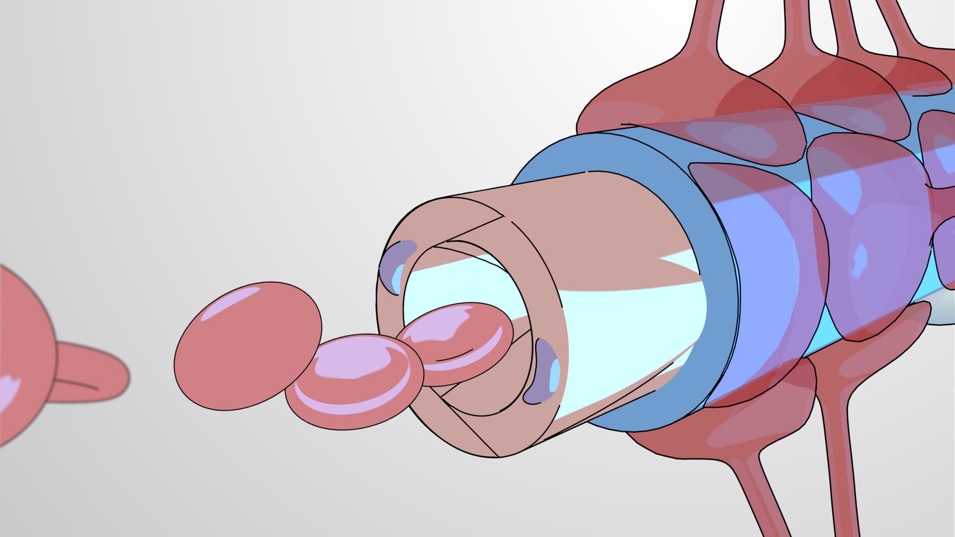
The axon represents the neuron's output pathway, transmitting action potentials from the axon hillock to distant targets. The myelin sheath, produced by oligodendrocytes in the central nervous system and Schwann cells in peripheral nerves, acts like electrical insulation around a wire. This neuron structure overview wouldn't be complete without understanding saltatory conduction-how action potentials "jump" between nodes of Ranvier, increasing transmission speed up to 120 meters per second in heavily myelinated axons.
Multiple sclerosis, affecting over 400,000 Americans, demonstrates the critical importance of myelin integrity. When autoimmune attacks damage myelin sheaths, neural transmission slows dramatically, causing the characteristic symptoms of muscle weakness and cognitive impairment that medical students study extensively.
Related Micro-courses