Loading subject page…

15,445 views

Video Summary: What Is Two Dimensional Microscopy
Ever wondered how pathologists at Mayo Clinic can diagnose cancer from tissue samples that appear invisible to the naked eye? Two dimensional microscopy makes this possible by visualizing structures within a single optical plane, creating detailed flat representations of specimens. This fundamental technique includes bright-field, dark-field, phase-contrast, and fluorescence microscopy methods that reveal cellular structures and microorganisms. Watch the full video on JoVE Coach to master this concept with expert-led visuals and step-by-step explanations.
Two dimensional microscopy represents the cornerstone of biological visualization, capturing images within a single focal plane to create flat, detailed representations of specimens. Unlike three-dimensional techniques that build depth perception, two-dimensional methods excel at revealing fine structural details, making them indispensable in medical diagnostics, research laboratories, and educational settings across American universities and hospitals.
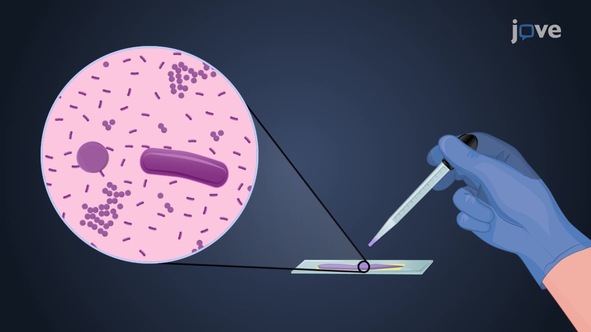
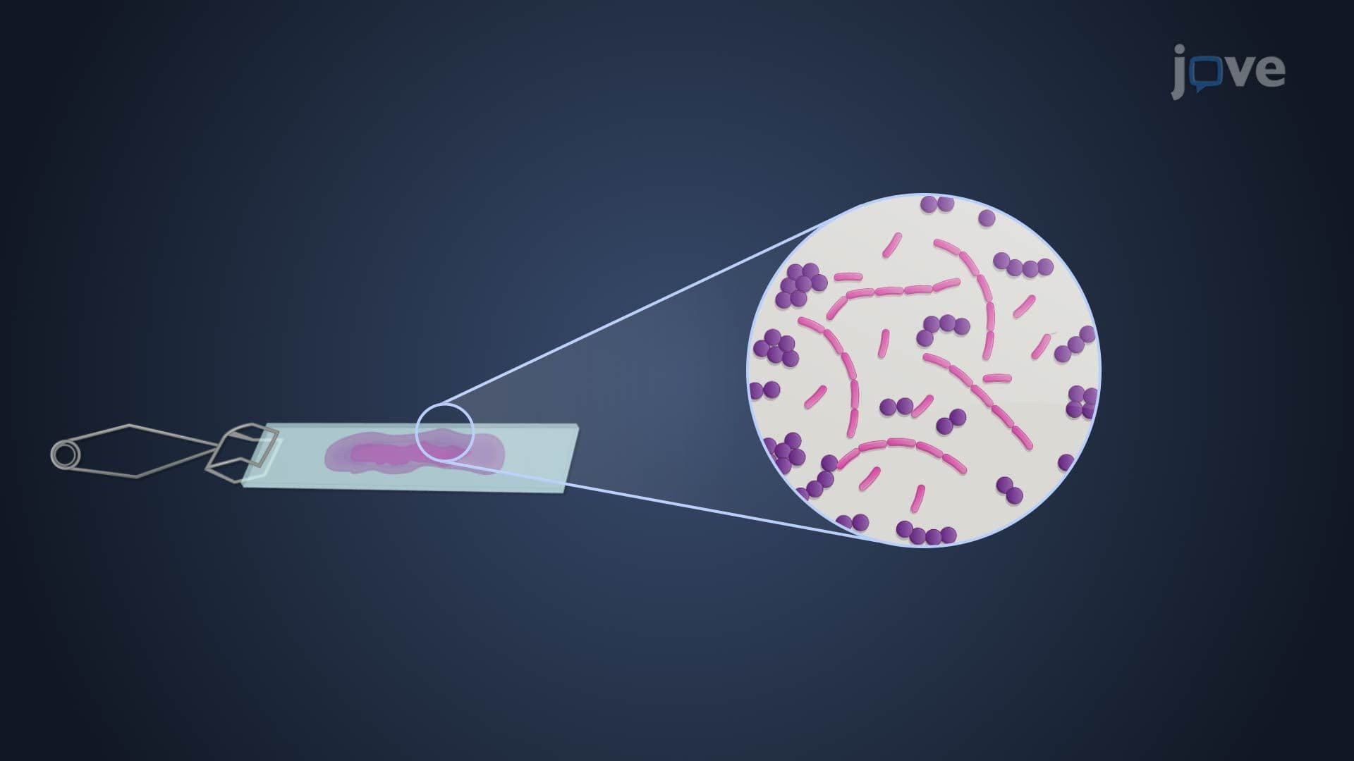
Bright-field microscopy serves as the most widely used technique in clinical laboratories nationwide. This method transmits light directly through specimens, creating uniformly illuminated images against bright backgrounds. Stained tissue sections, commonly prepared in pathology departments at institutions like Johns Hopkins and Cleveland Clinic, reveal cellular structures through differential dye absorption. The technique proves essential for routine blood smears, bacterial identification, and histopathological diagnosis. Students preparing for MCAT biology sections frequently encounter bright-field microscopy applications, particularly in microbiology and cell biology contexts.
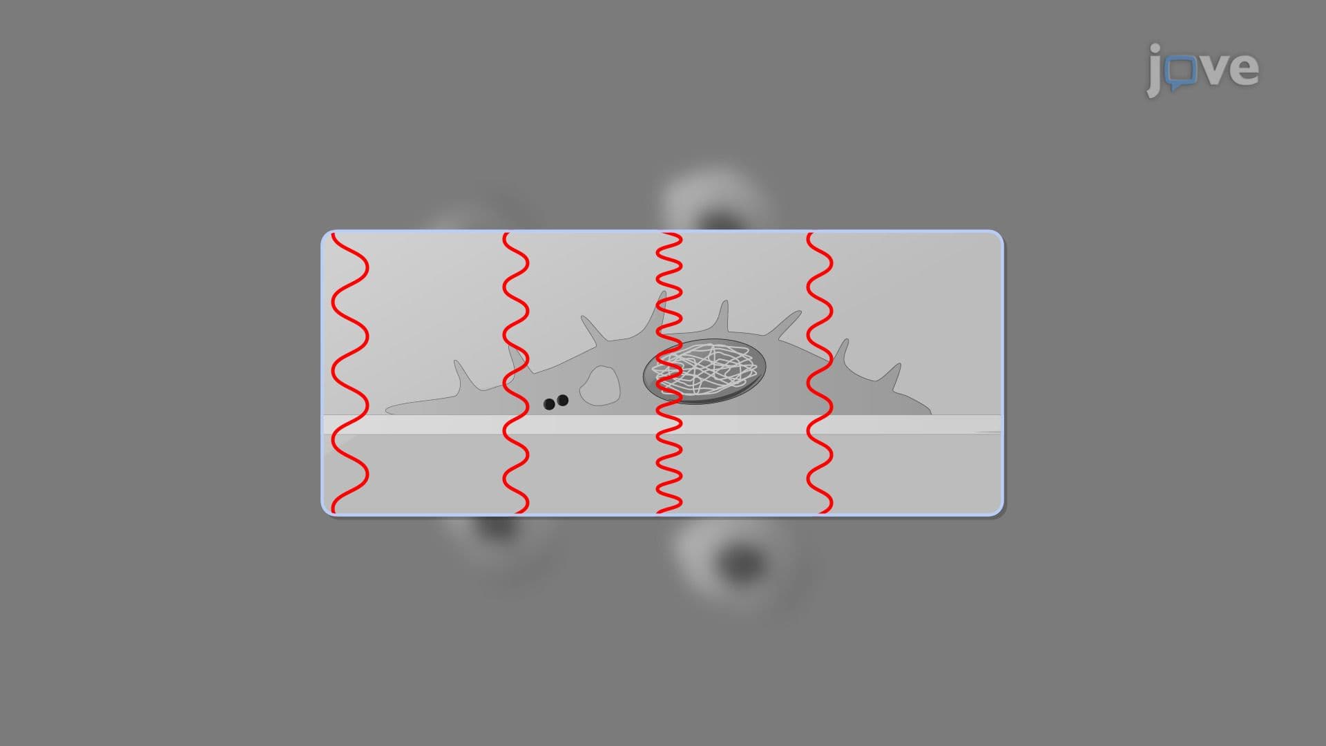
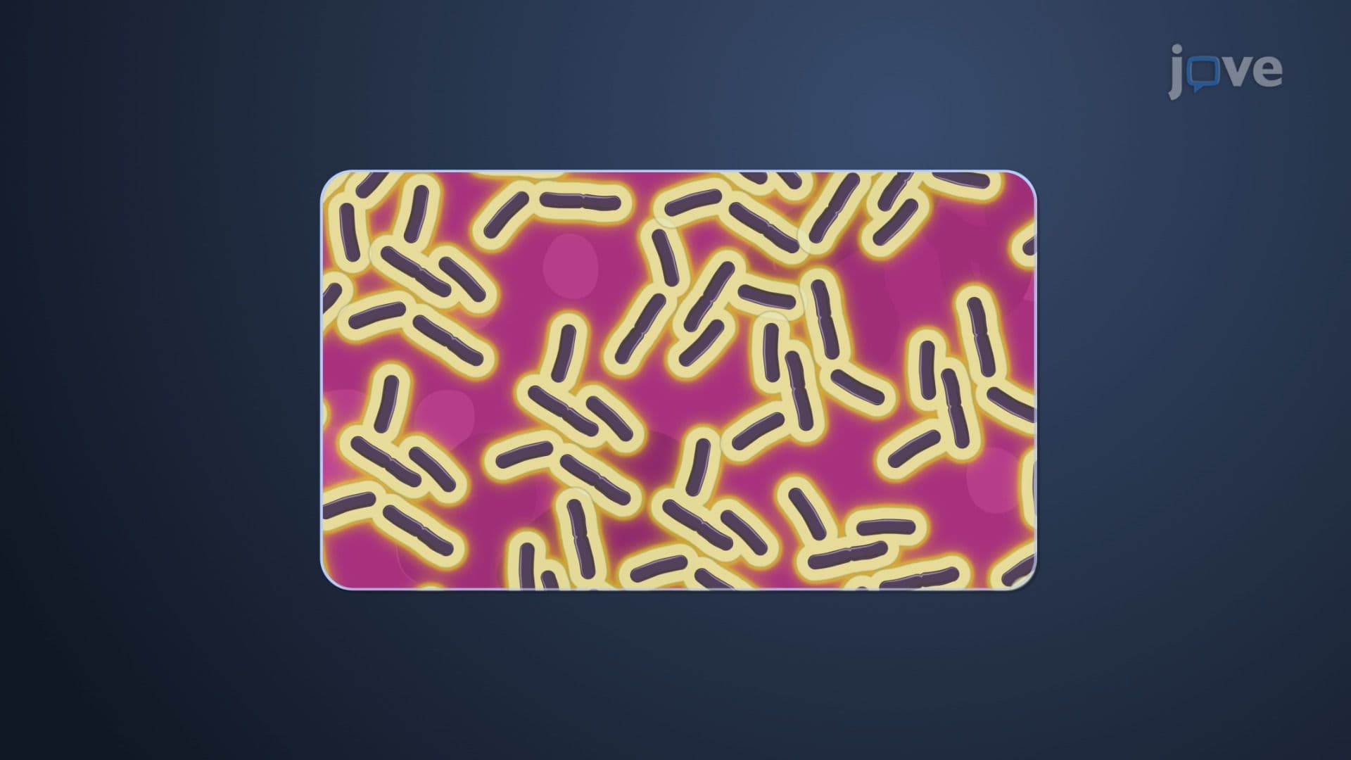
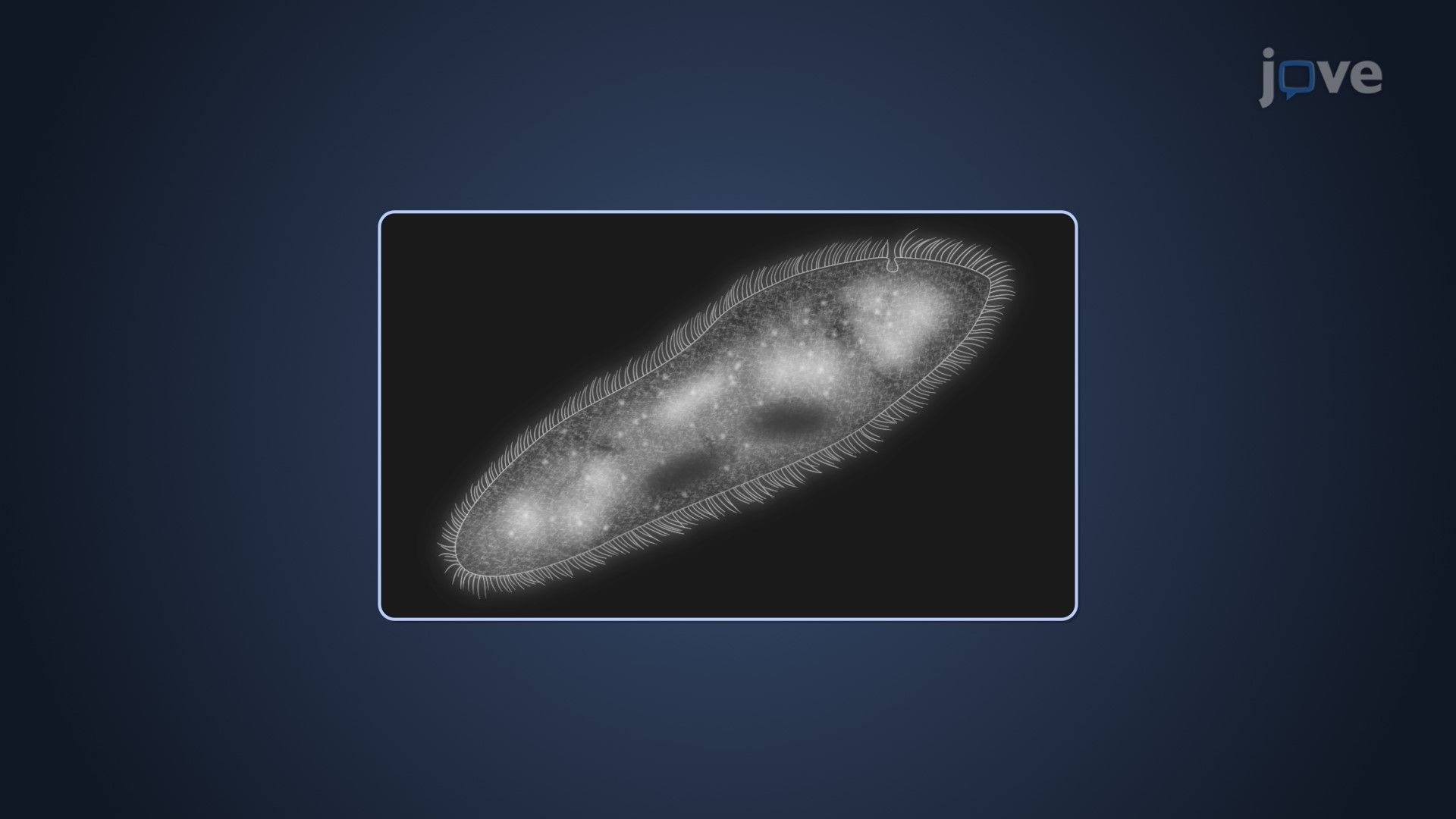
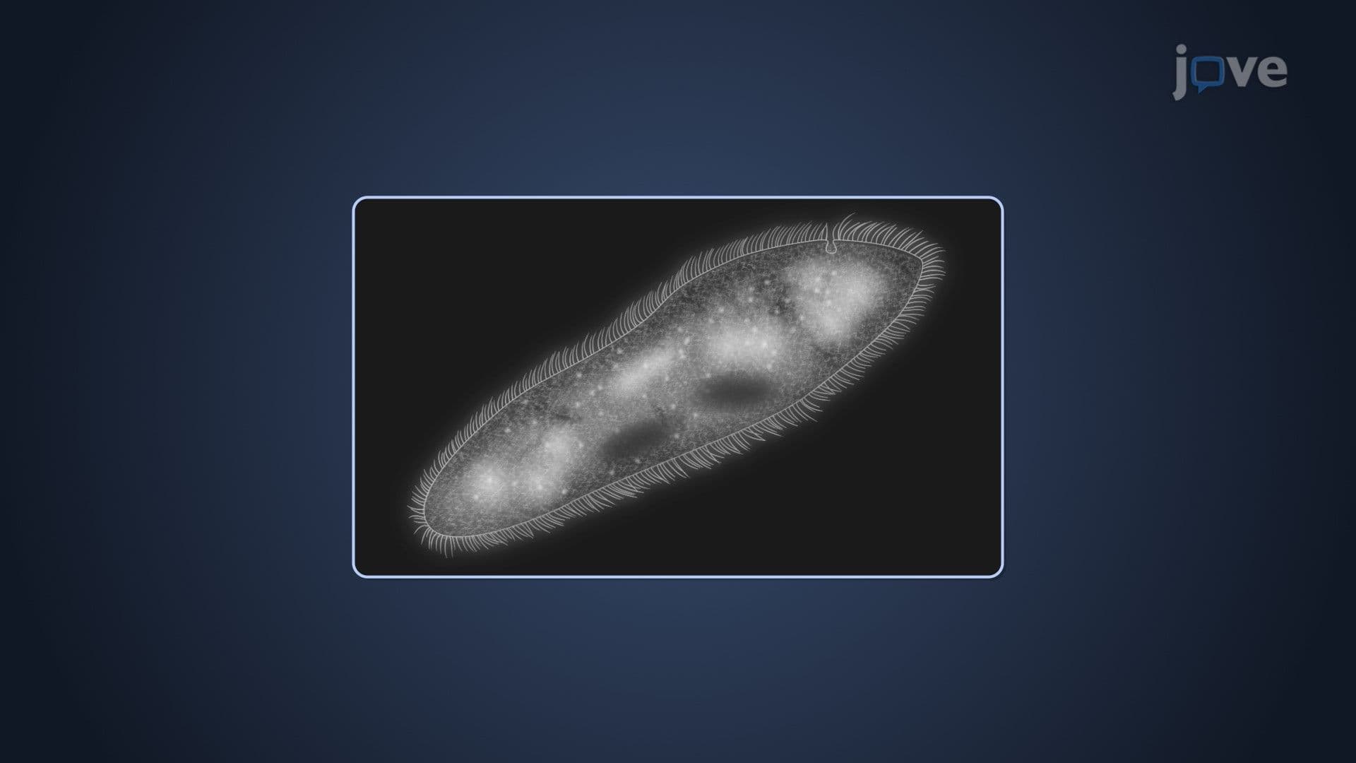
Dark-field microscopy revolutionizes specimen visualization by illuminating objects with scattered light, creating brilliant images against dark backgrounds. This technique excels at detecting live microorganisms without staining, making it invaluable for diagnosing syphilis through spirochete observation in clinical settings. Phase-contrast microscopy takes a different approach, converting phase shifts caused by refractive index variations into brightness differences. This allows researchers at pharmaceutical companies like Pfizer and Moderna to observe live cell division, organelle movement, and protein trafficking without killing specimens.
Fluorescence microscopy employs fluorophores-specialized dyes and naturally occurring molecules-that emit light when excited by specific wavelengths. This technique provides exceptional contrast and protein-level resolution, making it crucial for immunology research and cancer studies. Major research universities like MIT and Stanford utilize fluorescence microscopy for drug development and genetic research. Students encounter this technique in AP Biology courses and college biochemistry labs, where it helps visualize DNA, proteins, and cellular processes in real-time.
Understanding these microscopy fundamentals prepares students for advanced coursework in cell biology, microbiology, and medical school prerequisites, while providing practical knowledge applicable to future careers in healthcare and biological research.
Related Micro-courses