Loading concepts…


11 Concepts
13 Concepts
26 Concepts
13 Concepts
6 Concepts
31 Concepts
37 Concepts
10 Concepts
7 Concepts
22 Concepts
11 Concepts
11 Concepts
22 Concepts
16 Concepts
11 Concepts
15 Concepts
14 Concepts
12 Concepts
9 Concepts
 13 Concepts
 14.7K Views
 Popular topics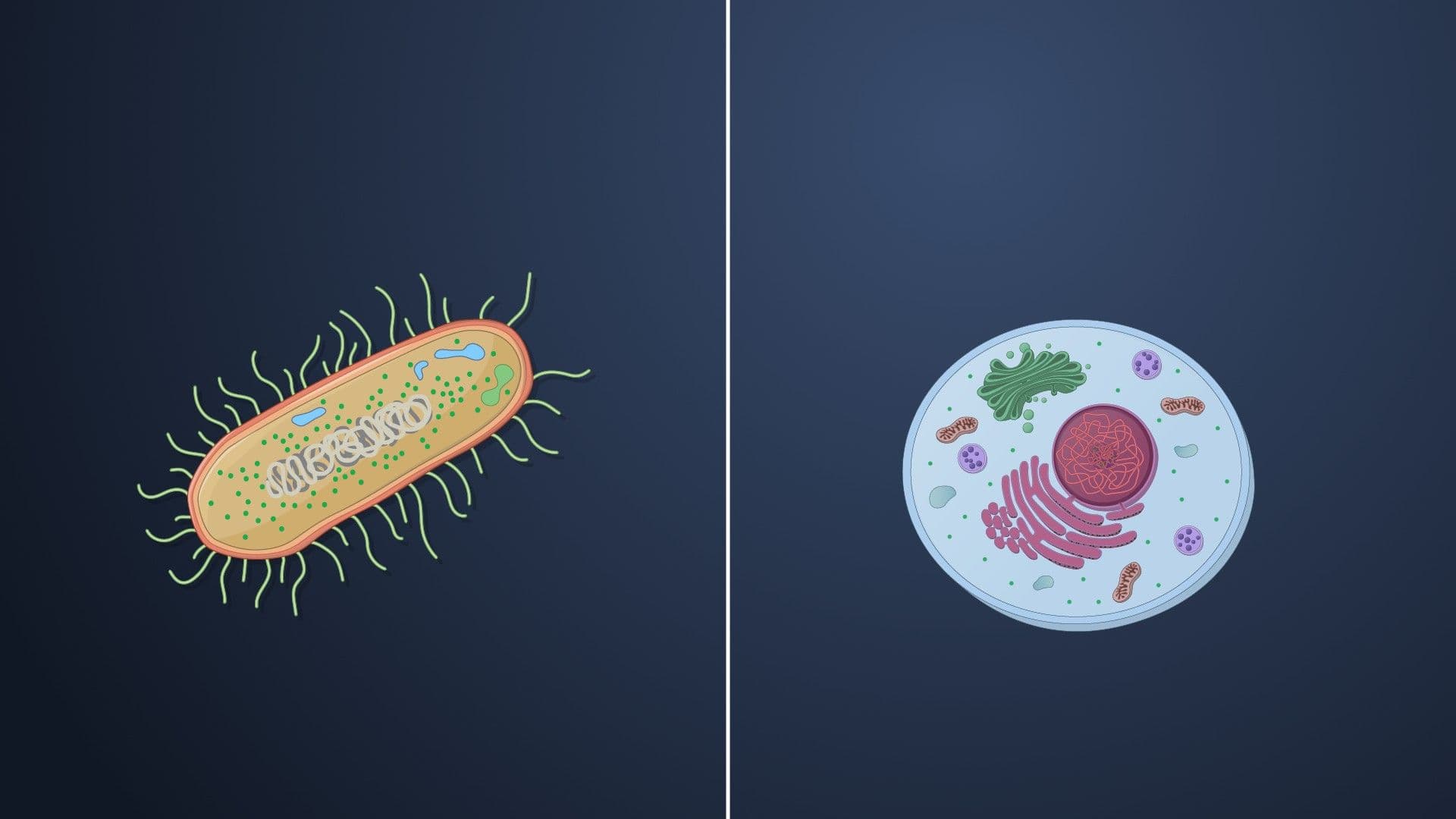
Microbial cell structure forms the foundation for understanding how bacteria and archaea survive in diverse environments. From the protective peptidoglycan layers in *Streptococcus* causing strep throat to the specialized flagella enabling *E. coli* motility in your intestines, prokaryotic cellular components directly impact human health and disease. JoVE Coach's comprehensive exploration reveals how these microscopic structures enable essential functions like antibiotic resistance, pathogen virulence, and environmental adaptation across American healthcare and research settings.
1. Prokaryotic vs. Eukaryotic Cell Architecture The fundamental distinction between prokaryotic and eukaryotic cells lies in organizational complexity and size. Prokaryotic cells like *E. coli* measure 0.1-5.0 micrometers and lack membrane-bound organelles, storing their circular DNA in the nucleoid region. Their 70S ribosomes enable rapid protein synthesis supporting quick reproduction through binary fission. This simplified structure allows pathogenic bacteria to rapidly colonize human hosts, as seen in urinary tract infections where *E. coli* can double every 20 minutes under optimal conditions.
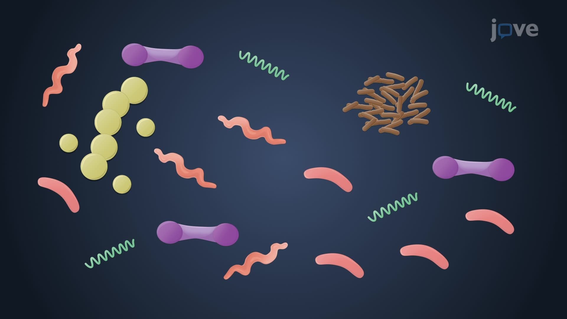
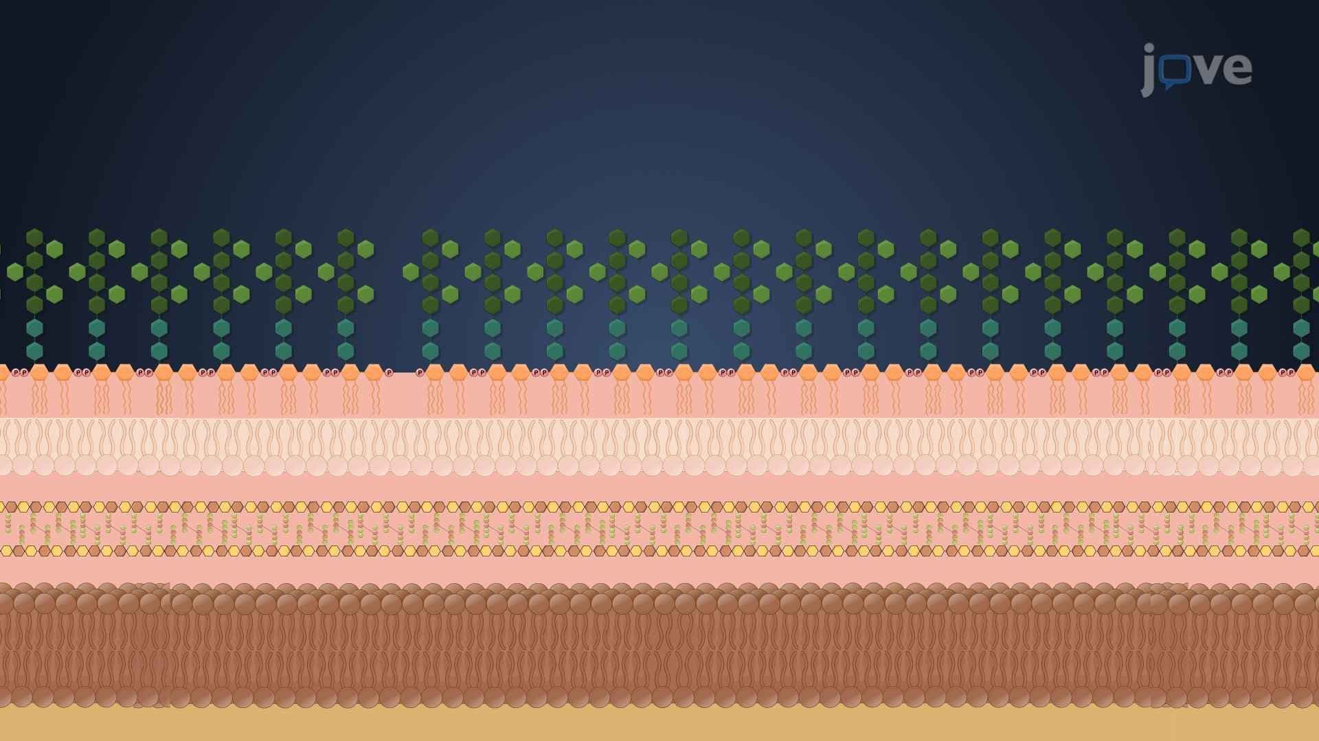
2. Bacterial Cell Wall Structure and Gram Staining The bacterial cell anatomy centers around peptidoglycan, a mesh-like polymer of N-acetylglucosamine and N-acetylmuramic acid cross-linked by amino acids. Gram-positive bacteria like *Staphylococcus epidermidis* possess thick peptidoglycan layers with embedded teichoic acids, retaining crystal violet in Gram staining and appearing purple. Gram-negative pathogens such as *Pseudomonas aeruginosa* have thinner peptidoglycan surrounded by lipopolysaccharide-containing outer membranes, appearing pink after counterstaining. This structural difference determines antibiotic susceptibility patterns crucial for clinical treatment decisions.
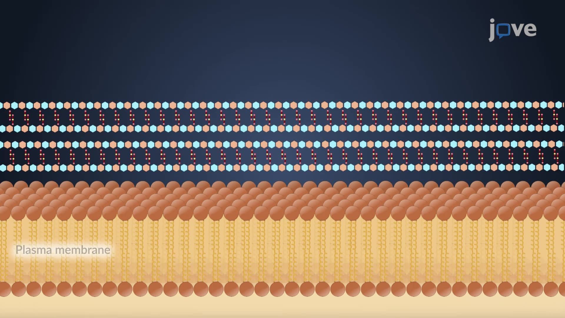
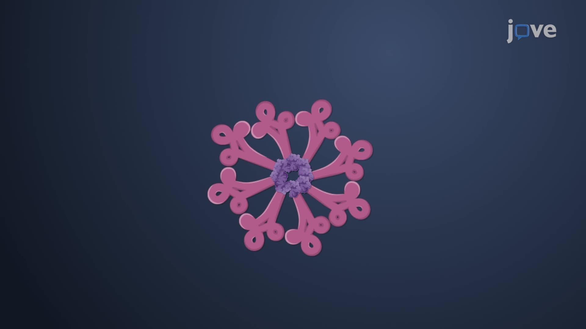
3. Archaeal Cell Wall Diversity and Extremophile Adaptations Archaeal cell walls completely lack peptidoglycan, instead featuring S-layer proteins that self-assemble into crystalline lattices. Species like *Methanosarcina* add polysaccharide layers of methanochondroitin for extra protection. Methanogens utilize pseudomurein containing N-acetyltalosaminuronic acid linked by β-1,3 bonds, making them resistant to lysozyme and penicillin. Some extreme thermophiles like *Ignicoccus* lack cell walls entirely, relying on specialized outer membranes for structural integrity in hydrothermal vents.
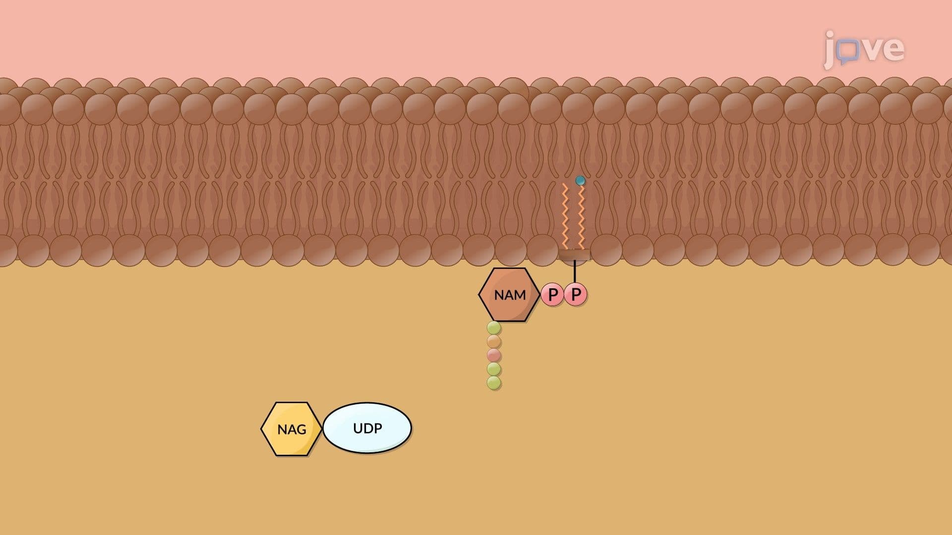
4. Peptidoglycan Synthesis Pathway and Antibiotic Targets Peptidoglycan synthesis occurs through three coordinated phases beginning in the cytoplasm where UDP-NAG converts to UDP-NAM-pentapeptide. The membrane phase creates Lipid II complexes that flip across to the periplasm via flippase enzymes. Periplasmic transpeptidases form final cross-links between glycan chains. This pathway provides multiple antibiotic targets: penicillin inhibits transpeptidases, vancomycin blocks pentapeptide binding, and bacitracin prevents bactoprenol recycling, explaining why understanding microbial cell structure is essential for antimicrobial therapy development.
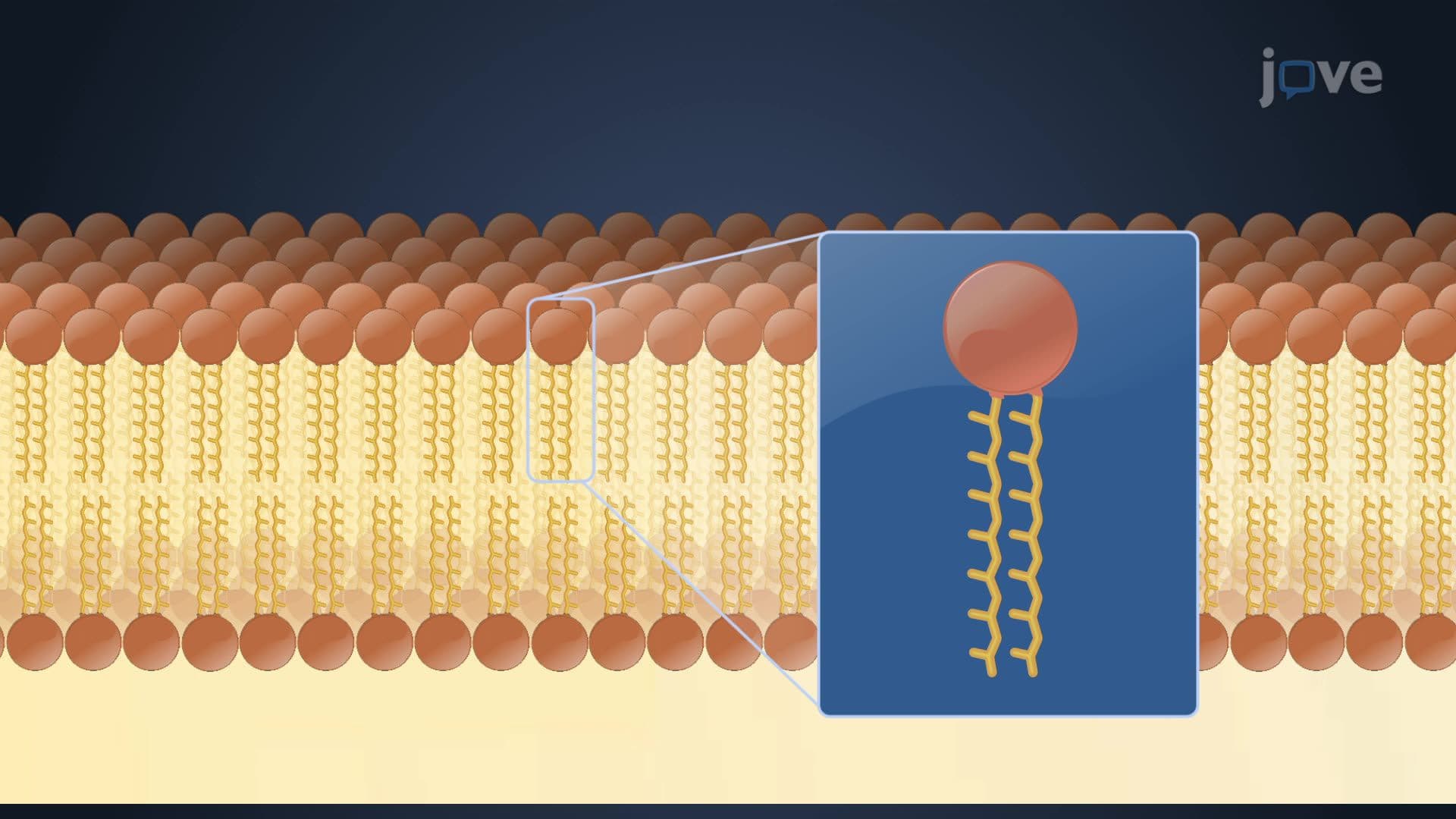
5. Plasma Membrane Composition and Transport Systems Bacterial membrane structures consist of phospholipid bilayers with fatty acids attached to glycerol through ester bonds, while archaeal membranes contain unique ether-linked isoprene derivatives. Archaeal glycerol diethers form bilayers, but diglycerol tetraethers create monolayers providing exceptional stability for hyperthermophiles. Both membrane types contain integral and peripheral proteins facilitating nutrient transport through passive diffusion and active transport systems, essential for maintaining cellular homeostasis in pathogens colonizing human hosts.
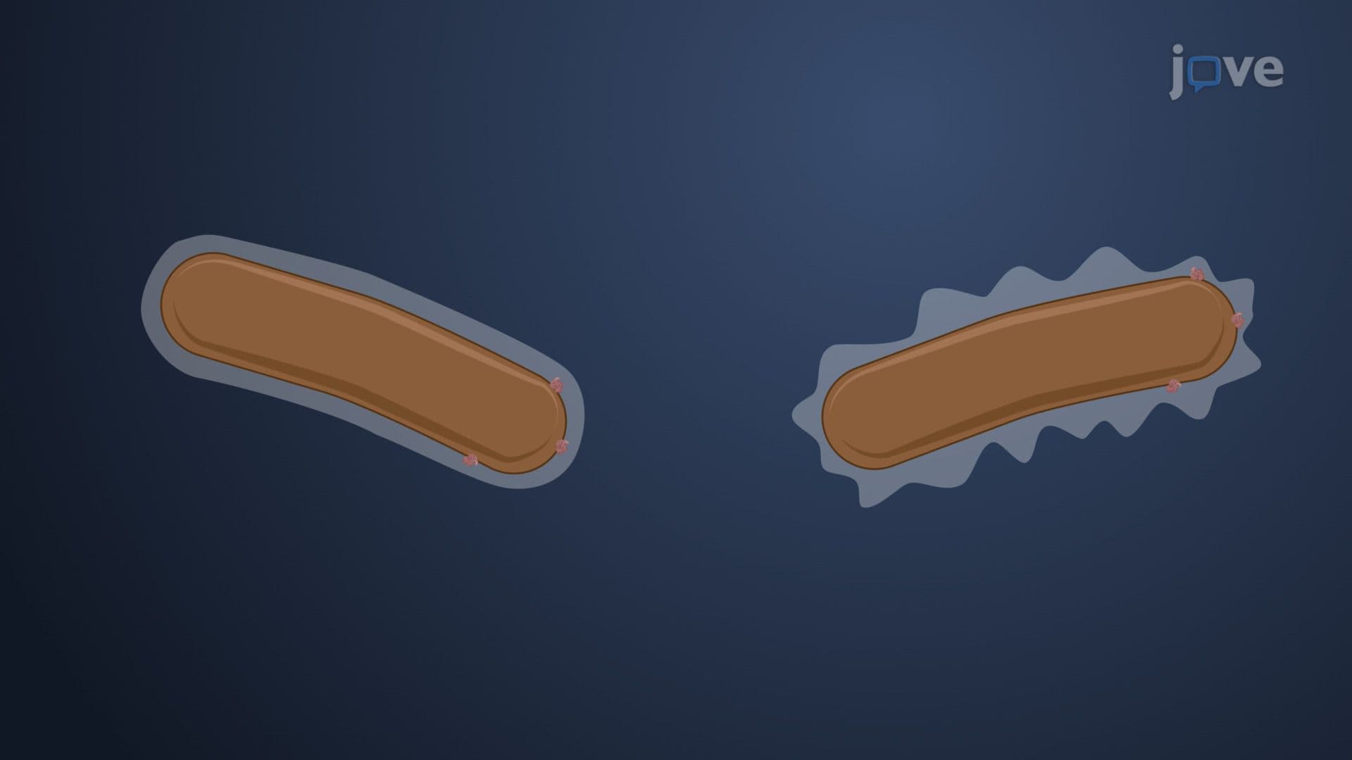
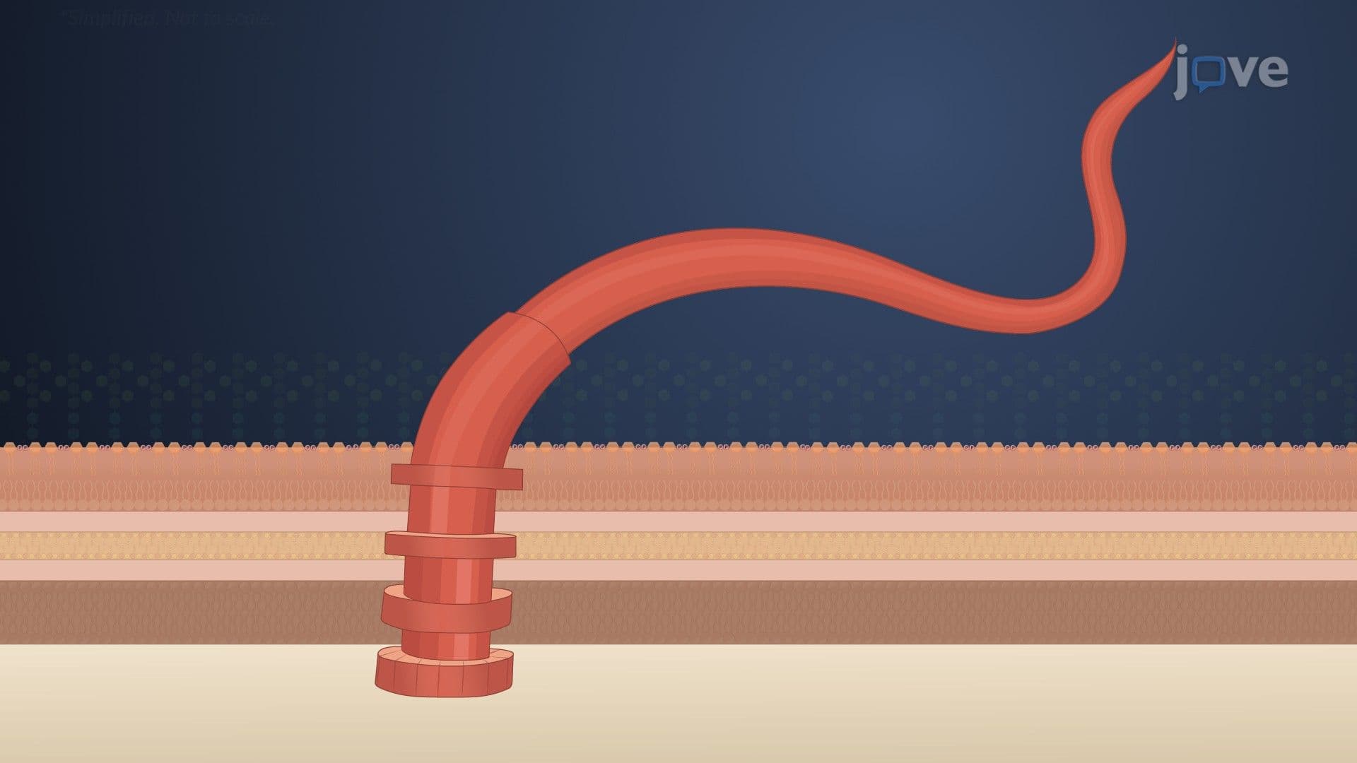
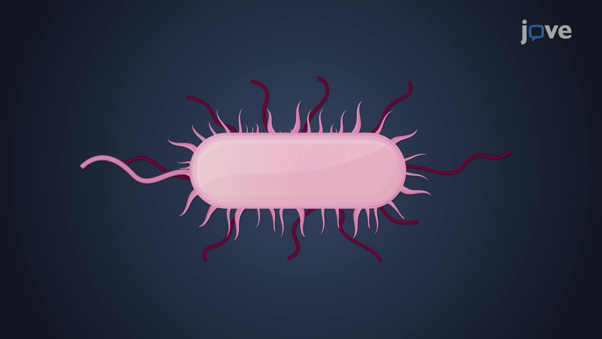
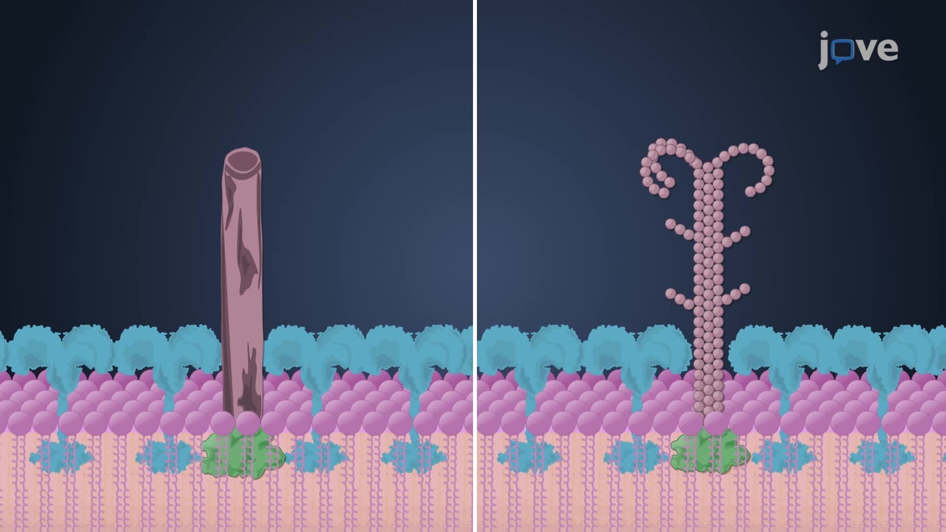
6. Surface Appendages and Bacterial Motility Flagella enable directional movement through rotating helical filaments powered by proton motive force. The basal body contains four rings in Gram-negative bacteria (*E. coli*) but only two in Gram-positive species (*Bacillus subtilis*). Fimbriae and pili facilitate surface adhesion and genetic exchange, with type IV pili enabling twitching motility in *Pseudomonas*. Sex pili transfer plasmids carrying antibiotic resistance genes between bacteria. Spirochetes like *Treponema pallidum* use axial filaments for corkscrew motion through viscous tissues, enabling syphilis transmission.
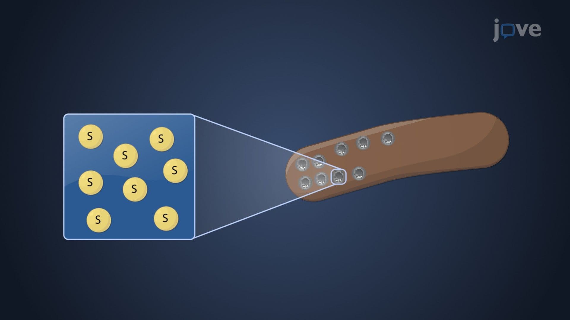
7. Cellular Inclusions and Storage Mechanisms Microbial organelles include specialized inclusion bodies storing carbon as poly-β-hydroxybutyric acid granules or glycogen. Metachromatic granules in *Corynebacterium diphtheriae* store polyphosphate for nucleic acid synthesis. Sulfur globules provide energy reserves for chemolithotrophic bacteria. Carboxysomes contain RuBisCO enzymes for CO₂ fixation in cyanobacteria. Gas vacuoles maintain buoyancy in aquatic microbes, while magnetosomes containing magnetite crystals help magnetotactic bacteria navigate toward optimal oxygen concentrations using Earth's magnetic field.
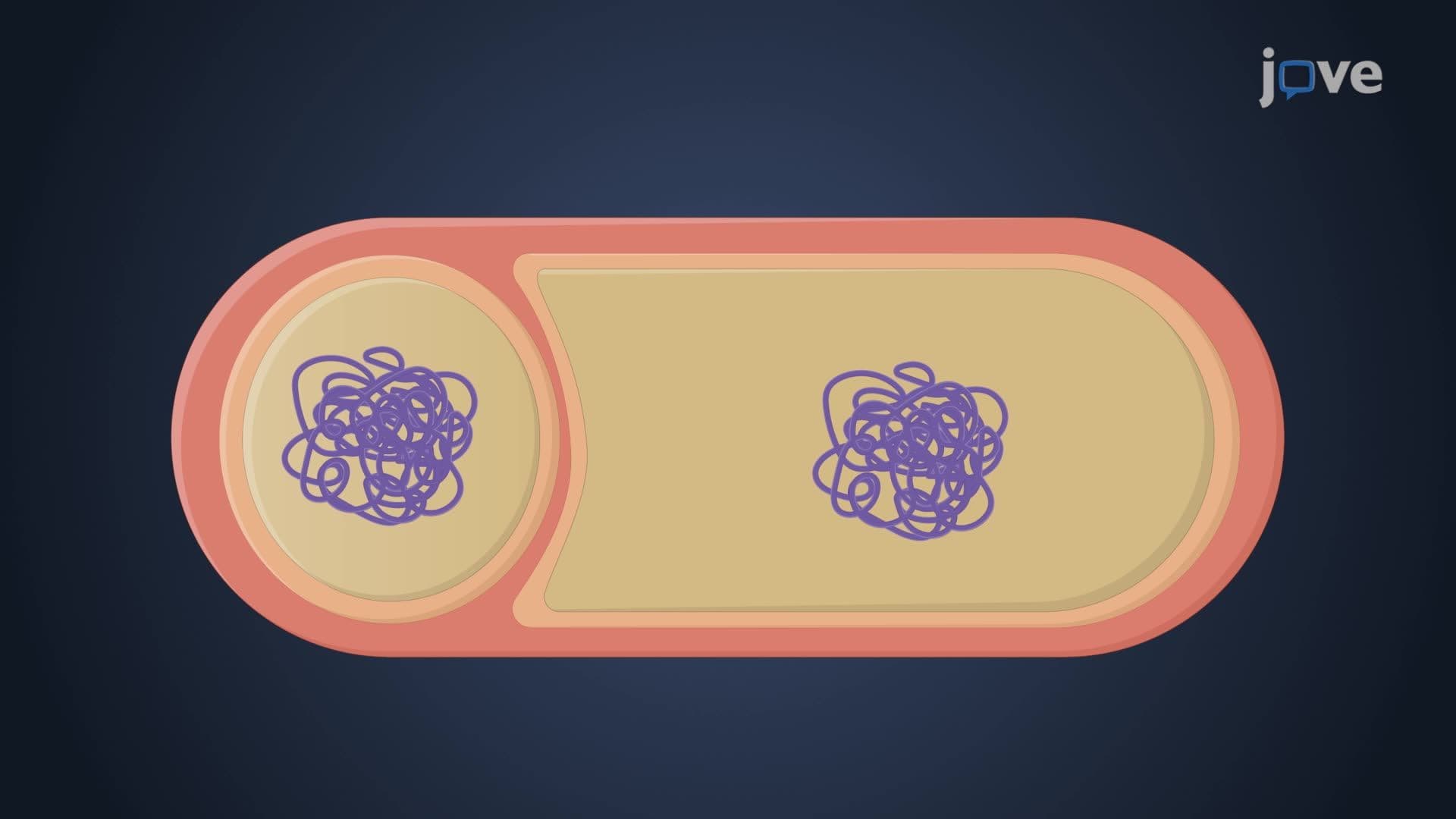
8. Endospore Formation and Clinical Significance Endospore formation represents the ultimate survival strategy for Gram-positive bacteria facing environmental stress. *Bacillus anthracis* spores can survive decades in soil, while *Clostridium difficile* spores resist alcohol-based sanitizers, contributing to healthcare-associated infections. The sporulation process involves asymmetric division, forespore engulfment, cortex formation, and coat protein deposition. Calcium-dipicolinic acid complexes dehydrate spores and protect DNA from heat damage. Understanding spore resistance mechanisms is crucial for hospital sterilization protocols and biodefense preparedness.