Loading subject page…

14,638 views
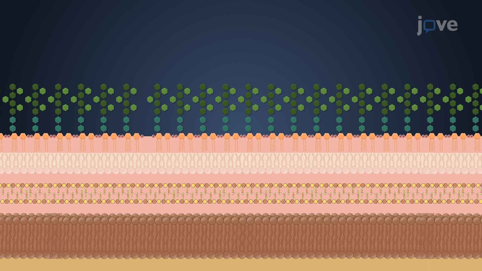

Video Summary: What Is Bacterial Cell Wall
Why do bacteria survive extreme conditions that would destroy human cells? The bacterial cell wall acts as molecular armor, providing structural integrity that allows these microscopic organisms to thrive everywhere from Yellowstone's hot springs to hospital surfaces. This rigid structure surrounding the plasma membrane determines how bacteria respond to antibiotics-explaining why MRSA infections in US hospitals resist certain treatments. Understanding the bacterial cell wall composition reveals the key differences between Gram-positive and Gram-negative bacteria. Watch the full video on JoVE Coach to master this concept with expert-led visuals and step-by-step explanations.
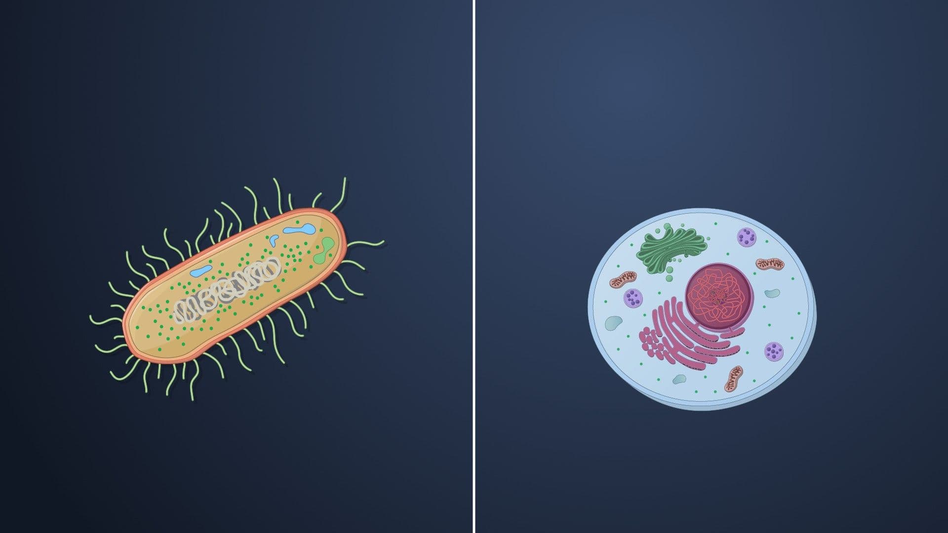
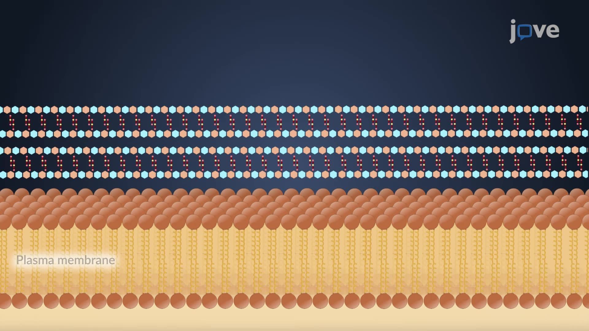
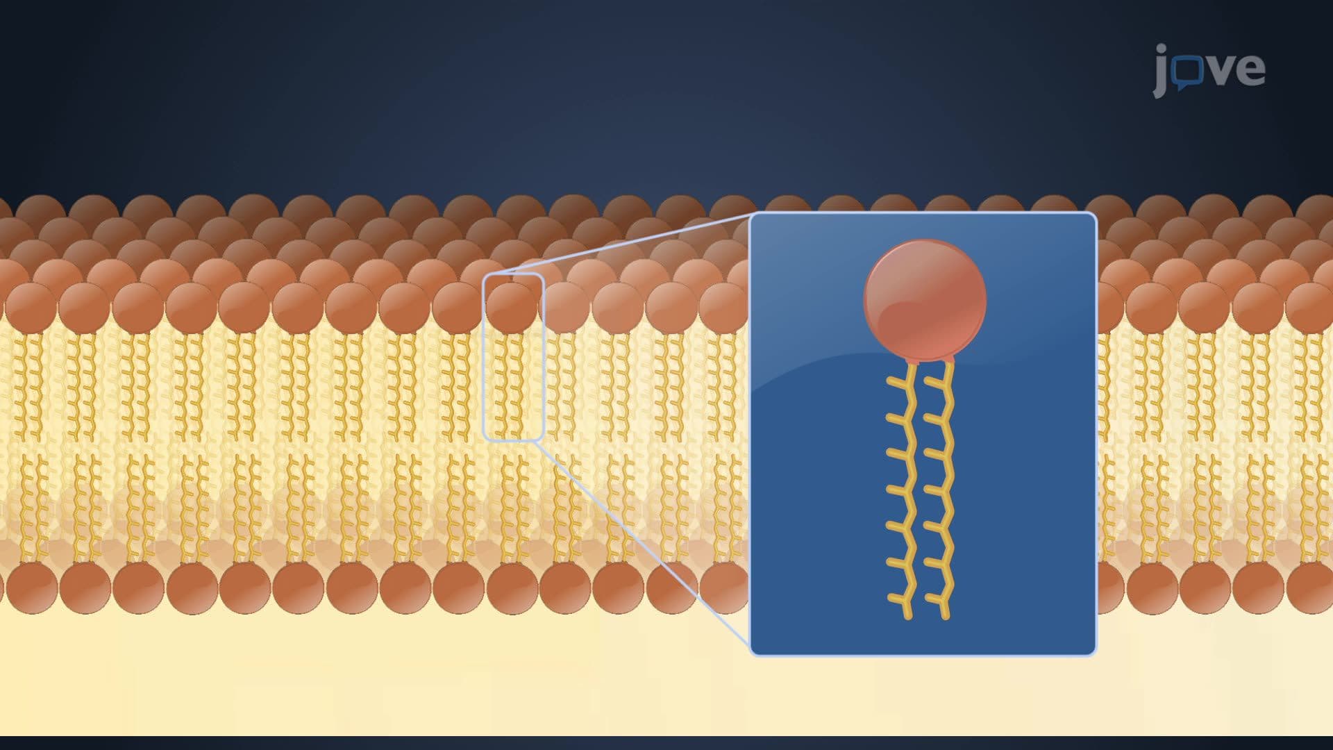
The bacterial cell wall represents one of biology's most sophisticated protective systems, functioning as a molecular fortress that maintains cellular integrity. This rigid structure encases the plasma membrane, providing essential shape maintenance and osmotic protection that allows bacteria to survive in diverse environments-from the acidic conditions in your stomach to the salt-rich surfaces of medical equipment.
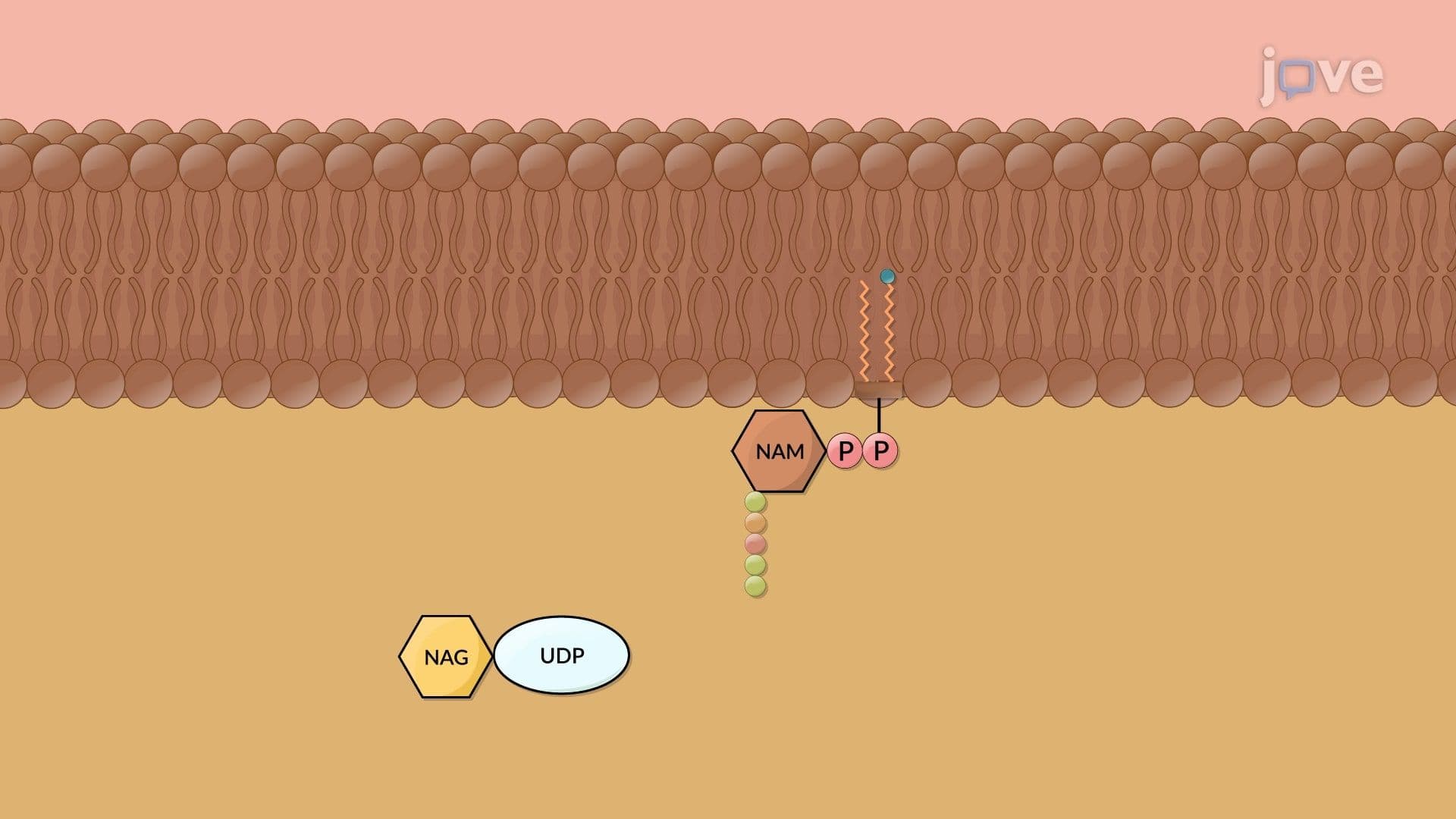
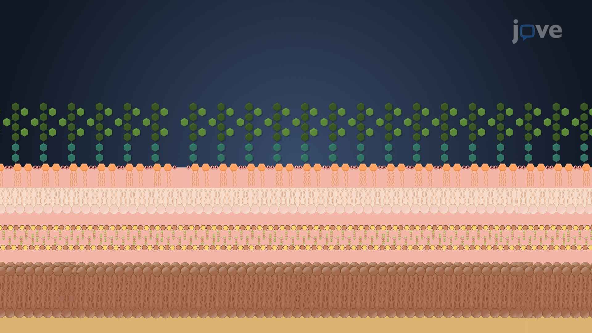
At the heart of every bacterial cell wall lies peptidoglycan, a unique polymer absent in human cells. This mesh-like structure consists of alternating sugar molecules-N-acetylglucosamine (NAG) and N-acetylmuramic acid (NAM)-cross-linked by short amino acid chains. Think of peptidoglycan as a flexible chain-link fence that can bend without breaking, allowing bacteria to expand during growth while maintaining structural integrity.
This peptidoglycan layer varies dramatically between bacterial types, creating the fundamental distinction that drives medical treatment decisions. For students preparing for the MCAT or AP Biology exams, understanding this structural variation proves crucial for predicting bacterial behavior and antibiotic effectiveness.
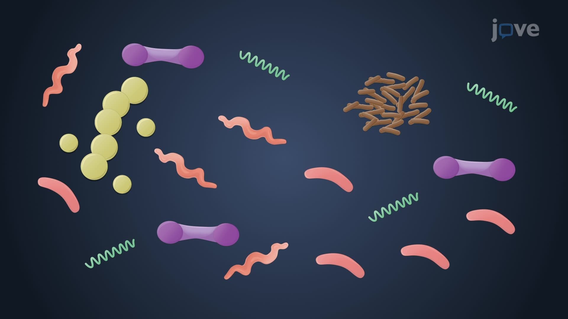
Gram-positive bacteria, including *Staphylococcus aureus* (responsible for staph infections in US hospitals), possess thick peptidoglycan layers comprising up to 90% of their cell wall. These bacteria also contain teichoic acids-unique polymers that facilitate metal ion uptake and provide additional protection against antimicrobial compounds. This robust structure explains why Gram-positive bacteria retain the purple crystal violet dye during Gram staining procedures.
Conversely, Gram-negative bacteria like *E. coli* feature thinner peptidoglycan layers sandwiched between two membranes. Their outer membrane contains lipopolysaccharides (LPS), molecules that trigger intense immune responses in humans-explaining why Gram-negative infections often cause severe inflammatory reactions. This complex structure makes Gram-negative bacteria inherently more resistant to many antibiotics, presenting ongoing challenges in clinical settings.
Understanding bacterial cell wall differences directly impacts treatment strategies in US healthcare systems. Penicillin effectively targets peptidoglycan synthesis, making it highly effective against Gram-positive infections but less useful against Gram-negative bacteria. This knowledge guides physicians in selecting appropriate antibiotics and helps explain the rise of multidrug-resistant organisms in hospital environments.
Related Micro-courses