Loading subject page…

- Anatomy and Physiology
- The Appendicular Skeleton
- Bones of the Upper Limb: Humerus
Micro-course: The Appendicular Skeleton
27,600 views
Learn key concepts and earn your certificate.

Bones of the Upper Limb: Humerus

00:00
01:19
Video Summary: What Is Bones Upper Limb Humerus
Ever wonder why baseball pitchers like Jacob deGrom can throw 100+ mph fastballs without their arm bones shattering? The secret lies in humerus bone anatomy – specifically how this remarkable upper arm bone transfers tremendous forces from shoulder to elbow. Understanding what bones upper limb humerus encompasses reveals how the humerus's unique head, shaft, and condyles work together to create both mobility and stability. From anchoring rotator cuff muscles to housing nerve pathways, this single bone orchestrates complex arm movements that power everything from tennis serves to surgical procedures. Watch the full video on JoVE Coach to master this concept with expert-led visuals and step-by-step explanations.
- Understand what bones upper limb humerus includes and identify the humerus as the sole long bone connecting shoulder to elbow
- Identify the three main regions of humerus bone anatomy: proximal head, middle shaft, and distal condyles
- Analyze how the humeral head and anatomical neck facilitate shoulder joint articulation with the scapula's glenoid cavity
- Recognize the functional roles of greater and lesser tubercles in anchoring rotator cuff muscles for shoulder stability
- Explore how the deltoid tuberosity serves as the critical attachment site for the deltoid muscle's powerful arm movements
- Compare the distal humerus condyles (trochlea and capitulum) and their specific articulations with ulna and radius bones
- Apply knowledge of radial groove anatomy to understand nerve pathway protection and potential injury sites
- Evaluate the clinical significance of medial and lateral epicondyles in common sports-related injuries like tennis elbow
What Bones Upper Limb Humerus: Structural Foundation
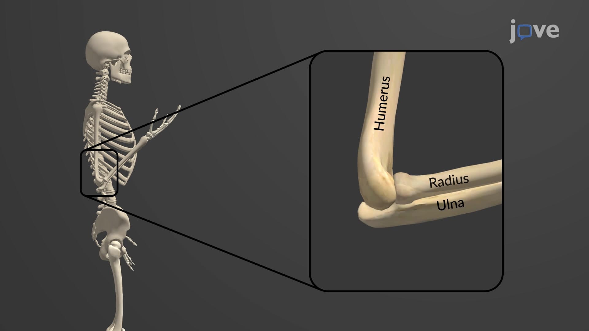
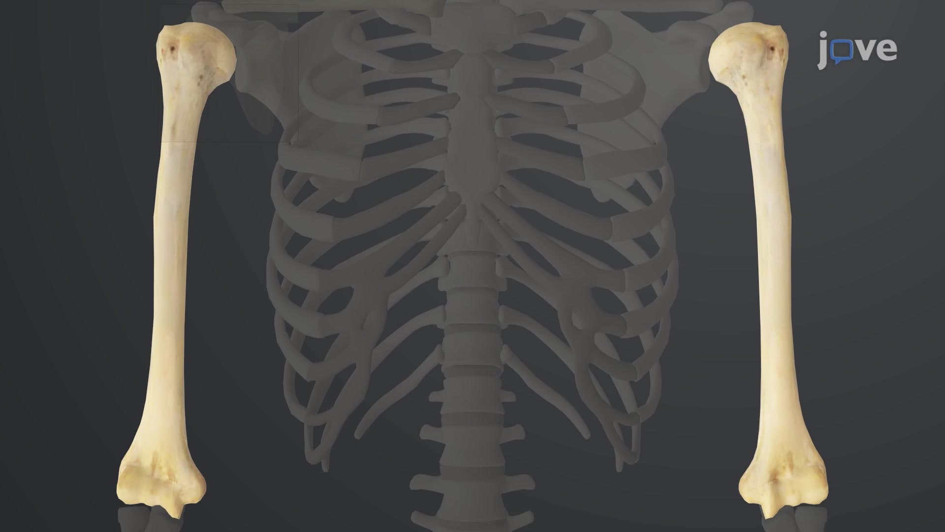
The humerus bone anatomy represents one of the most mechanically sophisticated structures in the human skeletal system. As the only long bone in the upper arm, the humerus must simultaneously provide structural support for lifting heavy objects while maintaining the flexibility needed for precise movements like writing or throwing. This 12-14 inch bone connects the highly mobile shoulder joint to the stable elbow joint, creating a biomechanical lever system that amplifies muscle forces.
Proximal Humerus: The Humeral Head and Anatomical Neck
The upper arm humerus explained begins with understanding its proximal end, where the spherical humeral head forms a ball-and-socket joint with the scapula. This smooth, rounded surface allows for the arm's remarkable 360-degree range of motion. The anatomical neck, a slight constriction below the head, marks the original growth plate location – crucial information for orthopedic surgeons treating fractures in young athletes.
The greater and lesser tubercles serve as anchor points for the rotator cuff muscles, which are essential for shoulder stability in overhead activities. Baseball pitchers, swimmers, and tennis players rely heavily on these muscle attachments. Between these tubercles runs the bicipital groove, housing the long head of the biceps tendon – a structure frequently injured in weightlifters and rock climbers.
Mid-Shaft Features: The Deltoid Tuberosity
The deltoid tuberosity humerus appears as a rough, triangular elevation on the lateral shaft, serving as the primary attachment site for the deltoid muscle. This powerful muscle, responsible for shoulder abduction, generates forces exceeding 500 pounds during heavy lifting. Understanding this anatomy helps explain why deltoid injuries can severely limit arm function and why proper warm-up is crucial before athletic activities.
Distal Humerus: Condyles and Clinical Significance
The distal humerus condyles form the elbow joint's upper portion, with the trochlea articulating with the ulna and the capitulum connecting to the radius. The medial and lateral epicondyles serve as attachment sites for forearm muscles controlling wrist and finger movements. "Tennis elbow" (lateral epicondylitis) affects the lateral epicondyle, while "golfer's elbow" involves the medial epicondyle – conditions frequently seen in emergency departments across the United States.
For students preparing for the MCAT or AP Biology exams, understanding these anatomical features of the humerus bone provides essential foundation knowledge for questions about lever systems, joint mechanics, and injury mechanisms that appear regularly on standardized tests.
Frequently Asked Questions
The humerus is the single long bone of the upper arm, extending from shoulder to elbow and serving as the primary structural support for arm function. It's crucial because it connects the highly mobile shoulder joint to the stable elbow joint, creating a lever system that amplifies muscle forces for activities ranging from lifting groceries to throwing baseballs. Without the humerus, complex arm movements requiring both strength and precision would be impossible.
MCAT questions frequently test humerus anatomy through lever mechanics, joint classifications, and muscle attachment sites. Focus on understanding how the humeral head forms a ball-and-socket joint, how tubercles anchor rotator cuff muscles, and how condyles create hinge joints at the elbow. Practice identifying bone markings on diagrams and connecting anatomical features to their functional roles in movement and injury patterns.
Essential features include the humeral head (shoulder articulation), greater and lesser tubercles (rotator cuff attachments), deltoid tuberosity (deltoid muscle insertion), radial groove (nerve pathway), and distal condyles including trochlea and capitulum (elbow joint formation). Understanding how these structures relate to muscle attachments and joint mechanics is crucial for AP exam success, particularly in questions about skeletal system function and biomechanics.
Healthcare professionals use humerus anatomy knowledge daily when treating fractures, dislocations, and sports injuries. For example, knowing that the radial nerve runs in the radial groove helps doctors assess potential nerve damage in mid-shaft fractures. Understanding tubercle locations guides physical therapists in rotator cuff rehabilitation, while condyle anatomy is essential for emergency physicians evaluating elbow injuries in young athletes.
Not at all – humerus anatomy follows logical patterns that make sense when connected to everyday movements. Start by relating bone features to familiar activities like throwing a ball or lifting weights. The bone's structure directly reflects its function, so understanding why each feature exists makes memorization much easier. Most students successfully master this concept within a few focused study sessions using visual aids and hands-on models.
Use the "function-first" approach by learning what each marking does before memorizing its location. For example, understand that the deltoid tuberosity anchors the muscle that lifts your arm sideways, then locate it on models or diagrams. Create flashcards pairing bone features with their muscle attachments and movements. Practice with unlabeled diagrams and time yourself identifying structures to simulate exam conditions.
Epicondyle injuries represent some of the most common overuse conditions in sports medicine, affecting millions of Americans annually. Tennis elbow (lateral epicondylitis) and golfer's elbow (medial epicondylitis) require understanding of forearm muscle attachments to these bony prominences. This knowledge directly applies to careers in physical therapy, athletic training, orthopedic medicine, and sports rehabilitation, making it valuable beyond academic requirements.
Build on this foundation by studying scapular anatomy and the shoulder joint complex, then progress to forearm bones (radius and ulna) and elbow joint mechanics. Understanding muscle origins and insertions on the humerus leads naturally to rotator cuff anatomy and common shoulder pathologies. This progression prepares students for advanced topics in biomechanics, physical therapy, and orthopedic medicine.
Related Micro-courses